D3.3 Homeostasis
Explain how negative feedback keeps blood glucose, body temperature and (HL) the water content of the blood within narrow limits, and how diabetes disrupts it.
- IB Biology
- Part of D Continuity and change
- SL and HL
Key terms introduced here
Each is in the Biology glossary, with every other term in the subject.
- Antidiuretic hormone
A hormone made in the hypothalamus and secreted by the pituitary gland when the blood is too concentrated; it causes aquaporins to be inserted into collecting duct cells so more water is reabsorbed.
- Bowman's capsule
The cup-shaped start of a nephron that surrounds the glomerulus; its inner wall of podocytes allows filtrate to pass into the tubule during ultrafiltration.
- Brown adipose tissue
Fat tissue whose cells contain many mitochondria with thermogenin, specialised for producing heat by uncoupled respiration; abundant in newborn babies and present in the neck of many adults.
- Collecting duct
The tube that receives filtrate from many nephrons and carries it through the medulla, where water is reabsorbed by osmosis to an extent controlled by ADH.
- Filtrate
The fluid formed in Bowman's capsule by ultrafiltration, containing water, glucose, amino acids, ions and urea at about plasma concentrations but no blood cells or plasma proteins.
- Glomerulus
A knot of capillaries inside Bowman's capsule, supplied by a wide afferent arteriole and drained by a narrower efferent arteriole, where blood plasma is filtered under high pressure.
- Glucagon
A hormone secreted by the alpha cells of the islets of Langerhans when blood glucose concentration falls; it stimulates liver cells to break down glycogen to glucose and release it into the blood.
- Glycogenesis
The synthesis of glycogen from glucose, carried out in liver and muscle cells and stimulated by insulin.
- Glycogenolysis
The hydrolysis of glycogen to glucose, carried out in liver cells when stimulated by glucagon (and in muscle cells for their own use).
- Homeostasis
The maintenance of the internal environment of an organism within preset limits, despite fluctuations in the external environment.
- Insulin resistance
Reduced sensitivity of target cells, such as liver, muscle and adipose cells, to insulin, so they take up less glucose; the physiological change underlying type 2 diabetes.
- Islets of Langerhans
Groups of endocrine cells in the pancreas; their beta cells secrete insulin and their alpha cells secrete glucagon in response to changes in blood glucose concentration.
- Kidney
One of a pair of organs that excrete metabolic waste such as urea and regulate the osmotic concentration of the blood by forming urine in about a million nephrons each.
- Loop of Henle
The hairpin part of the nephron in the medulla; its water-impermeable ascending limb actively transports sodium ions out, making the medulla concentrated so water can be reabsorbed from collecting ducts.
- Nephron
The functional unit of the kidney: a tubule consisting of Bowman's capsule, proximal convoluted tubule, loop of Henle and distal convoluted tubule, draining into a collecting duct.
- Osmoreceptor
A receptor cell in the hypothalamus that detects the osmotic concentration of the blood and causes ADH secretion to change.
- Osmoregulation
The control of the osmotic concentration (water and solute content) of the blood and other body fluids; in mammals, mainly by the kidneys.
- Proximal convoluted tubule
The coiled part of the nephron after Bowman's capsule, lined with cells with microvilli and many mitochondria, where all glucose and amino acids and most ions and water are reabsorbed.
- Selective reabsorption
The reabsorption of useful substances, such as glucose, amino acids, ions and water, from the filtrate back into the blood, mainly in the proximal convoluted tubule, while waste remains in the filtrate.
- Set point
The value of a regulated variable that homeostasis maintains, such as a core body temperature of about 37 °C; the variable fluctuates within a narrow range around it.
- Shivering
Rapid, involuntary contraction and relaxation of skeletal muscles, stimulated by the hypothalamus when the body is cold; the respiration supplying ATP for contraction releases heat.
- Sweating
The secretion of a watery salt solution by sweat glands onto the skin surface; evaporation of the water removes heat from the skin and cools the body.
- Thermoreceptor
A sensory receptor that detects temperature; peripheral thermoreceptors in the skin and central thermoreceptors in the hypothalamus send information to the hypothalamus.
- Thermoregulation
The maintenance of body temperature within narrow limits by negative feedback, balancing heat gained from metabolism and the surroundings with heat lost to the surroundings.
- Thyroxine
An iodine-containing hormone secreted by the thyroid gland, under control of the hypothalamus and pituitary gland, that raises the metabolic rate of body cells and so increases heat production.
- Type 1 diabetes
A form of diabetes in which the beta cells of the islets of Langerhans are destroyed by the body's own immune system, so little or no insulin is secreted.
- Type 2 diabetes
A form of diabetes in which target cells become less sensitive to insulin (insulin resistance), so blood glucose is not lowered effectively even though insulin is secreted.
- Ultrafiltration
The filtration of blood plasma under high pressure in the glomerulus into Bowman's capsule, which removes water and small solutes but retains blood cells and plasma proteins.
- Uncoupled respiration
Cell respiration in which the energy released is given out as heat instead of being used to make ATP, because protons flow back across the inner mitochondrial membrane through thermogenin instead of ATP synthase.
- Urea
The main nitrogenous waste product of mammals, made in the liver from the excess amino groups of amino acids and excreted by the kidneys in urine.
- Vasoconstriction
The narrowing of arterioles when the smooth muscle in their walls contracts, reducing blood flow through the capillaries they supply; in the skin it reduces heat loss.
- Vasodilation
The widening of arterioles when the smooth muscle in their walls relaxes, increasing blood flow through the capillaries they supply; in the skin it increases heat loss.
Notes on D3.3 Homeostasis
Homeostasis is the maintenance of a stable internal environment in the body, despite changes in the conditions outside it. Every cell in the human body depends on it: enzymes, membranes and the transport of substances all work properly only within narrow ranges of temperature, pH, glucose concentration and water content.
The same principle, negative feedback, regulates each of these variables. Blood glucose is controlled mainly by hormones, body temperature by a combination of nerves and hormones, and (at HL) the water content of the blood by the kidneys. Each one shows how receptors, a control centre and effectors work together across several organ systems.
Homeostasis and the internal environment
The internal environment of a multicellular organism is the fluid that surrounds its cells: tissue fluid, which forms from the blood plasma that flows through the capillaries. Cells exchange substances with tissue fluid all the time, taking in oxygen, glucose and ions and releasing carbon dioxide, urea and heat. If the composition or temperature of this fluid changed as much as the external environment does, cells could not function.
Organisms therefore keep the conditions of their internal environment within narrow limits. A person can move from a room at 20 °C into snow at −10 °C, or eat a meal rich in sugar after a night without food, and the temperature and glucose concentration of the blood change only slightly. The process that achieves this is homeostasis.
Set points and normal ranges
Each regulated variable has a set point, the value around which it is kept. The variable is never held exactly at this value. It fluctuates slightly above and below it, within a normal range, because corrections only begin once a change has been detected. Homeostasis keeps these fluctuations small and stops them growing.
Four variables regulated by homeostasis in humans are especially important. The table gives the normal value or range for each and the reason it must be controlled.
Variable | Normal value in a healthy adult | Why it must be kept within limits |
|---|---|---|
Core body temperature | About 37 °C | Enzyme activity depends on temperature; too low slows metabolism, too high denatures enzymes and other proteins. |
Blood pH | 7.35–7.45 | pH affects the charges on amino acid side chains, so it changes the shape of enzymes and other proteins. |
Blood glucose concentration | About 4–6 mmol L⁻¹ (fasting) | Glucose is the main respiratory substrate for brain cells; too much lowers the water potential of the blood and damages blood vessels. |
Osmotic concentration of the blood | About 0.3 osmol L⁻¹ | Controls the movement of water into and out of cells by osmosis, and so cell volume. |
Other variables are also kept stable by homeostasis, including the concentrations of oxygen, carbon dioxide, sodium ions and calcium ions in the blood, and blood pressure. The same principles apply to all of them.
Why maintaining constant conditions benefits organisms
Keeping the internal environment stable allows cells to work at a steady, predictable rate. Enzymes catalyse reactions fastest near their optimum temperature and pH, so a stable body temperature and blood pH keep metabolism running efficiently. A stable glucose supply means that the brain, which uses glucose as its main respiratory substrate, never runs short of energy. A stable osmotic concentration prevents cells from swelling or shrinking as water moves by osmosis.
Homeostasis also gives organisms independence from their surroundings. Mammals and birds, which keep their body temperature high and constant, remain active on cold nights and in cold climates, when the body temperature and activity of a lizard fall with the air temperature. Maintaining a high, constant body temperature costs energy, because it needs a high metabolic rate and therefore a large intake of food. This is an example of interaction and interdependence between organ systems and the environment, because the body must continually exchange energy and materials with its surroundings to stay the same inside.
Negative feedback in homeostasis
All homeostatic control systems use negative feedback. A change in a variable away from its set point triggers responses that reverse the change and return the variable towards the set point. The feedback is called negative because the response is in the opposite direction to the change: a rise causes a fall, and a fall causes a rise.
Receptors, control centre and effectors
A negative feedback loop has three components, connected by nerves or hormones:
Receptors detect the value of the variable and any change in it. Thermoreceptors in the skin detect temperature; beta cells in the pancreas detect blood glucose concentration.
A control centre receives information from the receptors, compares the value with the set point, and sends signals to effectors. In many homeostatic systems in humans, the control centre is the hypothalamus in the brain.
Effectors are the muscles or glands that carry out the response. Sweat glands, skeletal muscles and the liver are examples.
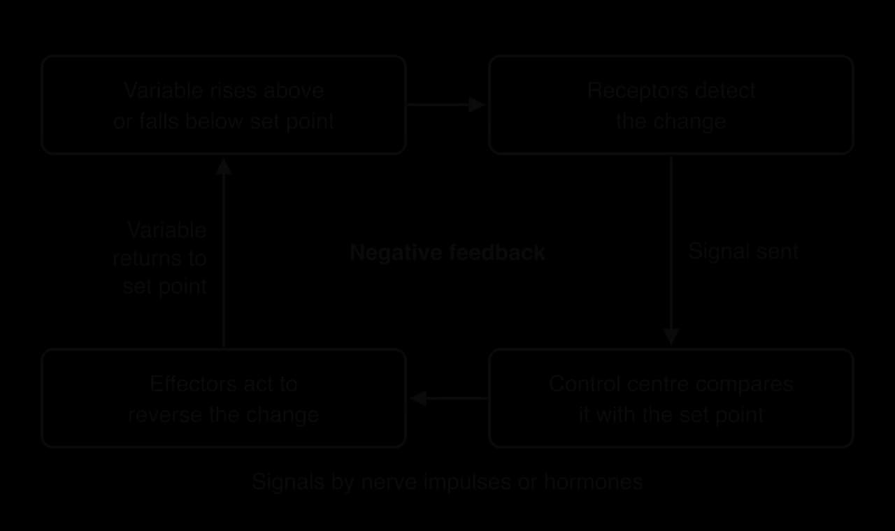
The diagram of the negative feedback loop shows one cycle. As soon as the variable returns towards the set point, the receptors detect the smaller deviation and the control centre reduces the signals to the effectors. The response therefore switches itself off as the variable is corrected. Negative feedback works in both directions: a rise above the set point and a fall below it are corrected, usually by different effectors or by the same effector working in opposite ways.
Why homeostasis uses negative rather than positive feedback
In positive feedback, a change in a variable triggers responses that increase the change further. Each response amplifies the next, so the variable moves further and further from its starting value until some outside event ends the cycle. Positive feedback is useful for processes that must be completed quickly once they have started, such as the contractions of the uterus during childbirth, which are strengthened by the hormone oxytocin until the baby is born (D3.1 Reproduction).
Positive feedback cannot maintain a stable internal environment, because it moves a variable away from its set point instead of back towards it. A body temperature controlled by positive feedback would rise faster and faster once it started to rise, which is why every homeostatic system uses negative feedback.
Feature | Negative feedback | Positive feedback |
|---|---|---|
Effect of the response on the change | Reverses it | Increases it |
Effect on the variable | Returned towards the set point | Moves further from its starting value |
Stability | Keeps conditions stable | Destabilising; ended by an outside event |
Examples | Body temperature, blood glucose, blood osmotic concentration | Uterine contractions in childbirth, ripening of fruit by ethylene |
 YouTube
Homeostatic systems often have more than one effector working in opposite directions, so that a variable can be pushed back from either side. Blood glucose has insulin to lower it and glucagon to raise it. This arrangement gives finer control than a single effector that can only be switched on or off.
Regulation of blood glucose by hormones
Blood glucose concentration is controlled by two hormones, insulin and glucagon, secreted by the pancreas. Hormones are chemical messengers secreted by endocrine glands directly into the blood. They travel in the blood plasma to every part of the body but only affect target cells, which have receptors for that hormone. Blood glucose regulation is the clearest example of hormones working in homeostasis.
Why blood glucose must be controlled
Glucose enters the blood from the small intestine after a meal, so the concentration rises sharply after eating carbohydrates. Between meals, during fasting overnight and during exercise, cells keep using glucose for cell respiration, so the concentration falls. Both extremes are harmful:
Too low (hypoglycaemia). Brain cells depend on a continuous supply of glucose. Low blood glucose causes confusion, loss of consciousness and, if severe, death.
Too high (hyperglycaemia). Excess glucose lowers the water potential of the blood, so water leaves cells by osmosis. Over months and years, high blood glucose damages blood vessels and nerves, especially in the eyes, kidneys and feet.
The pancreas and the islets of Langerhans
Most of the pancreas is exocrine tissue that secretes digestive enzymes into the small intestine through a duct. Scattered through it are small groups of endocrine cells called the islets of Langerhans. The islets contain two main types of cell involved in glucose control:
Beta (β) cells secrete insulin when blood glucose concentration rises above the set point.
Alpha (α) cells secrete glucagon when blood glucose concentration falls below the set point.
The alpha and beta cells are both the receptors and the control centre in this system: they detect the glucose concentration of the blood flowing through the islets and respond by changing their rate of hormone secretion. The islets have a dense network of capillaries, so the hormones pass straight into the blood and are carried throughout the body.
When blood glucose rises: insulin
After a meal, glucose absorbed from the small intestine raises the blood glucose concentration. Beta cells detect the rise and increase their secretion of insulin, while alpha cells reduce their secretion of glucagon. Insulin travels in the blood and binds to insulin receptors on its target cells. The main target cells are liver cells, skeletal muscle cells and adipose (fat storage) cells. Insulin causes:
Increased uptake of glucose. In muscle and adipose cells, insulin causes more glucose transporter proteins to be inserted into the plasma membrane, so glucose enters the cells faster by facilitated diffusion.
Glycogenesis. Liver and muscle cells convert glucose to glycogen, a branched polysaccharide of α-glucose, and store it in granules in the cytoplasm.
More use of glucose. Cells increase their rate of cell respiration, using more glucose.
Conversion of glucose to fat. Adipose cells and liver cells convert excess glucose to triglycerides.
Each of these effects removes glucose from the blood, so the concentration falls back towards the set point. As it falls, beta cells secrete less insulin, and the response switches off by negative feedback.
When blood glucose falls: glucagon
During fasting or prolonged exercise, the blood glucose concentration falls. Alpha cells detect this and secrete more glucagon, while beta cells secrete less insulin. Glucagon binds to receptors on liver cells and stimulates glycogenolysis, the hydrolysis of glycogen to glucose. Liver cells release this glucose into the blood, so the concentration rises back towards the set point. Glucagon also stimulates the liver to make new glucose from other substances, such as amino acids.
Muscle cells store glycogen too, but they use it only for their own respiration and do not release glucose into the blood. Only liver cells can supply glucose to the rest of the body, which makes the liver the main effector organ when blood glucose is low.
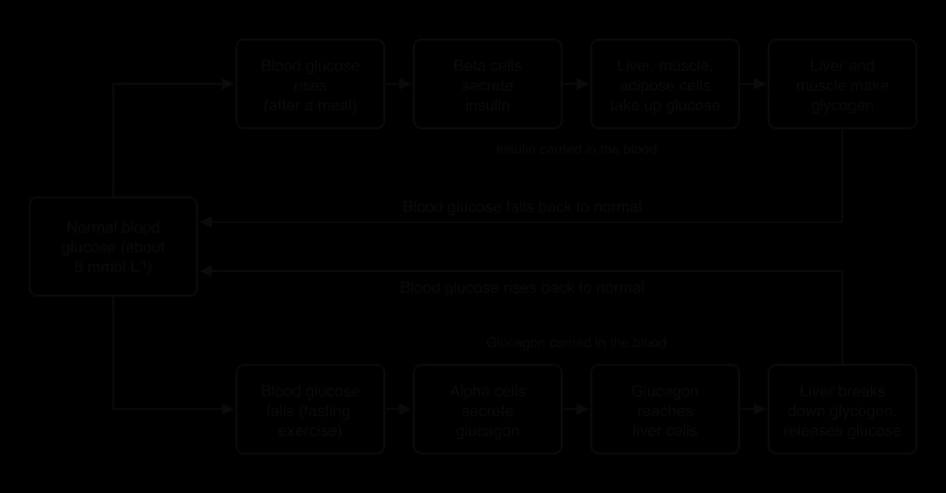
The flow chart of blood glucose control shows the two halves of the system. Each hormone is secreted in response to a change in one direction and causes a change in the opposite direction, which is negative feedback. Because both hormones are secreted at some rate all the time, it is the balance between them that sets the blood glucose concentration at any moment.
Feature | Insulin | Glucagon |
|---|---|---|
Secreted by | Beta cells of the islets of Langerhans | Alpha cells of the islets of Langerhans |
Stimulus for secretion | Rise in blood glucose | Fall in blood glucose |
Main target cells | Liver, skeletal muscle and adipose cells | Liver cells |
Effect on glycogen | Glycogenesis (glucose → glycogen) | Glycogenolysis (glycogen → glucose) |
Effect on glucose uptake by cells | Increases | No increase |
Effect on blood glucose | Lowers it | Raises it |
The glucose tolerance test
A glucose tolerance test shows how well the body controls blood glucose. After fasting overnight, a person drinks a solution containing 75 g of glucose, and the blood glucose concentration is measured at intervals over two or three hours. In a healthy person, insulin secretion rises quickly and the concentration returns to near the fasting value within about two hours. In a person with diabetes, the concentration starts higher, rises further and stays high for longer.
- Healthy person
- Person with diabetes
In the graph of glucose tolerance test results, the healthy curve peaks within about half an hour and falls back by two hours, because the beta cells respond quickly and the target cells respond to insulin. Diabetes is diagnosed when the concentration two hours after the drink is 11.1 mmol L⁻¹ or higher, or when the fasting concentration is 7.0 mmol L⁻¹ or higher.
Type 1 and type 2 diabetes
Diabetes mellitus is a condition in which blood glucose concentration is not controlled and remains too high for long periods. There are two main types, which have different physiological causes. In both, glucose stays in the blood instead of entering cells, and when the concentration in the blood is very high, glucose appears in the urine because the kidneys cannot reabsorb all of it.
Type 1 diabetes: loss of insulin production
Type 1 diabetes is an autoimmune disease. The immune system produces T lymphocytes that attack and destroy the body's own beta cells. As beta cells are lost, insulin secretion falls until little or none is produced. Without insulin, muscle and adipose cells take up little glucose and liver and muscle cells do not convert it to glycogen, so blood glucose rises after every meal and stays high.
Type 1 diabetes usually begins in childhood or early adulthood, often quite suddenly. Typical symptoms are thirst, passing large volumes of urine, tiredness and weight loss, as cells cannot use glucose and the body breaks down fat and protein for energy. It cannot be prevented, because it is not caused by lifestyle; there is a genetic predisposition, and an environmental trigger such as a viral infection may start the autoimmune response.
Treating type 1 diabetes
Type 1 diabetes is treated by replacing the missing hormone. People with the condition inject insulin, or receive it continuously from an insulin pump, and match their doses to what they eat and how active they are. They monitor their blood glucose with finger-prick tests or with a continuous glucose monitor, a sensor under the skin. Insulin must be injected rather than swallowed because it is a protein and would be digested in the stomach and small intestine.
Before insulin was available, type 1 diabetes killed most patients within months or a few years of diagnosis. Frederick Banting and Charles Best extracted insulin from dog pancreases at the University of Toronto in 1921. In January 1922 Leonard Thompson, a 14-year-old boy close to death from diabetes, became the first person to be treated with an insulin injection; his blood glucose fell and he lived for another 13 years. Banting and John Macleod received the Nobel Prize in Physiology or Medicine in 1923.
Type 2 diabetes: reduced response to insulin
In type 2 diabetes, the pancreas still secretes insulin, at least in the early stages, but the target cells respond to it less. This reduced sensitivity of target cells to insulin is called insulin resistance. Fewer insulin receptors, or faults in the signalling inside cells after insulin binds, mean that liver, muscle and adipose cells take up less glucose and the liver keeps releasing glucose even when the blood concentration is high.
At first the beta cells compensate by secreting more insulin. Over years, they may become exhausted and secrete less, so some people with long-standing type 2 diabetes need insulin injections too. Type 2 diabetes develops gradually, usually in adults over 40, though it is increasingly seen in younger people and children as obesity becomes more common. Many people have no symptoms for years before diagnosis.
Risk factors for type 2 diabetes
Obesity, especially fat stored around the abdomen, which is strongly linked to insulin resistance.
Physical inactivity. Active muscle takes up glucose even without insulin, and regular exercise increases the sensitivity of cells to insulin.
Diet high in sugars, refined carbohydrates and energy-dense foods, which leads to weight gain and repeated high blood glucose.
Genetic factors and family history. Type 2 diabetes is more common in some ethnic groups, including people of South Asian and African-Caribbean descent.
Age. The risk increases with age.
Prevention and treatment of type 2 diabetes
Because most risk factors are linked to lifestyle, type 2 diabetes can often be prevented or delayed. Losing weight, eating a diet low in sugar and refined carbohydrates and rich in fibre, and taking regular exercise all improve insulin sensitivity. Treatment starts with the same changes. If they are not enough, drugs are added: metformin, the most widely used, reduces the release of glucose by the liver and increases its uptake by muscle. Insulin injections are used if the beta cells can no longer secrete enough insulin.
Feature | Type 1 diabetes | Type 2 diabetes |
|---|---|---|
Physiological change | Beta cells destroyed by an autoimmune response | Target cells less sensitive to insulin (insulin resistance) |
Insulin secretion | Little or none | Normal or raised at first; may fall later |
Usual age of onset | Childhood or early adulthood | Usually after 40, but increasingly younger |
Onset | Rapid | Gradual, often without symptoms |
Main risk factors | Genetic predisposition; possible viral trigger | Obesity, inactivity, diet, family history, age |
Prevention | Not currently possible | Healthy diet, exercise, weight control |
Treatment | Insulin injections or pump; glucose monitoring | Diet and exercise, then drugs such as metformin, then insulin if needed |
Share of all cases | Under 10% | Over 90% |
Source: International Diabetes Federation, IDF Diabetes Atlas, 10th edition, 2021 (share of cases).
Diabetes is now one of the most common diseases in the world. In 2021 an estimated 537 million adults aged 20–79 were living with diabetes, more than 90% of them with type 2. Most of this increase is linked to changes in diet and physical activity, which is why prevention focuses on lifestyle.
Case study: preventing type 2 diabetes in the Diabetes Prevention Program
Evidence that lifestyle change prevents type 2 diabetes comes from randomised controlled trials, in which people are assigned to treatments by chance so that the groups differ only in the treatment. The largest early trial was the Diabetes Prevention Program in the United States. It enrolled 3,234 adults whose blood glucose was raised, but not yet high enough for a diagnosis of diabetes. These people were already insulin resistant to some degree, so they were at high risk.
Participants were assigned at random to one of three groups:
Placebo: standard advice on diet and exercise, plus a placebo tablet twice a day.
Metformin: standard advice plus 850 mg of metformin twice a day.
Intensive lifestyle programme: a programme of coaching aimed at losing at least 7% of body mass and at least 150 minutes of moderate physical activity, such as brisk walking, each week.
The participants were followed for an average of 2.8 years, and the number of new cases of diabetes was recorded in each group. Results are given as cases per 100 person-years, which allows groups followed for different lengths of time to be compared: 100 person-years could be 100 people followed for one year or 50 people for two years.
Group | New cases of diabetes per 100 person-years | Reduction in incidence compared with placebo |
|---|---|---|
Placebo | 11.0 | – |
Metformin | 7.8 | 31% |
Intensive lifestyle programme | 4.8 | 58% |
Source: Diabetes Prevention Program Research Group, New England Journal of Medicine 346:393, 2002.
The lifestyle programme reduced the incidence of type 2 diabetes by 58%, almost twice the effect of metformin. Weight loss and physical activity reduce insulin resistance: less abdominal fat improves the response of liver and muscle cells to insulin, and active muscle takes up more glucose. The result supports the explanation of type 2 diabetes as a disease of reduced sensitivity of target cells, which can be reversed at least partly before beta cells fail. No comparable prevention exists for type 1 diabetes, where the cause is the loss of beta cells.
Thermoregulation as negative feedback
Thermoregulation is the control of body temperature. Humans, like other mammals and birds, are endotherms: they generate heat from their own metabolism and keep the core of the body, the brain and the organs of the chest and abdomen, close to 37 °C. Heat is produced mainly by cell respiration in the liver, the muscles and the brain, and it is lost mainly through the skin.
Body temperature stays constant when the rate of heat production equals the rate of heat loss. Thermoregulation adjusts both. When the body is too cold, heat production is increased and heat loss is reduced; when it is too hot, heat loss is increased.
Thermoreceptors detect changes in temperature
Changes in temperature are detected by thermoreceptors, which are sensory receptors that respond to temperature. Peripheral thermoreceptors are free nerve endings in the skin. Some respond to warmth and some to cold, and they send nerve impulses through sensory neurons to the hypothalamus. Because they are in the skin, they detect changes in the external environment early, before the core temperature has changed.
The hypothalamus also contains its own thermoreceptors, which detect the temperature of the blood flowing through the brain and therefore the core temperature directly. Together, the two kinds of thermoreceptor allow the body to respond both to changes in the surroundings and to changes in its own core.
The hypothalamus as the control centre
The hypothalamus is the thermoregulation centre. It compares the information from the thermoreceptors with the set point of about 37 °C. If the temperature deviates, it sends nerve impulses through motor neurons to effectors in the skin and skeletal muscles. It also controls heat production through hormones.
The pituitary gland and thyroxine
When the body is cold, the hypothalamus secretes a releasing hormone (TRH) that stimulates the pituitary gland, just below it, to secrete thyroid-stimulating hormone (TSH). TSH travels in the blood to the thyroid gland in the neck, which responds by secreting thyroxine. Thyroxine is carried in the blood to almost all body cells and raises their metabolic rate, increasing their rate of cell respiration. Because cell respiration releases energy as heat, more heat is produced. When the body warms, the hypothalamus reduces its signals and less thyroxine is secreted.
Thyroxine contains iodine, so a diet low in iodine reduces its production. People with too little thyroxine (hypothyroidism) often feel cold and tired, which shows how much this hormone contributes to normal heat production.
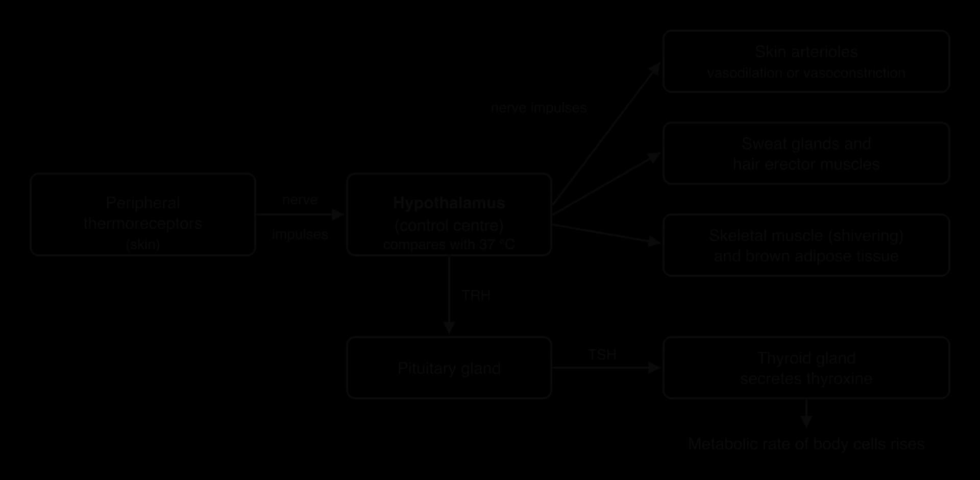
The flow chart of thermoregulation shows how one control centre uses two communication systems. Nerve impulses produce fast responses within seconds, such as shivering and changes in blood flow to the skin. Thyroxine acts more slowly but for longer, adjusting heat production over hours to days. This integration of the nervous and endocrine systems is typical of homeostasis.
Muscle and adipose tissue as effectors
Two tissues act as effectors that increase heat production. Skeletal muscle produces heat by shivering, and also produces heat during ordinary activity. Adipose tissue acts in two ways: white adipose tissue under the skin insulates the body and reduces heat loss, and brown adipose tissue produces heat directly. Shivering and brown adipose tissue are the two main sources of extra heat when the body is cold.
Thermoregulation mechanisms in humans
Birds and mammals regulate their body temperature by physiological means, such as sweating and shivering, and by behavioural means, such as seeking shade, huddling together or changing posture. Humans add clothing, buildings and heating. The physiological mechanisms in humans are of two kinds: those that change the rate of heat loss through the skin, and those that change the rate of heat production.
Responses when the body is too hot
Vasodilation
Blood reaching the skin flows through arterioles, small arteries with smooth muscle in their walls. When the body is too hot, the hypothalamus reduces the nerve impulses to this muscle, so it relaxes and the arterioles supplying the capillaries near the skin surface widen. This is vasodilation. More warm blood flows close to the surface, and more heat is lost from the skin to the surroundings by radiation and convection. The skin looks flushed, especially in people with pale skin.
The skin of the hands, feet, ears and nose also has arteriovenous anastomoses, short vessels that connect arterioles directly to venules. When the body is hot they dilate, carrying a large volume of warm blood into the veins just under the skin surface, where it loses heat.
Sweating
Sweating is the secretion of sweat, a watery solution of salts, by sweat glands in the dermis onto the skin surface. Heat from the skin is used to evaporate the water in sweat. Changing liquid water to water vapour needs a lot of energy (the latent heat of vaporisation, about 2.4 kJ for every gram evaporated at skin temperature), so evaporation removes heat from the skin and cools the blood flowing through it. Sweating is the main way humans lose heat when the air is as warm as the skin or warmer, because radiation can then no longer remove heat.
Sweat only cools the body if it evaporates. In humid air, the air already holds a lot of water vapour, so evaporation is slow and sweat drips off the skin without cooling it. Very hot, humid weather is therefore more dangerous than hot, dry weather.
Responses when the body is too cold
Vasoconstriction
When the body is too cold, the hypothalamus sends more nerve impulses to the smooth muscle of the skin arterioles. The muscle contracts and the arterioles narrow: this is vasoconstriction. Less blood flows through the capillaries near the skin surface, and the arteriovenous anastomoses in the hands and feet also constrict. Warm blood is kept deeper in the body, under the insulating layer of fat, and less heat is lost. The skin looks pale, and the hands and feet feel cold because they receive less warm blood.
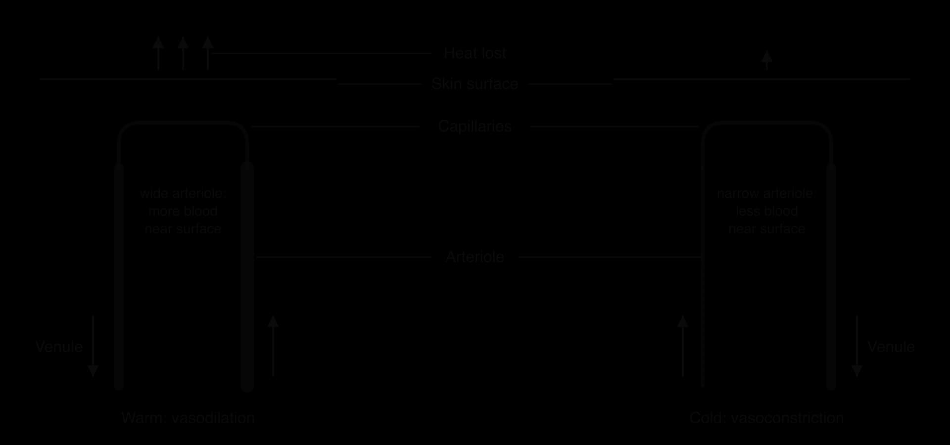
The diagram of skin in warm and cold conditions shows the capillaries in the same position in both panels. The diameter of the arteriole that supplies them is what changes, and with it the volume of blood flowing close to the surface.
Shivering
Shivering is rapid, involuntary contraction and relaxation of skeletal muscles. The hypothalamus sends nerve impulses to the muscles, which contract many times a second without producing coordinated movement. Muscle contraction uses ATP, and the cell respiration that regenerates the ATP releases a large amount of energy as heat. Shivering can increase heat production several times over within minutes.
Uncoupled respiration in brown adipose tissue
Brown adipose tissue (brown fat) is a type of fat tissue specialised for producing heat. Its cells contain many small droplets of fat and very large numbers of mitochondria, whose iron-containing cytochromes give the tissue its brown colour. It also has a dense supply of capillaries, which carry the heat produced to the rest of the body. Newborn babies have a large amount of brown fat, especially between the shoulder blades, because they cannot shiver effectively and lose heat quickly through their large surface area relative to volume.
In most cells, the energy released by cell respiration in the mitochondria is used to make ATP. In brown adipose cells, the inner mitochondrial membrane contains a protein called thermogenin (uncoupling protein 1, UCP1). Thermogenin allows protons to flow back across the inner membrane without passing through ATP synthase. The oxidation of fatty acids and glucose continues, but much less ATP is made: the energy is released as heat instead. This is called uncoupled respiration, because respiration is uncoupled from ATP production. Sympathetic nerves from the hypothalamus activate brown fat when the body is cold.
Brown fat was long thought to disappear soon after infancy. In 2009, three studies using PET-CT scans, which show tissues that take up glucose rapidly, found active brown adipose tissue in the neck and above the collarbones of many adults, especially when they were exposed to cold. Adults therefore also produce some heat by uncoupled respiration.
Hair erection
Each hair in the skin has a small erector muscle (arrector pili) attached to its follicle. When the body is cold, the hypothalamus sends impulses that make these muscles contract, pulling the hairs upright. In mammals with fur, the erect hairs trap a thicker layer of still air next to the skin. Air is a poor conductor of heat, so this layer insulates the body and reduces heat loss. Humans have very little body hair, so hair erection produces only "goose bumps" and has almost no effect on heat loss; it is a mechanism that is much more effective in furry mammals such as cats and arctic foxes.
Mechanism | Effector | When it is used | How it changes body temperature |
|---|---|---|---|
Vasodilation | Smooth muscle of skin arterioles (relaxes) | Too hot | More blood near the skin surface, so more heat lost by radiation |
Sweating | Sweat glands | Too hot | Evaporation of water from sweat removes heat from the skin |
Vasoconstriction | Smooth muscle of skin arterioles (contracts) | Too cold | Less blood near the skin surface, so less heat lost |
Shivering | Skeletal muscle | Too cold | Respiration for repeated contractions releases heat |
Uncoupled respiration | Brown adipose tissue | Too cold | Respiration releases energy as heat instead of making ATP |
Hair erection | Erector muscles of hair follicles | Too cold | Traps an insulating layer of air (effective in furry mammals) |
Increased thyroxine secretion | Thyroid gland, then most body cells | Too cold (longer term) | Raises metabolic rate, so more heat is produced |
These responses are usually combined. On a cold day, vasoconstriction is the first response, because it costs no energy. If the core temperature still falls, shivering and brown fat increase heat production, and over a longer period thyroxine secretion rises. Behaviour adds to all of them: people put on clothes, curl up to reduce their exposed surface area and move to warmer places.
The kidney in osmoregulation and excretion (HL only)
The two kidneys lie at the back of the abdomen and receive blood through the renal arteries. They have two separate homeostatic roles, which use the same structures but should be kept distinct: excretion and osmoregulation.
Excretion and osmoregulation compared
Excretion is the removal from the body of the waste products of metabolism. The main nitrogenous waste in mammals is urea, made in the liver from the excess amino groups of amino acids that are not needed for protein synthesis. Amino groups would otherwise form ammonia, which is very toxic. Urea is much less toxic and very soluble, so it is carried in the blood plasma to the kidneys and removed in urine. Carbon dioxide from cell respiration is also an excretory product, removed by the lungs.
Osmoregulation is the control of the osmotic concentration of the blood and tissue fluid, which means controlling their water content and solute concentration. If the blood becomes too concentrated, water leaves cells by osmosis and they shrink; if it becomes too dilute, water enters cells and they swell. The kidneys osmoregulate by changing the volume and concentration of the urine.
Excretion | Osmoregulation | |
|---|---|---|
Definition | Removal of the waste products of metabolism | Control of the osmotic concentration of body fluids |
Substances involved | Urea, excess ions, toxins, drugs; CO₂ (lungs) | Water and solutes, especially sodium ions |
Main parts of the nephron | Glomerulus, Bowman's capsule, proximal convoluted tubule | Loop of Henle, collecting duct |
Hormone involved | None needed | Antidiuretic hormone (ADH) |
Measuring osmotic concentration
Osmotic concentration is the total concentration of dissolved solute particles in a solution, measured in osmoles per litre (osmol L⁻¹). One osmole is one mole of solute particles. Because ionic compounds separate into ions in solution, a 0.1 mol L⁻¹ solution of sodium chloride (Na⁺ and Cl⁻) has an osmotic concentration of about 0.2 osmol L⁻¹, while 0.1 mol L⁻¹ glucose, which does not separate, has 0.1 osmol L⁻¹. Human blood plasma has an osmotic concentration of about 0.3 osmol L⁻¹. Urine ranges from about 0.05 osmol L⁻¹ when a person has drunk a lot of water to about 1.2 osmol L⁻¹ when they are dehydrated.
The structure of the nephron
Each kidney contains about a million nephrons, the tubules that make urine. A nephron begins in the outer region of the kidney (the cortex) as a cup-shaped Bowman's capsule surrounding a knot of capillaries, the glomerulus. The capsule leads into the proximal convoluted tubule, which runs into the loop of Henle, a hairpin loop that dips into the inner region (the medulla). The loop returns to the cortex as the distal convoluted tubule, which empties into a collecting duct shared by many nephrons. Collecting ducts run back down through the medulla and empty into the renal pelvis, from which urine passes along the ureter to the bladder.
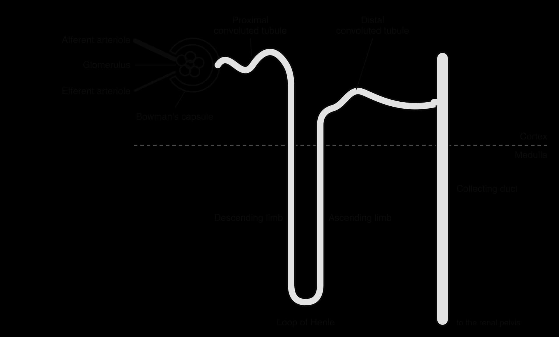
The diagram of the nephron shows the order in which fluid passes through it. Blood arrives in the afferent arteriole, which is wider than the efferent arteriole that carries blood away from the glomerulus. After the efferent arteriole, the blood flows through a second network of capillaries around the tubules, which takes up the substances that the nephron reabsorbs.
Ultrafiltration in the glomerulus and Bowman's capsule
Ultrafiltration is the first stage in forming urine. Blood in the glomerulus is at high pressure, because the efferent arteriole leaving it is narrower than the afferent arteriole entering it. This pressure forces water and small solutes out of the blood plasma through three layers into the space inside Bowman's capsule:
The walls of the glomerular capillaries, which are fenestrated: their endothelium has many small pores.
The basement membrane, a layer of protein fibres and glycoproteins that covers the capillaries. It is the actual filter, and it holds back large molecules.
The inner wall of Bowman's capsule, made of cells called podocytes with many foot-like extensions. The narrow gaps between the extensions (filtration slits) let the fluid through to the capsule space.
The fluid that collects in Bowman's capsule is the filtrate (glomerular filtrate). It contains water, glucose, amino acids, ions, urea and other small molecules at about the same concentrations as in the plasma. Blood cells and plasma proteins are too large to pass through the basement membrane, so they stay in the blood. Filtration is not selective by usefulness: useful and waste molecules pass equally, as long as they are small enough.
The kidneys of an adult form about 180 litres of filtrate a day, yet only about 1.5 litres of urine are excreted. About 99% of the filtrate is therefore reabsorbed into the blood, most of it in the proximal convoluted tubule. Filtering such a large volume means that waste products are removed quickly from all of the plasma, many times a day.
Substance | Blood plasma | Filtrate in Bowman's capsule | Urine |
|---|---|---|---|
Water | Present | Present | Present (volume depends on ADH) |
Plasma proteins | Present | Absent | Absent |
Blood cells | Present | Absent | Absent |
Glucose | Present | Present (same concentration as plasma) | Absent in a healthy person |
Amino acids | Present | Present | Absent |
Sodium and other ions | Present | Present | Present (amount varies) |
Urea | Present | Present | Present at a much higher concentration |
Selective reabsorption in the proximal convoluted tubule
Selective reabsorption is the return of useful substances from the filtrate to the blood. It happens mainly in the proximal convoluted tubule (PCT), where the following are reabsorbed into the surrounding capillaries:
All of the glucose and amino acids, by co-transport with sodium ions. The sodium ions are pumped out of the other side of the cell by the sodium–potassium pump, which keeps their concentration inside the cell low so that more Na⁺ diffuses in from the filtrate, bringing glucose or amino acids with it.
Most of the sodium ions and other ions, by active transport and co-transport.
About two-thirds of the water, by osmosis, because the reabsorption of solutes raises the solute concentration in the tissue around the tubule.
The cells of the PCT wall are adapted for reabsorption, a clear example of form and function. Their surface facing the filtrate has a brush border of microvilli, which increases the surface area for absorption. They contain many mitochondria to supply the ATP needed for active transport. They are joined by tight junctions, and the tubule is surrounded by many capillaries, so reabsorbed substances are carried away quickly and a concentration gradient is maintained.
Toxins and other unwanted solutes are not reabsorbed, or only partly. They remain in the filtrate and pass out in the urine. About half of the urea diffuses back into the blood, but the rest stays in the filtrate, and its concentration rises as water is reabsorbed. Some substances, including certain drugs, are also actively secreted from the blood into the tubule, which speeds their removal.
The loop of Henle and the medulla
The role of the loop of Henle is to create a high solute concentration in the tissue fluid of the medulla, which later allows water to be reabsorbed from the collecting ducts. The two limbs of the loop have different properties:
The ascending limb actively transports sodium ions (with chloride ions) out of the filtrate into the tissue fluid of the medulla. Its walls are impermeable to water, so water cannot follow the ions by osmosis. Solutes therefore accumulate in the medulla.
The descending limb is permeable to water. As filtrate flows down it, water moves out by osmosis into the concentrated medulla, so the filtrate becomes more concentrated towards the bottom of the loop.
Because the filtrate flows in opposite directions in the two limbs, the effect is multiplied along the loop. Concentrated filtrate from the bottom of the descending limb enters the ascending limb, which pumps more sodium ions out at each level. The result is a gradient of osmotic concentration in the medulla, from about 0.3 osmol L⁻¹ near the cortex to about 1.2 osmol L⁻¹ deep in the inner medulla in humans. This arrangement is called a countercurrent multiplier.
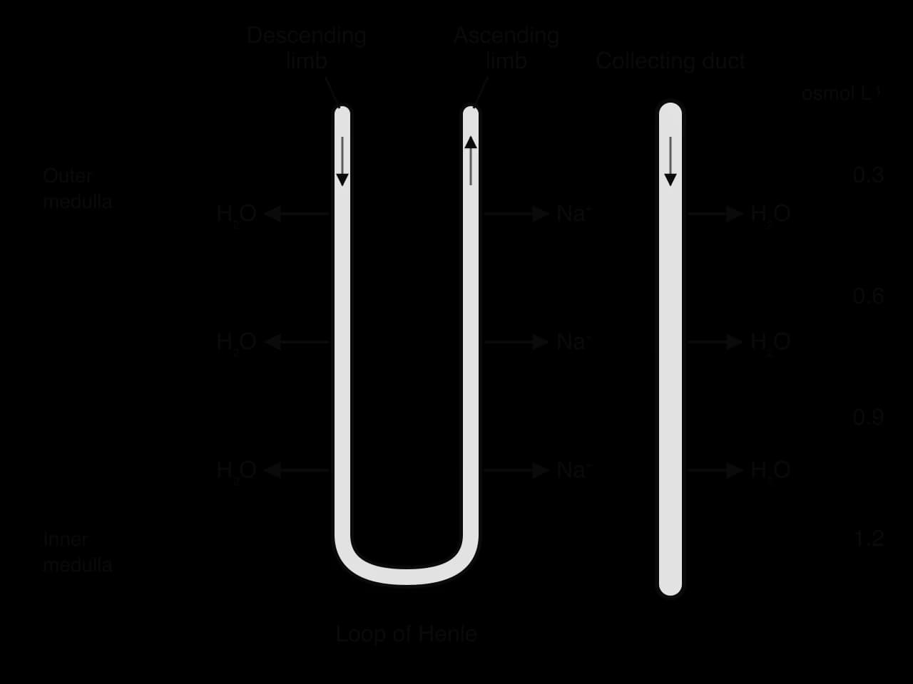
The diagram of the loop of Henle shows why the gradient matters. The collecting duct runs through the same concentrated medulla, so water can leave it by osmosis if its walls are permeable. The deeper the duct goes, the more concentrated the surrounding tissue, so water continues to leave along its whole length.
The length of the loop of Henle determines how concentrated the medulla, and therefore the urine, can become. Desert mammals such as kangaroo rats (Dipodomys) have exceptionally long loops of Henle and a deep medulla. They produce urine several times more concentrated than the most concentrated human urine, which allows them to survive on the water in their food and the water made by their own cell respiration, without drinking.
Osmoregulation by water reabsorption in the collecting ducts
The volume and concentration of the urine are adjusted in the collecting ducts by antidiuretic hormone (ADH, also called vasopressin). ADH controls how permeable the walls of the collecting ducts are to water, and so how much water is reabsorbed from the filtrate by osmosis into the concentrated medulla.
Detecting the osmotic concentration of the blood
The receptors in this system are osmoreceptors in the hypothalamus, cells that detect the osmotic concentration of the blood. When the blood becomes too concentrated, for example after sweating heavily or eating salty food without drinking, water leaves the osmoreceptor cells by osmosis and they shrink. This stimulates nerve impulses, and the hypothalamus increases the secretion of ADH. ADH is made by neurosecretory cells in the hypothalamus and released into the blood from the posterior pituitary gland. Thirst is stimulated at the same time.
How ADH changes the collecting duct
ADH travels in the blood to the kidneys and binds to receptors on the cells of the collecting duct walls. Binding triggers a signalling pathway inside the cells that causes vesicles containing aquaporins (water channel proteins) to move to the plasma membrane facing the filtrate and fuse with it. The aquaporins are inserted into the membrane, so it becomes much more permeable to water. Water moves by osmosis out of the filtrate, through the cells and into the concentrated tissue fluid of the medulla, and from there into the blood. A small volume of concentrated urine is produced.
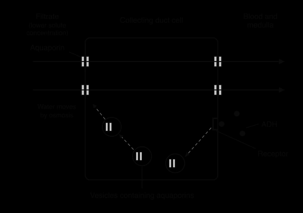
When the blood becomes too dilute, for example after drinking a large volume of water, the osmoreceptors swell and the hypothalamus reduces ADH secretion. Without ADH, aquaporins are removed from the membrane by endocytosis and stored again in intracellular vesicles. The collecting duct becomes much less permeable to water, little water is reabsorbed, and a large volume of dilute urine is produced. In both directions the change is corrected by negative feedback, so the osmotic concentration of the blood returns to about 0.3 osmol L⁻¹.
Condition of the blood | Osmoreceptors | ADH secretion | Aquaporins in collecting duct membrane | Urine produced |
|---|---|---|---|---|
Too concentrated (dehydration) | Shrink; stimulated | Increased | More inserted from vesicles | Small volume, concentrated |
Too dilute (excess water) | Swell; less stimulated | Decreased | Removed into vesicles | Large volume, dilute |
Recycling aquaporins between the membrane and intracellular vesicles allows a fast response. The cell does not have to make new proteins when ADH arrives or destroy them when it leaves, so the permeability of the collecting duct can change within minutes.
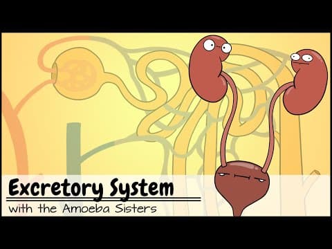 YouTube
Changes in blood supply to organs (HL only)
The amount of blood pumped by the heart each minute is the cardiac output. It is about 5 litres per minute at rest in an adult and can rise to about 20 litres per minute during maximal exercise in a young adult who is not trained, and to far more in endurance athletes. The share of this blood that each organ receives is not fixed. It changes with the body's activity, so that the organs working hardest get the most oxygen and glucose.
How arterioles control the distribution of blood
Blood flow to each organ is controlled by its arterioles. The walls of arterioles contain a thick layer of smooth muscle, which the sympathetic nervous system and local chemical signals can make contract or relax. When the smooth muscle contracts, the arteriole narrows (vasoconstriction) and less blood flows to the capillaries it supplies. When it relaxes, the arteriole widens (vasodilation) and more blood flows.
During exercise, active skeletal muscle releases chemicals such as carbon dioxide, H⁺ ions and adenosine, and its oxygen concentration falls. These local changes make the muscle's own arterioles dilate, even though sympathetic nerves are causing vasoconstriction in the arterioles of the gut and kidneys. The combination diverts blood away from the inactive organs and towards the active muscles.
Blood supply in sleep, wakeful rest and vigorous activity
Organ | Sleep | Wakeful rest | Vigorous physical activity |
|---|---|---|---|
Skeletal muscle | Low: muscles relaxed, metabolic rate at its lowest | About one-fifth of cardiac output | Greatly increased: over 80% of a much larger cardiac output at maximal effort |
Gut (stomach and intestines) | Maintained; digestion continues | High, especially after a meal | Reduced by vasoconstriction |
Kidneys | Slightly reduced; filtration continues, and less urine is made at night | High: about one-fifth of cardiac output | Reduced by vasoconstriction |
Brain | Remains high; falls in deep (non-REM) sleep and rises again in dreaming (REM) sleep | About 750 mL per minute | About the same (about 750 mL per minute); a smaller share of cardiac output |
Sources: Joyner, M. J. and Casey, D. P., Physiological Reviews 95: 549–601, 2015 (exercise); typical values for a healthy adult from human physiology textbooks (sleep and rest).
The brain is protected from large changes in blood flow. Its arterioles adjust automatically to keep flow almost constant over a wide range of blood pressures, because brain cells have very small energy reserves and depend on a continuous supply of glucose and oxygen. During exercise, the volume of blood reaching the brain stays about the same, but because cardiac output rises it becomes a much smaller percentage of the total.
Skin blood flow changes with both activity and temperature. During prolonged exercise, heat production by the muscles raises body temperature, so arterioles in the skin dilate for heat loss even though the muscles need blood too. This competition between thermoregulation and the supply of muscle is one reason that endurance exercise in hot weather is especially tiring.
Exam practice
Homeostasis is assessed in every paper. Paper 1A multiple-choice questions often ask which cells secrete insulin or glucagon, which effector is involved in a response, or which statement distinguishes type 1 from type 2 diabetes. Paper 1B and Paper 2 Section A use data such as glucose tolerance curves, the results of diabetes trials, body temperature recordings and, at HL, the composition of plasma, filtrate and urine or blood flow to organs. Extended-response questions in Section B commonly ask candidates to explain the control of blood glucose or body temperature, to compare and contrast the two types of diabetes, or (HL) to describe ultrafiltration and explain the role of ADH.
The command terms used most often are outline, explain, distinguish, calculate and suggest. Answers about feedback loops earn marks for naming each component in order (receptor, control centre, effector) and the direction of each change.
Questions on thermoregulation and osmoregulation are often worded in terms of a scenario, such as a swimmer, a marathon runner or a person lost in a desert. Marks are given for applying the general mechanism to the situation described, for example linking heavy sweating to a rise in the osmotic concentration of the blood and therefore to more ADH secretion.
Homeostasis connects many other subtopics, including enzyme activity and temperature (C1.1), membrane transport by aquaporins and pumps (B2.1), insulin as a protein hormone (B1.2) and water potential (D2.3).
Self-check
- To do:
I can define homeostasis and give four variables regulated in humans with their normal values.
- To do:
I can explain negative feedback in terms of receptors, a control centre and effectors, and why homeostasis does not use positive feedback.
- To do:
I can explain how insulin and glucagon from the beta and alpha cells regulate blood glucose, including glycogenesis and glycogenolysis.
- To do:
I can compare and contrast the physiological changes, risk factors, prevention and treatment of type 1 and type 2 diabetes.
- To do:
I can interpret glucose tolerance curves and the results of a diabetes prevention trial.
- To do:
I can explain the roles of thermoreceptors, the hypothalamus, the pituitary gland and thyroxine in thermoregulation.
- To do:
I can explain how vasodilation, vasoconstriction, sweating, shivering, uncoupled respiration in brown adipose tissue and hair erection change body temperature.
- To do:
I can distinguish between excretion and osmoregulation and use osmol L⁻¹ for osmotic concentration. (HL)
- To do:
I can explain ultrafiltration in the glomerulus and selective reabsorption in the proximal convoluted tubule. (HL)
- To do:
I can explain how the loop of Henle makes the medulla concentrated and how ADH and aquaporins control water reabsorption in the collecting ducts. (HL)
- To do:
I can describe and explain changes in blood supply to skeletal muscle, the gut, the brain and the kidneys during sleep, wakeful rest and vigorous activity. (HL)
Questions on D3.3
46 exam-style questions on this subtopic, written in the style of the IB papers. The first has its markscheme open. Counted on
Question 1 · BIO-00071HL only · 15 marks
Organisms must control the rates of the chemical reactions in their cells and bodies.
- (a)
Describe the control of blood glucose concentration by hormones.
4 marks
Markscheme (a)
a. blood glucose concentration is monitored by (endocrine cells in) the pancreas / islets of Langerhans;
b. when glucose concentration is high, β cells secrete insulin;
c. insulin stimulates uptake of glucose by liver / muscle / adipose cells;
d. (insulin stimulates) conversion of glucose to glycogen in liver and muscle cells;
e. when glucose concentration is low, α cells secrete glucagon;
f. glucagon stimulates breakdown of glycogen to glucose in the liver, which is released into the blood;
g. hormones are transported in the blood to target cells;
h. negative feedback returns glucose concentration to the set point;[4 max]
- (b)
Compare and contrast competitive and non-competitive inhibition of enzymes.
7 marks
Markscheme (b)
Similarities:
a. both reduce the rate of the enzyme-catalysed reaction;
b. in both, the inhibitor binds reversibly to the enzyme;
c. both are specific: only particular substances can bind;
d. both reduce the number of enzyme–substrate complexes formed;Differences:
e. competitive inhibitor binds to the active site whereas non-competitive inhibitor binds to an allosteric site;
f. competitive inhibitor has a shape similar to the substrate whereas non-competitive inhibitor does not;
g. competitive inhibitor competes with the substrate for the active site whereas non-competitive inhibitor does not;
h. non-competitive inhibitor causes a conformational change that alters the active site whereas competitive inhibitor blocks the active site without changing it;
i. competitive inhibition is reduced / overcome by increasing substrate concentration whereas non-competitive inhibition is not;
j. with a competitive inhibitor the maximum rate can still be reached at high substrate concentrations, whereas a non-competitive inhibitor lowers the maximum rate;
k. example of competitive inhibition: statins inhibiting HMG-CoA reductase;
l. example of non-competitive / allosteric inhibition: isoleucine inhibiting threonine deaminase;[7 max]
Answers must include at least one similarity and one difference for full marks. Differences may be presented in a table. - (c)
Explain why a large number of different enzymes is needed to carry out and control metabolism.
4 marks
Markscheme (c)
a. metabolism is a complex network of many interdependent reactions;
b. enzymes are specific / each active site binds only one substrate or a small group of substrates;
c. so each reaction (or type of reaction) needs its own enzyme;
d. without catalysis reactions would be too slow at body / cell temperature;
e. each step of a pathway can be controlled separately by controlling its enzyme;
f. by regulating how much of each enzyme is synthesized / which genes are expressed;
g. by inhibiting or activating particular enzymes to switch pathways on or off;
h. anabolic and catabolic pathways (for the same molecule) use different enzymes, so they can be controlled independently;[4 max]
Markscheme notes
One additional mark is available for the construction of the answer.
Question 2 · BIO-00072HL only · 15 marks
Living organisms release energy from food and use it in many ways.
- (a)
Outline the role of ATP in cells.
4 marks
- (b)
Explain how heat is produced by metabolism and how it is used to maintain a constant body temperature in humans.
7 marks
- (c)
Distinguish between linear and cyclical pathways in metabolism, using named examples.
4 marks
Answer it, then compare with the markscheme.Check this one with a free account
Question 3 · BIO-00252HL only · 15 marks
Proteins must be delivered to the correct place inside or outside a cell.
- (a)
Outline how vesicles are formed and used to move materials into and out of cells.
4 marks
- (b)
Describe how insulin and glucagon regulate the concentration of glucose in the blood.
4 marks
- (c)
Explain how a protein hormone such as insulin is synthesized, processed and secreted by a cell.
7 marks
Answer it, then compare with the markscheme.Check this one with a free account
Flashcards for D3.3
The official deck for this part of the syllabus.
D3.3 Homeostasis · front
Define homeostasis.
D3.3 Homeostasis · front
Homeostasis is the maintenance of the […] of an organism within preset limits, despite fluctuations in the external environment.
- D3.3 Homeostasis251 cards
Carry on with D3.3 in Augment
With a free account, Augment remembers where you stopped and what you have practiced.