Explain how brain structures, neurotransmitters, stress hormones and sleep affect memory, evaluate the research from H.M. to sleep studies through the concepts, and apply biological explanations to learning scenarios.
Each is in the Psychology glossary, with every other term in the subject.
A loss of the ability to form new long-term memories after brain damage, while memories from before the damage remain largely available.
A physical characteristic of the body, such as a brain structure, neurotransmitter, hormone, gene, sleep or age, that influences behaviour or a cognitive process.
A vivid, detailed and confidently held memory of the circumstances in which a person learned of a surprising, emotional and important event.
The process by which a newly encoded, fragile memory trace is stabilised into a lasting long-term memory, first at synapses within hours and then across brain systems over months or years.
The proposal that stress hormones such as adrenaline, acting through the amygdala, strengthen the consolidation of memories formed during emotional arousal rather than creating them.
A loss of memories formed before brain damage, usually worst for the period just before the damage and less severe for older memories.
The pattern in retrograde amnesia in which recent memories are lost more than older ones, because older memories have been consolidated outside the hippocampus.
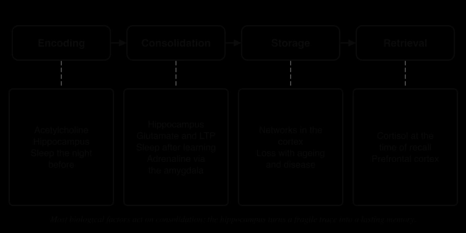
Describe the stages of memory and explain which biological factors act on each stage.
Explain the role of the hippocampus in memory consolidation, with reference to amnesia.
Describe and evaluate the case of Henry Molaison (H.M.) as evidence for a biological factor in memory.
Explain how neurotransmitters, especially acetylcholine and glutamate, affect the formation of memories.
Explain how adrenaline and cortisol can strengthen or weaken memory, and evaluate research on flashbulb memories.
Explain the role of sleep in memory consolidation and evaluate the evidence that the relationship is causal.
Evaluate research on biological factors in memory with reference to causality, measurement, bias and responsibility.
Apply biological explanations of memory to an unfamiliar situation in learning and cognition.
Every memory that a person forms is stored as a physical change in the brain. Biological factors, such as which brain structures are intact, which chemicals pass between neurons and how much a person sleeps, therefore affect how well information is learned, kept and later recalled. They help to explain why some brain injuries remove the ability to learn new facts, why frightening events are remembered so vividly, why stress can make well-learned material hard to recall, and why a night without sleep harms learning.
Memory is the main cognitive process discussed, with attention mentioned where the same factor matters. Explanations draw on the biological approach, especially localisation of function, neurotransmission and chemical messengers, and on the
With a free account, Augment remembers where you stopped and what you have practiced.
Cognitive psychologists describe memory in terms of stores and processes. The multi-store model describes information passing from sensory memory to short-term memory and, with rehearsal, to long-term memory, and the working memory model divides short-term memory into several components. These models describe what memory does. A biological explanation asks how the brain does it: which structures, cells and chemicals carry out each step.
A biological factor is any physical characteristic of the body that affects behaviour or cognition. For memory, the main biological factors are brain structures (such as the hippocampus and amygdala), neurotransmitters, hormones, sleep, genes, age and general physical health. Each factor tends to act on one or two stages of memory, so naming the stage is the quickest way to explain how a factor matters.
Encoding is the process of turning information that has been attended to into a form that can be stored. A newly encoded memory is fragile and easily lost. Over the following hours, days and even years it is stabilised by memory consolidation, so that it no longer depends on the conditions of learning. Storage is the holding of the memory in networks of neurons, mainly in the cortex, and retrieval is the reactivation of the stored memory so that it can be used.
The figure shows why a single question such as “Does stress harm memory?” has no single answer. Stress hormones released while an event is happening can strengthen its consolidation, while the same hormones at the moment of recall can block retrieval. The stage at which a factor acts decides its effect.
Two levels of consolidation are usually distinguished. Synaptic consolidation is the strengthening of connections between the neurons that were active during learning, and it is complete within hours. Systems consolidation is a slower reorganisation in which a memory that at first needs the hippocampus to be retrieved is gradually stored in networks of the cortex, so that it can later be retrieved without the hippocampus.
The hippocampus is a curved structure inside the temporal lobe of each hemisphere, part of a group of structures called the medial temporal lobe. It has three main roles in memory. It binds the separate parts of an experience (what happened, where, when and with whom) into a single episodic memory; it consolidates new declarative memories (facts and events) into long-term storage; and it supports spatial memory, the mental map used to find the way.
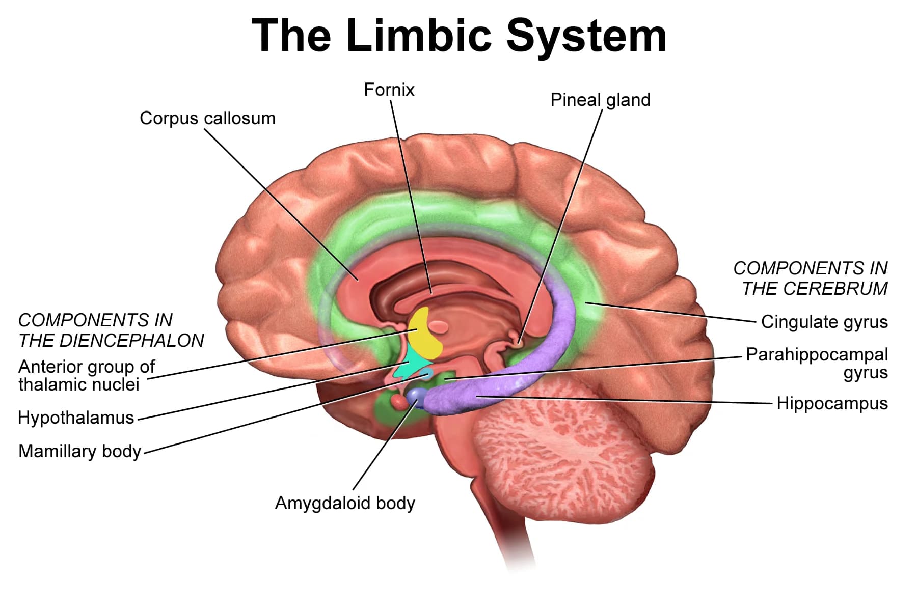
The amygdala, at the front of the hippocampus, is best known for its role in emotion, especially fear. In memory, it tags events as emotionally important and increases the consolidation of those events by the hippocampus. The prefrontal cortex organises encoding and retrieval, for example by choosing a strategy or searching for a memory, and the cerebellum and basal ganglia support procedural memory, the memory for skills.
Structure | Main role in memory | Typical evidence |
|---|---|---|
Hippocampus | Consolidation of new declarative memories; binding the parts of an episode; spatial memory | Amnesia after damage (H.M.); activity during encoding in fMRI studies |
Amygdala | Strengthening the consolidation of emotional events | Activation when vivid emotional memories are recalled; drugs that block noradrenaline |
Prefrontal cortex | Strategies for encoding and searching memory; working memory | Activity during effortful recall; poorer source memory after frontal damage |
Cerebellum and basal ganglia | Procedural memory (skills and habits) | Skills learned normally by people with hippocampal amnesia |
This pattern is evidence for localisation of function: different memory abilities depend on different structures, so damage to one structure removes one ability and leaves others. The pattern also shows the limits of localisation. No single structure “contains” a memory; a memory of an event is stored across the cortex, and the hippocampus is the structure that links the pieces together while the memory is new.
Amnesia is a loss of memory caused by brain damage, illness or psychological trauma. Two forms are distinguished by the time of the memories that are lost, measured from the moment of the damage. Most people with damage to the hippocampus show both forms, but in very different amounts.
A person with severe anterograde amnesia can hold a conversation, because short-term memory is intact, but forgets it within minutes once attention moves elsewhere. They can read the same magazine repeatedly as if for the first time and must be told again and again of events such as the death of a relative. Causes include surgery or injury to the medial temporal lobes, infections such as herpes encephalitis, lack of oxygen to the brain, and Korsakoff’s syndrome caused by long-term alcohol misuse.
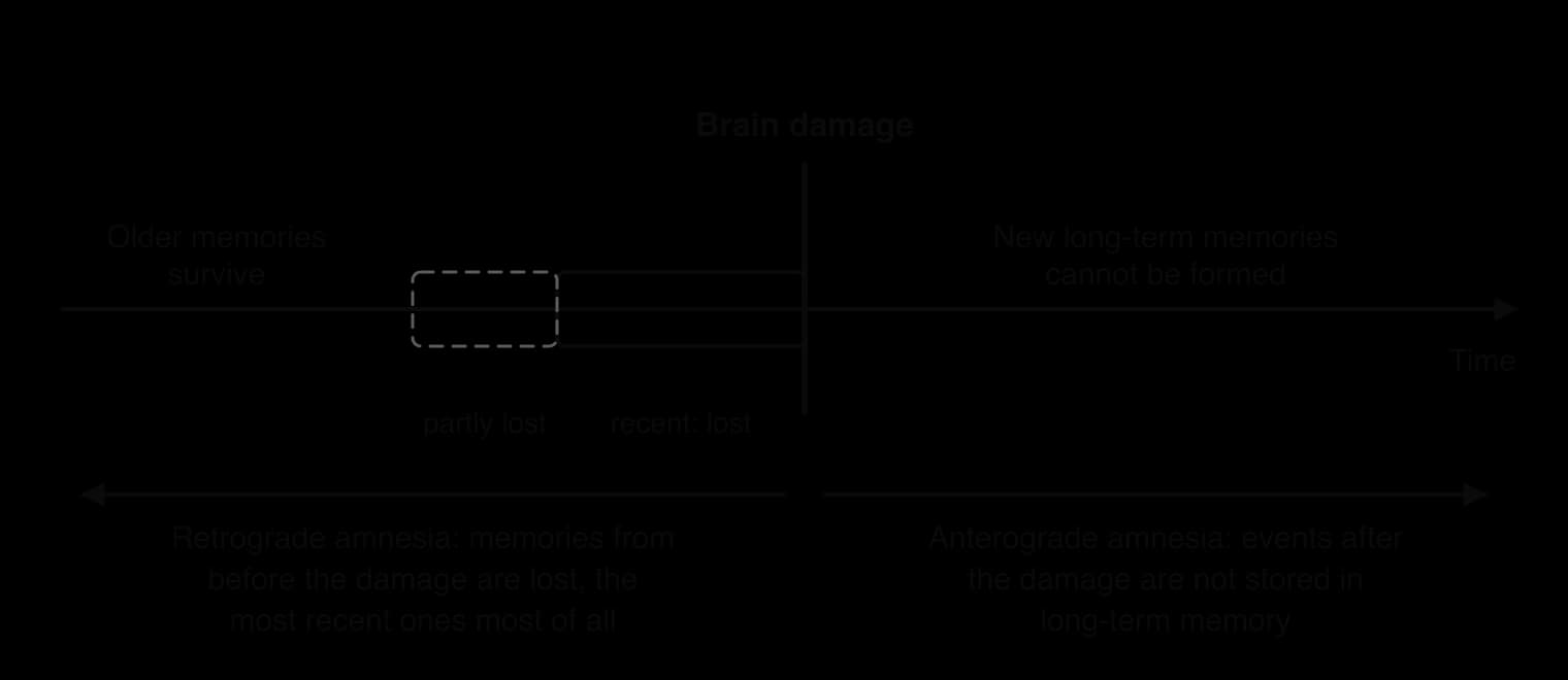
The pattern in retrograde amnesia is called a temporal gradient. Memories from years before the damage usually survive, while memories from the months or years just before it are lost. This is direct evidence for systems consolidation. Recent memories still depend on the hippocampus and are lost with it; older memories have already been consolidated into the cortex and can be retrieved without it.
A second well-known case shows the same pattern. Clive Wearing, a British musician, developed severe anterograde and retrograde amnesia in 1985 after herpes simplex encephalitis, a viral infection of the brain. He could still play the piano and conduct, a sign that procedural memory was spared, while he could not keep new events in mind for more than a short time.
The case of Henry Molaison is the most studied case in the history of memory research. Because his surgery removed known structures in an otherwise healthy brain, it showed for the first time which parts of memory depend on the medial temporal lobes and which do not.
The case was not an experiment planned in advance. After the surgery left Molaison with severe memory loss, neurosurgeon William Scoville and psychologist Brenda Milner aimed to describe the memory loss caused by removing parts of the medial temporal lobes on both sides of the brain, and to find out which structures were responsible. Later researchers, led by Suzanne Corkin, aimed to map which kinds of memory he had lost and which he had kept.
Molaison had epilepsy from childhood, and by the age of 27 his seizures, despite high doses of medication, stopped him from working. On 1 September 1953 Scoville removed the medial temporal lobes of both hemispheres, including the front part of the hippocampus, most of the amygdala and almost all of the entorhinal cortex, which carries information into the hippocampus. Scoville and Milner (1957) reported formal psychological testing of nine patients who had had similar operations (eight with psychosis and Molaison with epilepsy), carried out between one and a half and four years after surgery.
Molaison was then studied from 1957 until his death in 2008, first at the Montreal Neurological Institute and from 1966 at the Massachusetts Institute of Technology; Corkin (2002) reported that nearly 100 investigators had worked with him. As a case study, the research combined many methods: intelligence tests, memory tests, learning tasks, interviews, observation of his daily life and, from the 1990s, magnetic resonance imaging. His identity was protected by his initials until his death.
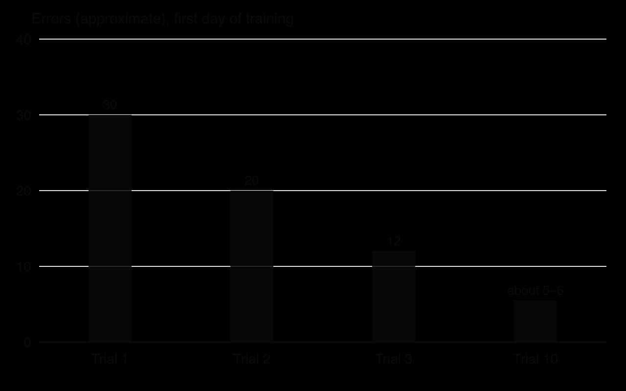
One of the best-known tasks was Milner’s mirror-drawing task, carried out in the early 1960s. Molaison had to trace a star shape by looking only at the reflection of his hand and paper in a mirror, which reverses the movements needed. He did the task ten times a day over three days, and the number of times his line crossed the edges of the star was counted.
After the operation Molaison had profound anterograde amnesia: he could not form new long-term declarative memories, either of events or of new facts. He also had a temporally graded retrograde amnesia, losing most memories from the one or two years before surgery and some from up to eleven years before, while his childhood memories were intact. His intelligence was normal or above average, his language was largely intact and his short-term memory span for numbers was no worse than that of control participants.
Lost after surgery | Kept after surgery |
|---|---|
Forming new declarative memories of events and facts (anterograde amnesia) | Short-term memory span, for example for digits |
Most memories from the one to two years before surgery | Childhood and early adult memories |
Recognising people met after 1953 | Intelligence, language and personality |
Learning new motor skills (procedural memory) |
In mirror drawing, his performance improved like that of a healthy person, even though he did not remember having done the task before. Scoville and Milner (1957) also found that the degree of memory loss depended on how much of the hippocampus had been removed: removing only the uncus and amygdala did not seem to cause memory loss, while the most extensive removals caused the most severe loss.
Some learning outside the hippocampus was also found. Molaison could draw a floor plan of the house he moved into five years after his surgery, and with cues he could recall some facts about famous people who became well known after 1953. Corkin (2002) explained these as the work of medial temporal tissue that had survived the operation and of structures outside it.
The case supports four conclusions. First, the hippocampus and nearby medial temporal structures are needed to consolidate new declarative memories. Second, short-term memory and long-term memory depend on different brain systems, because one was intact and the other was not. Third, long-term memory is not a single system: procedural memory was spared while declarative memory was lost. Fourth, old memories are stored outside the hippocampus, because his childhood memories survived.
The conclusions depend on knowing which structures were damaged, and for decades this was based on the surgeon’s description. MRI scans from the 1990s showed that the damage was more widespread than had been assumed, and after his death his brain was cut into 2,401 slices and reconstructed in three dimensions. Annese et al. (2014) found a significant amount of surviving hippocampal tissue, mainly at the back of the structure, and a small, previously unknown lesion in the left orbitofrontal cortex. Some of his abilities and difficulties may therefore reflect damage to areas other than the hippocampus.
Molaison’s amnesia had a clear cause: he had no memory problem before surgery and a severe one immediately afterwards, so the surgery caused it. This is stronger causal evidence than most case studies can offer. However, a single person is a sample of one, with a long history of epilepsy and anticonvulsant drugs that may have affected his brain. Conclusions about the hippocampus became convincing only when they were supported by other patients, by animal research and by brain imaging of healthy people.
Molaison agreed to take part in research for over fifty years, but because of his amnesia he could not remember agreeing, which makes informed consent in the usual sense impossible and places the duty to protect his interests on the researchers and on those who consented for him. In 2016 the journalist Luke Dittrich, the grandson of Molaison’s surgeon, raised questions about how consent was obtained and alleged that research files had been destroyed; MIT published a rebuttal and more than 200 neuroscientists signed a letter defending Corkin. The dispute shows that researchers who hold a vulnerable participant’s data for decades carry a lasting responsibility.
Sources: Scoville, W. B., & Milner, B. (1957). Loss of recent memory after bilateral hippocampal lesions. _Journal of Neurology, Neurosurgery and Psychiatry, 20, 11–21. Corkin, S. (2002). What’s new with the amnesic patient H.M.? Nature Reviews Neuroscience, 3, 153–160. Annese, J., et al. (2014). Nature Communications, 5, 3122._
 YouTube
Memories are formed and stored through changes in communication between neurons. A neurotransmitter is a chemical released by one neuron into the synapse that affects the next neuron by binding to its receptors. Two neurotransmitters are especially important for memory: acetylcholine, which affects how well new information is encoded, and glutamate, which is involved in strengthening the synapses that store a memory.
Neurons that release acetylcholine run from the base of the forebrain to the hippocampus and the cortex. High levels of acetylcholine during alert waking help the hippocampus to encode new information and sharpen attention to relevant stimuli; levels fall during deep sleep, when the hippocampus shifts from encoding to consolidating what has already been learned. Acetylcholine therefore links attention and memory: what is attended to is more likely to be encoded.
The evidence comes partly from drugs. Scopolamine is an antagonist that blocks one type of acetylcholine receptor. In a double-blind crossover study, Antonova et al. (2011) gave 20 healthy young men scopolamine or a placebo before a virtual-reality version of a spatial memory task; under scopolamine, activation of the hippocampus during the task was reduced. Further evidence comes from Alzheimer’s disease, in which neurons that release acetylcholine are lost early and new learning is among the first abilities to fail.
Glutamate is the brain’s main excitatory neurotransmitter. When a pathway into the hippocampus is stimulated strongly and repeatedly, the synapses in that pathway become more responsive for hours, days or longer. This lasting strengthening is called long-term potentiation (LTP), and it is the leading explanation of how experiences are stored at the level of synapses. Glutamate’s NMDA receptors are needed to start LTP, because they open only when the receiving neuron is already active and glutamate arrives at the same time.
Morris et al. (1986) tested whether LTP is needed for learning. Rats infused with AP5, a drug that blocks NMDA receptors, were impaired in learning the location of a platform hidden under water in a pool, a spatial task that depends on the hippocampus, but learned a visual discrimination task normally; the dose used was enough to block LTP in the hippocampus. Keith and Rudy (1990) argued that the drug also disturbed the rats’ movement and that the treated rats still learned a good deal, so the link between LTP and memory is strong but not proven.
Hormones are chemical messengers released into the blood by glands of the endocrine system. Two stress hormones have strong effects on memory: adrenaline, released quickly by the adrenal glands in an emotional or frightening situation, and cortisol, released more slowly through the HPA axis during stress. Their effects look contradictory at first, until the stage of memory and the timing and duration of the hormone release are taken into account.
According to the memory modulation hypothesis, developed by James McGaugh and colleagues, emotional arousal does not create memories but strengthens the consolidation of memories formed at the time. Adrenaline released during an emotional event acts, through nerve signals, on the amygdala, where noradrenaline is released; the amygdala in turn increases consolidation in the hippocampus. Emotional events are therefore remembered better than neutral ones, which may have survival value, because remembering a source of danger helps an animal to avoid it later.
Cahill et al. (1994) gave participants either propranolol, a drug that blocks the receptors through which adrenaline and noradrenaline act, or a placebo before they watched a slide show with a narrated story. Propranolol impaired memory of an emotionally arousing version of the story but not of a closely matched neutral version, which supports the claim that the emotional advantage depends on this hormone system.
Brown and Kulik (1977) introduced the term flashbulb memory for the vivid, detailed and confident memories that people have of the circumstances in which they heard shocking news, such as an assassination. They proposed a special biological mechanism that “prints” such events in memory. If emotional arousal strengthens consolidation, flashbulb memories should be both vivid and accurate.
Talarico and Rubin (2003) tested this the day after the attacks of 11 September 2001. On 12 September, 54 students at Duke University in the United States recorded their memory of hearing about the attacks and of a recent everyday event, and were tested again 1, 6 or 32 weeks later. The consistency of both kinds of memory declined at the same rate, but vividness and belief in accuracy declined only for the everyday memories. Flashbulb memories were special in how confident people felt, not in how accurate they were.
Sharot et al. (2007) used fMRI three years after the attacks to compare people who had been in Downtown Manhattan, close to the World Trade Center, with people who had been a few miles away in Midtown. When recalling 11 September, only the Downtown participants showed selective activation of the amygdala and reported more vivid recollection than for control events from the same year. Close personal experience, and the emotion it produced, seems to be needed for the amygdala to enhance a memory.
The hippocampus has many receptors for cortisol, which makes it sensitive to stress. Brief, moderate rises in cortisol around the time of learning can help consolidation, especially of emotional material. High levels at the moment of retrieval, and high levels over months or years, impair memory. This pattern is often summarised as an inverted U.
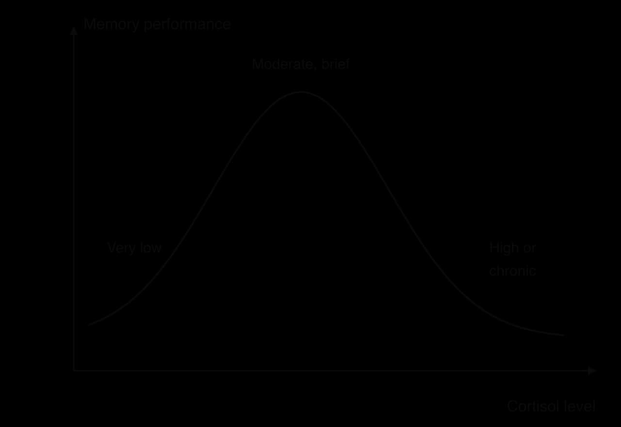
Newcomer et al. (1999) tested the effect of cortisol directly in a randomised, double-blind, placebo-controlled experiment with 51 healthy adults. Participants took a placebo, a lower dose of cortisol (40 mg a day, similar to mild stress) or a higher dose (160 mg a day, similar to major stress) for four days. The higher dose reduced verbal declarative memory, measured by immediate and delayed recall of a paragraph, but did not affect nonverbal memory, attention or executive function, and memory recovered after a six-day washout period.
Two further studies separate the effects of timing and duration. De Quervain et al. (2000) found that a single dose of cortisone impaired the retrieval of declarative memories that had already been learned, which may help to explain why stressed people sometimes cannot recall material they know well. Lupien et al. (1998) found that older adults whose cortisol had stayed high over several years had a smaller hippocampus and poorer hippocampus-dependent memory than older adults with normal cortisol, and the degree of shrinkage was related to how high cortisol had been.
 YouTube
Memory processing continues during sleep, and much consolidation happens then, when the brain is not taking in new information. Two main kinds of sleep alternate through the night: non-rapid eye movement (NREM) sleep, which includes deep slow-wave sleep, and rapid eye movement (REM) sleep, when most dreaming happens. Slow-wave sleep is thought to support the consolidation of declarative memories, and lighter NREM sleep and REM sleep the consolidation of skills.
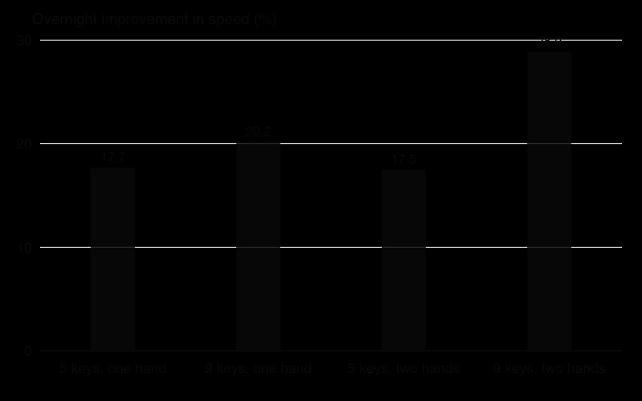
Walker et al. (2002) trained participants on a finger-tapping task in which they typed a sequence of keys as quickly and accurately as possible. After a night of sleep, speed increased by about 20% without any further practice and without a loss of accuracy, while an equal period of time awake gave no significant benefit. The size of the overnight improvement correlated with the amount of stage 2 NREM sleep, especially late in the night.
Kuriyama, Stickgold and Walker (2004) varied the difficulty of the sequence. The overnight improvement was similar for three sequences but much larger for the hardest one, which used nine keys and both hands, and within each sequence the transitions that had been slowest before sleep improved most. Sleep seems to give the most benefit to the parts of a skill that were hardest at the end of practice.
Correlations between sleep and memory could have other causes; for example, people who sleep more may differ in health or motivation. Rasch et al. (2007) manipulated what happened during sleep. Participants learned the locations of pairs of cards while a smell of roses was present. When the same smell was presented again during slow-wave sleep, their memory for the card locations next morning improved, but there was no effect when the smell was given during REM sleep or while awake, or when no smell had been present during learning. The smell did not improve a procedural task, and fMRI showed hippocampal activation when it was presented during slow-wave sleep.
These findings support the idea that memories are reactivated during slow-wave sleep and that reactivation causes consolidation. Sleep before learning matters too. Yoo et al. (2007) found that one night of sleep deprivation reduced hippocampal activity while participants encoded new pictures, and they remembered fewer of them later. Missing sleep therefore harms both the night after learning, when consolidation happens, and the day after the missed night, when encoding is weaker.
The hippocampus shrinks in late adulthood, and this shrinkage is linked to poorer memory and a higher risk of dementia. Biological factors such as age therefore interact with lifestyle. In a randomised controlled trial, Erickson et al. (2011) assigned 120 older adults to aerobic exercise training or a control group for a year. Aerobic exercise increased the volume of the front part of the hippocampus by about 2%, reversing one to two years of age-related loss, and spatial memory improved. Hippocampal volume declined in the control group.
The study shows that a biological factor in memory can be changed by behaviour, a clear example of the concept of change and of neuroplasticity in later life. It also shows how the approaches interact: the decision to exercise and keep exercising depends on motivation, habits and social support, not on biology alone.
Biological factors rarely act alone. Whether a stressful situation raises cortisol depends on how it is appraised, which is a cognitive factor; how much people sleep depends on school start times, working hours, screens and cultural norms about rest, which are environmental and sociocultural factors. What is encoded depends on attention, which is guided by schemas and by goals. A full explanation of a memory failure usually needs all three approaches.
A student revising late into the night before an important test is an example. The biological factor is sleep loss, which weakens hippocampal encoding and consolidation. The cognitive factors include the strategy used (rereading rather than testing oneself) and anxious thoughts about failing. The sociocultural factors include pressure from family and school and a culture that values long hours of study. Each factor makes the others worse, and changing any one of them may help.
Concept | How it applies to biological factors in memory |
|---|---|
Causality | Lesion cases and drug studies can show cause and effect, but single cases, animal models and correlations from imaging limit the conclusions. |
Measurement | Memory, hippocampal size and brain activity are measured indirectly: recall scores, MRI volume and the fMRI BOLD signal stand in for the processes themselves. |
Bias | Many studies use small samples of young Western university students or single patients; null results, such as failed replications, are less likely to be published. |
Perspective | A biological explanation alone is reductionist; cognitive and sociocultural factors decide when the biology is triggered. |
Responsibility | Patients with amnesia cannot give informed consent in the usual way; giving drugs or depriving sleep needs careful justification. |
Change | Exercise, sleep and stress management can change the biological factors that support memory. |
Different methods give different strengths of causal evidence. Lesion cases such as H.M. show what is lost when a structure is removed, and the timing of the loss can make the cause clear, but each case is unique and damage is rarely limited to one structure. Drug studies such as Newcomer et al. (1999) and Cahill et al. (1994) manipulate a chemical in healthy people with random allocation and double-blind designs, which gives stronger causal evidence, but the doses and short time spans may not match natural stress.
Experiments on rats, such as Morris et al. (1986), allow drugs to be placed directly in the brain and LTP to be measured, which is impossible in people. Their conclusions about human memory rest on the assumption that rat and human hippocampi work in similar ways, which is reasonable for spatial memory but less so for language-based memories. Converging evidence from several methods is what makes a biological explanation convincing.
Neither memory nor neurotransmitter activity can be observed directly. Memory is operationalised as recall of a word list, a paragraph or a card location, which may not capture memory for real events. Brain activity is inferred from fMRI, which measures changes in blood oxygen rather than the firing of neurons, and the amount of hippocampal activity says little about what the activity is doing. Sharot et al. (2007) could show that the amygdala was more active in Downtown participants, not that its activity caused their vivid memories.
Much of the research uses small samples: 20 men in Antonova et al. (2011), 54 students in Talarico and Rubin (2003), and single cases such as H.M. Most participants are young adults from Western universities, so effects of age, health and culture on the biology are rarely tested. Studies that find a clear effect of a drug or of sleep are more likely to be published than null results, a publication bias that may make biological effects look larger and more consistent than they are.
Biological explanations are precise and testable, and they have practical value, for example in explaining the memory effects of sleep loss, stress or brain injury. They become reductionist when they treat memory as nothing more than chemistry. The same cortisol rise may help one student and harm another depending on how each appraises the situation, and what a person attends to and encodes depends on goals, schemas and culture.
Questions on biological factors in cognitive processes appear in Paper 1. Section B sets a short unseen scenario in learning and cognition and asks you to apply a biological explanation to it; Section C asks for a concept-based essay on this area of study. Paper 1 does not reward the details of studies for their own sake, but a study is often the clearest way to show that an explanation is supported.
A Section C question names a concept and asks for an evaluation or discussion of this area of study. Causality is a natural concept for biological research, because its methods differ so much in how far they can show cause and effect.
Biological factors such as brain structures, neurotransmitters, hormones and sleep act on particular stages of memory: encoding, consolidation, storage and retrieval.
The hippocampus consolidates new declarative memories; old memories are stored across the cortex, which explains the temporal gradient in retrograde amnesia.
H.M. showed profound anterograde amnesia after removal of the medial temporal lobes on both sides, including the front of both hippocampi, with normal short-term memory and intact procedural learning, so memory is not a single system.
Acetylcholine supports encoding, and glutamate and long-term potentiation are the leading explanation of how synapses store memories.
Adrenaline acting through the amygdala strengthens the consolidation of emotional events, but flashbulb memories are high in confidence rather than in accuracy.
Cortisol has an inverted-U effect: brief moderate rises may help, high levels at recall impair retrieval, and prolonged high levels are linked to a smaller hippocampus.
Sleep before learning supports encoding and sleep after learning, especially slow-wave sleep, supports consolidation.
The strongest evidence combines lesion cases, drug experiments, sleep manipulations, imaging and animal research; each method alone has limits of causality, measurement and bias.
I can describe the four stages of memory and say which biological factors act on each.
I can explain the role of the hippocampus in memory consolidation, with reference to anterograde and retrograde amnesia.
I can describe the H.M. case and evaluate it through measurement, causality and responsibility.
I can explain how acetylcholine and glutamate contribute to forming memories.
I can explain how adrenaline and cortisol affect memory, and evaluate research on flashbulb memories.
I can explain the role of sleep in consolidation and evaluate whether the evidence is causal.
I can evaluate research on biological factors in memory through at least three of the concepts.
I can apply a biological explanation of memory to an unfamiliar learning scenario.