Draw and explain the structures of DNA and RNA, and show how base pairing lets genetic information be stored, copied and expressed, with the evidence behind it.
Each is in the Biology glossary, with every other term in the subject.
A purine base (A) found in DNA and RNA, which pairs with thymine in DNA and with uracil in RNA by two hydrogen bonds.
Running side by side in opposite directions; the two strands of DNA are antiparallel, so the 5′ end of one lies opposite the 3′ end of the other.
Two complementary bases on opposite strands joined by hydrogen bonds (A–T or C–G); DNA length is measured in base pairs (bp).
In double-stranded DNA the amount of adenine equals thymine and guanine equals cytosine, so purines equal pyrimidines, while the proportions of A + T and G + C vary between species.
Descent of different species from a shared ancestor; the near-universal genetic code is evidence that all living organisms share a common ancestor.
The specific pairing of bases by hydrogen bonds between nucleic acid strands: adenine with thymine (or uracil in RNA) and cytosine with guanine.
A pyrimidine base (C) found in DNA and RNA, which pairs with guanine by three hydrogen bonds.
The pentose sugar in DNA nucleotides, which has a hydrogen atom instead of an –OH group on carbon 2′.
The difference between the two ends of a nucleic acid strand, the 5′ end with a phosphate and the 3′ end with a free –OH; strands are built 5′ to 3′.
Deoxyribonucleic acid: a double-stranded nucleic acid with deoxyribose and the bases A, C, G and T, which is the genetic material of all living organisms.
The shape of a DNA molecule: two antiparallel strands of nucleotides coiled around each other, with the backbones outside and paired bases inside.
A section of DNA with a specific base sequence that codes for a polypeptide or a functional RNA molecule and influences a characteristic.
The set of rules by which the sequence of base triplets (codons) in mRNA specifies the sequence of amino acids in a polypeptide; it is almost universal.
A purine base (G) found in DNA and RNA, which pairs with cytosine by three hydrogen bonds.
The 1952 experiment that used T2 bacteriophages labelled with ³⁵S (protein) or ³²P (DNA) to show that DNA, not protein, enters bacteria during infection and is the genetic material.
The short stretch of DNA that connects one nucleosome to the next.
A nitrogen-containing ring molecule attached to carbon 1′ of the pentose in a nucleotide; the five bases are adenine, cytosine, guanine, thymine and uracil.
A polymer of nucleotides that stores or transmits genetic information; the two nucleic acids are DNA and RNA.
The unit of DNA packing in eukaryotes: about 147 base pairs of DNA wrapped about 1.65 turns around an octamer of eight histones, held in place by histone H1.
The monomer of nucleic acids, consisting of a pentose sugar, a phosphate group and a nitrogenous base.
A sugar with five carbon atoms; the pentose in DNA is deoxyribose and in RNA is ribose.
The negatively charged group in a nucleotide, attached to carbon 5′ of the pentose, that links to the next sugar in the sugar–phosphate backbone.
The covalent bond, formed by a condensation reaction, linking the phosphate of one nucleotide to carbon 3′ of the pentose of the next in a nucleic acid strand.
A nitrogenous base with a double-ring structure; adenine and guanine are purines, and each pairs with a pyrimidine.
A nitrogenous base with a single-ring structure; cytosine, thymine and uracil are pyrimidines, and each pairs with a purine.
A radioactive form of an element, such as ³²P or ³⁵S, whose radiation can be detected so that molecules containing it can be traced.
The pentose sugar in RNA nucleotides, which has an –OH group on carbon 2′.
Ribonucleic acid: a usually single-stranded nucleic acid with ribose and the bases A, C, G and U, involved in expressing genetic information.
The chain of alternating pentose sugars and phosphate groups, linked by covalent bonds, that forms the structural core of a strand of DNA or RNA, with the bases projecting from it.
The disproved hypothesis that DNA is a simple repeating unit of the four bases in equal amounts, which would make it unable to carry genetic information.
A pyrimidine base (T) found only in DNA, which pairs with adenine by two hydrogen bonds.
A pyrimidine base (U) found only in RNA, where it replaces thymine and pairs with adenine.
A non-cellular infectious particle made of genetic material (DNA or RNA) inside a protein coat, which can reproduce only inside a host cell.
Nucleic acids are the molecules that store and transmit hereditary information. Every living cell uses the same two nucleic acids, DNA and RNA, built from the same small set of components and read with the same code.
The structure of these molecules explains both of their main roles. A sequence of four different bases can encode an almost unlimited amount of information, and pairing between complementary bases allows that information to be copied accurately and turned into proteins.
42 exam-style questions on this subtopic, written in the style of the IB papers. The first has its markscheme open. Counted on
Hydrogen bonds are individually weak, but together they have major effects in living organisms.
Describe how hydrogen bonds form between water molecules.
4 marks
a. oxygen and hydrogen atoms in a water molecule are joined by covalent bonds;
b. electrons are shared unequally / oxygen is more electronegative;
c. (so) oxygen has a partial negative charge (δ−) and hydrogen a partial positive charge (δ+) / the molecule is polar;
d. a hydrogen bond is the attraction between a δ+ hydrogen of one molecule and the δ− oxygen of another molecule;
e. hydrogen bonds are weaker than covalent bonds;
f. in liquid water, hydrogen bonds constantly break and reform;
g. each water molecule can form hydrogen bonds with several others;
[4 max]
Marks may be awarded for information in a clearly annotated diagram.
Outline the role of hydrogen bonds in the structure of DNA.
4 marks
a. hydrogen bonds link the two (antiparallel) strands of the double helix;
b. they form between the bases of the two strands;
c. between complementary base pairs / adenine with thymine and cytosine with guanine;
d. pairing is specific because only complementary bases form hydrogen bonds with each other;
One of 2 short multiple-choice questions on A1.2. Pick an answer to see why it's right or wrong.
Which bases pair with each other in DNA?
The official deck for this part of the syllabus.
What is a nucleic acid?
Name the two nucleic acids found in every living cell.
With a free account, Augment remembers where you stopped and what you have practiced.
DNA (deoxyribonucleic acid) carries the genetic information of every living organism, from bacteria to blue whales. The information is held in genes, sections of DNA whose sequence of bases codes for a protein or a functional RNA molecule. When a cell divides, it copies its DNA and passes a copy to each daughter cell, and when organisms reproduce, DNA passes from parents to offspring.
RNA (ribonucleic acid) has several roles in reading the information in DNA and turning it into proteins. In all cells, though, the permanent store of genetic information is DNA.
Some viruses use RNA instead of DNA as their genetic material. Examples include SARS-CoV-2, which causes COVID-19, and the influenza viruses. This might seem to contradict the statement that DNA is the genetic material of all living organisms. However, viruses are not usually considered to be living. They have no metabolism of their own and can reproduce only inside a host cell, using the host's enzymes and ribosomes. They are therefore not cells, and RNA viruses do not contradict the generalisation. Viruses are studied further in A2.3 Viruses (HL).
RNA viruses do show that RNA is able to store genetic information. This supports the hypothesis that RNA, not DNA, was the genetic material of the earliest forms of life (A2.1 Origins of cells, HL).
Nucleic acids are polymers: long chains made of many repeating subunits called monomers. The monomers of nucleic acids are nucleotides. Each nucleotide has three parts, joined by covalent bonds:
A phosphate group, which is negatively charged and makes nucleic acids acidic.
A pentose sugar, a sugar with five carbon atoms. In DNA the pentose is deoxyribose; in RNA it is ribose.
A nitrogenous base, a ring-shaped molecule containing nitrogen. There are five bases in nucleic acids, and each nucleotide carries one of them.
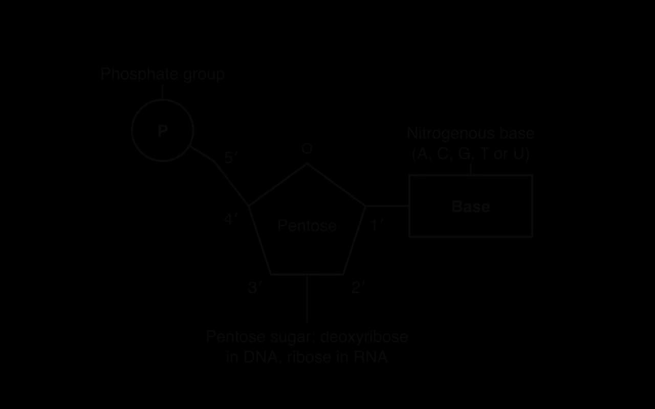
In diagrams of nucleic acids, the parts of a nucleotide are drawn with simple shapes rather than full chemical structures: a circle for the phosphate, a pentagon for the pentose and a rectangle for the base.
The five carbon atoms of the pentose are numbered 1′ to 5′ (read as "one prime" to "five prime"). The prime symbol distinguishes the carbon atoms of the sugar from the atoms of the base, which have their own numbers. Four of the carbons (1′ to 4′) form the ring together with an oxygen atom, and carbon 5′ sticks out of the ring.
The base is attached to carbon 1′.
The phosphate is attached to carbon 5′.
Carbon 3′ carries an –OH group, where the phosphate of the next nucleotide attaches in a strand.
Carbon 2′ carries an –OH group in ribose but only a hydrogen atom in deoxyribose. "Deoxy" means "missing an oxygen".
Four different bases occur in each nucleic acid. DNA contains adenine (A), cytosine (C), guanine (G) and thymine (T). RNA contains adenine, cytosine and guanine, but uracil (U) in place of thymine.
Base | Letter | Found in DNA | Found in RNA |
|---|---|---|---|
Adenine | A | Yes | Yes |
Cytosine | C | Yes | Yes |
Guanine | G | Yes | Yes |
Thymine | T | Yes | No |
Uracil | U | No | Yes |
The sugar and phosphate are the same in every nucleotide of a strand, so they carry no information. The bases differ, and it is the sequence of bases along a strand that stores genetic information, in the same way that the sequence of letters in a sentence carries its meaning. The bases are read in groups of three, and each group of three bases (a codon) specifies one amino acid in a protein or a signal to stop. The full set of rules linking codons to amino acids is the genetic code.
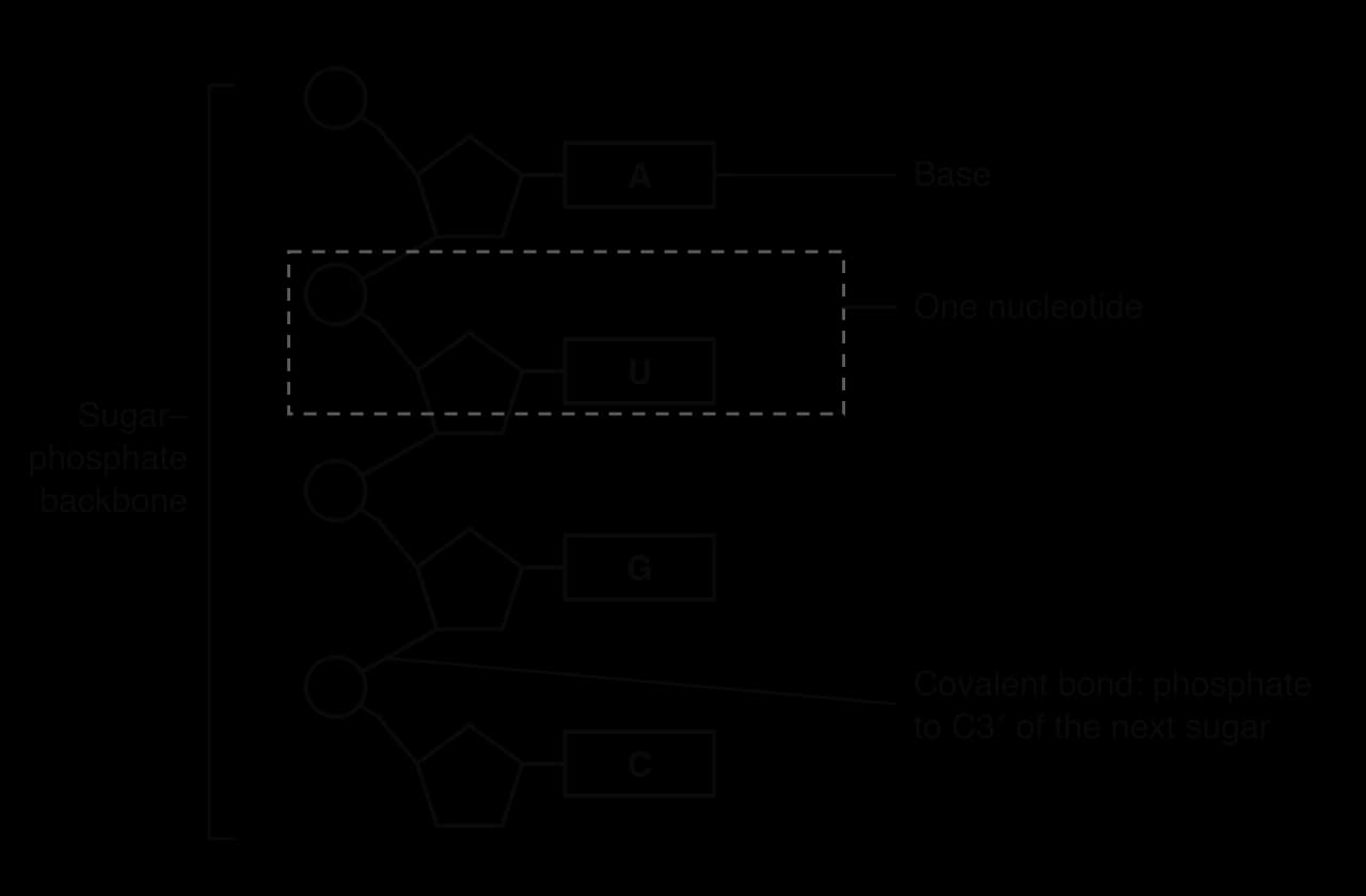
Nucleotides are joined into a strand by condensation reactions. In each reaction, the phosphate group of one nucleotide bonds to the –OH group on carbon 3′ of the pentose of the previous nucleotide, and a molecule of water is released. The reverse reaction, which breaks the strand apart by adding water, is hydrolysis.
The bond formed links the phosphate to two sugars, one on each side, so it is called a phosphodiester bond. Repeated condensation reactions produce a long chain in which sugar and phosphate alternate. This chain is the sugar–phosphate backbone, and the bases project from it, one on each sugar.
An RNA molecule is a single strand of nucleotides. A diagram of a short RNA strand shows nucleotides drawn with simple shapes, linked from the C3′ of one pentose to the phosphate of the next.
A DNA molecule consists of two strands of nucleotides twisted around each other to form a double helix. Each strand has its own sugar–phosphate backbone on the outside of the helix, and the bases point inwards, towards the bases of the other strand. The structure was published in 1953 by James Watson and Francis Crick, using X-ray diffraction images produced by Rosalind Franklin and Maurice Wilkins.
The two strands run in opposite directions. In one strand the sugars all point one way, and in the other strand they point the opposite way, so one strand is "upside down" relative to the other. Strands arranged like this are antiparallel. In diagrams the pentagons of the two strands are drawn the opposite way up to show this.
The two strands are held together by hydrogen bonds between the bases. Base pairing is specific: adenine always pairs with thymine, and cytosine always pairs with guanine. These are the complementary bases, and the rule is called complementary base pairing. Each pair of bonded bases is a base pair (bp), and the length of a DNA molecule is often given in base pairs.
The pairing is specific because of the shape of the bases and the positions of the atoms that form hydrogen bonds. Adenine and thymine form two hydrogen bonds; cytosine and guanine form three. Each hydrogen bond is weak, but a DNA molecule has millions of them, so the double helix is stable. Because hydrogen bonds are much weaker than covalent bonds, the two strands can be separated by enzymes without breaking the backbones.
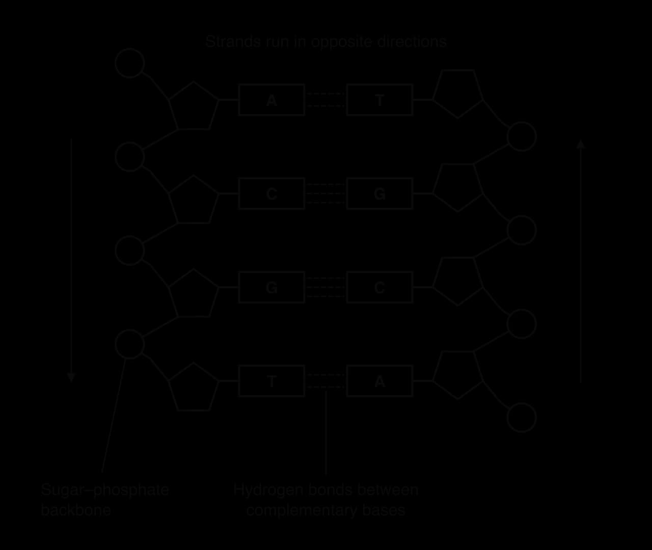
The helix itself does not have to be drawn: a "ladder" diagram like the one of four base pairs shows all the features that matter. The sugar–phosphate backbones are the sides of the ladder, and the base pairs are the rungs.
DNA and RNA are built in the same way, but they differ in three structural features: the number of strands, the bases they contain and the type of pentose sugar.
Feature | DNA | RNA |
|---|---|---|
Number of strands | Two strands, forming a double helix | Usually one strand |
Bases | Adenine, cytosine, guanine and thymine | Adenine, cytosine, guanine and uracil |
Pentose sugar | Deoxyribose (H on carbon 2′) | Ribose (–OH on carbon 2′) |
These differences suit the two molecules to different jobs. DNA is double-stranded and its sugar lacks the reactive –OH group on carbon 2′, so it is chemically stable: a good long-term store of information that can be copied using either strand. RNA is single-stranded and less stable. It is made when needed, for example as a short-lived copy of a gene, and its single strand can fold into shapes such as transfer RNA. This is an example of form and function at the level of molecules.
The Amoeba Sisters animation compares the structures and roles of DNA and RNA.
 YouTube
Complementary base pairing means that the sequence of one strand determines the sequence of the other. If one strand reads ATGC, its partner must read TACG. This property allows genetic information to be copied (replicated) and to be used to make proteins (expressed). In each case an existing strand acts as a template for building a new one.
In DNA replication, the two strands of a DNA molecule separate and each one acts as a template. Free nucleotides pair with the exposed bases by complementary base pairing, A with T and C with G, and are linked into a new strand. The result is two DNA molecules, each identical to the original. Each new molecule contains one strand from the original molecule and one newly built strand, so replication is semi-conservative.
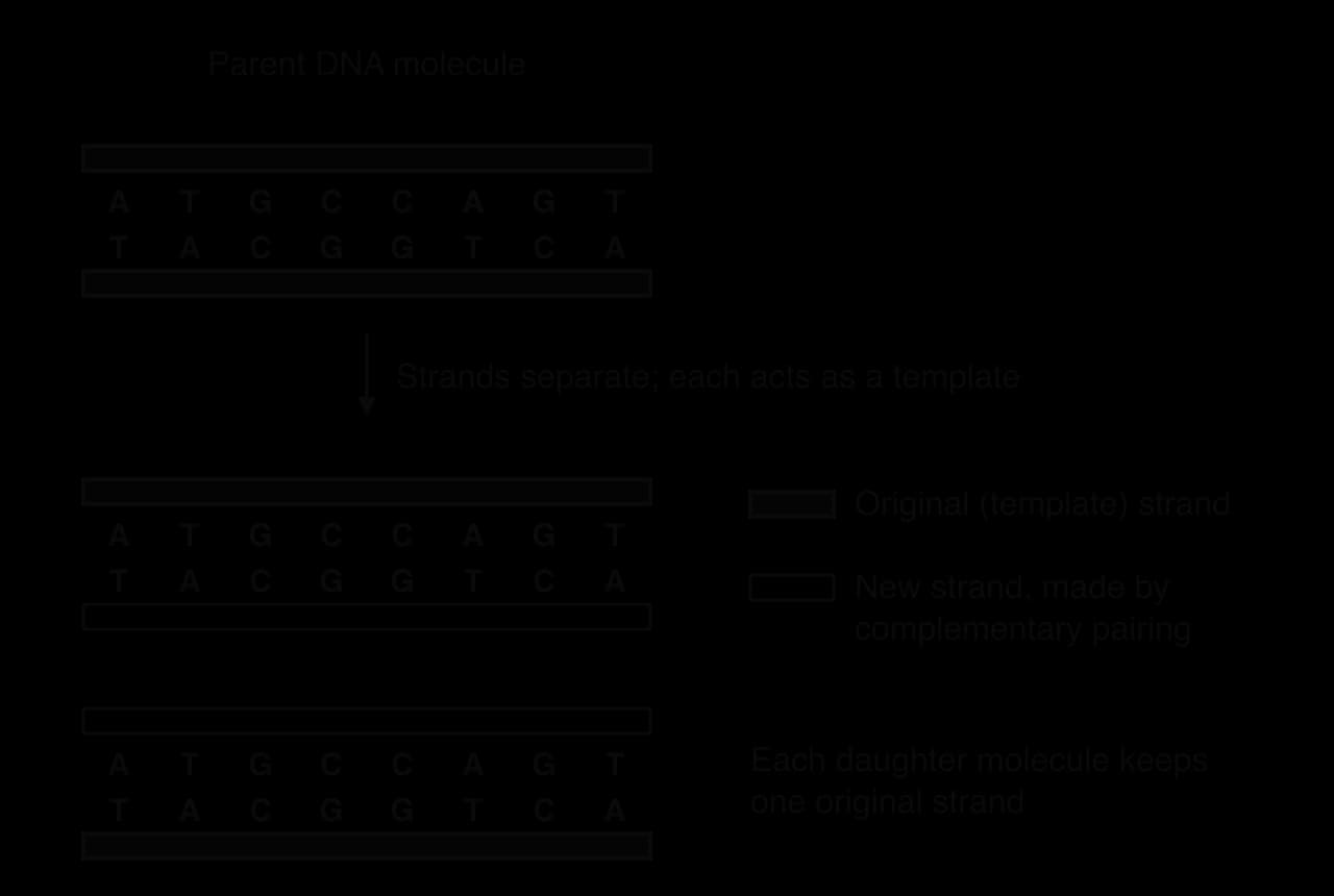
Because pairing is so specific, replication is extremely accurate. The mechanism, including the enzymes involved, is studied in D1.1 DNA replication.
Base pairing is also used in gene expression, studied in D1.2 Protein synthesis.
In transcription, one strand of a gene acts as a template for a molecule of messenger RNA (mRNA). RNA nucleotides pair with the DNA bases: A on DNA pairs with U on RNA, T with A, C with G and G with C. The mRNA carries a copy of the gene's base sequence out of the nucleus.
In translation, transfer RNA (tRNA) molecules bring amino acids to the ribosome. Each tRNA has an anticodon, three bases that pair with a complementary codon on the mRNA. Base pairing between codon and anticodon makes sure that amino acids are added in the order the mRNA specifies.
In each of these processes the template fixes the sequence of the new molecule through complementary base pairing, so genetic information flows reliably from DNA to RNA to protein.
There is no restriction on which base can follow another along a DNA strand. Any of the four bases can occupy each position, whatever bases are next to it. This freedom makes the number of possible sequences enormous.
For a sequence one base long, there are 4 possibilities (A, C, G or T). For two bases there are 4 × 4 = 16 (AA, AC, AG, AT, CA and so on). Each extra base multiplies the number of possibilities by 4. The number of possible sequences of length n is therefore:
Genomes are very long. The first complete, gap-free sequence of a human genome, published in 2022, has about 3.055 billion base pairs. The number of possible sequences of that length, , is far larger than the number of atoms in the universe. For practical purposes, the capacity of DNA for storing information is limitless, which is why DNA can encode the instructions for organisms as different as bacteria, oak trees and humans. This is the molecular basis of the diversity of life.
Because DNA holds so much information in so little space, it has been tested as a medium for storing digital data. Computers store data as binary digits (bits), 0s and 1s. With four bases, each nucleotide can in principle encode two bits (for example A = 00, C = 01, G = 10, T = 11), since .
In 2012, George Church, Yuan Gao and Sriram Kosuri encoded a whole book in DNA: Regenesis, containing 53,426 words, 11 images and a JavaScript program, totalling 5.27 megabits. They used a simple code of one bit per base (A or C for 0, G or T for 1), synthesised short DNA strands carrying the data, and then read the book back by DNA sequencing. Only 10 bits out of more than five million were wrong.
In 2017, Yaniv Erlich and Dina Zielinski developed a method called "DNA Fountain" that stored about 1.6 bits per nucleotide. That is close to the practical maximum of about 1.8 bits, which is below the theoretical 2 bits because some sequences, such as long runs of the same base, are hard to synthesise and sequence accurately and are avoided. They estimated that their method could store about 215 petabytes (215 million gigabytes) in one gram of DNA.
Study | Data stored | Bits per nucleotide | Key result |
|---|---|---|---|
Church, Gao and Kosuri (2012) | Book Regenesis: 53,426 words, 11 images, a JavaScript program (5.27 megabits) | 1 | Read back with 10 bit errors |
Erlich and Zielinski (2017) | Files including an operating system and a film | About 1.6 | Density about 215 petabytes per gram |
Sources: Church, Gao and Kosuri, Science, 2012; Erlich and Zielinski, Science 355:950, 2017.
These experiments make the idea of "limitless capacity" concrete. A cell stores the information for a whole organism in a nucleus a few micrometres across, and DNA in a cool, dry place stays readable for thousands of years. The same features that make DNA a good store of hereditary information make it attractive for archives that must last a long time.
The genetic code is almost the same in every organism. The codon GGC codes for the amino acid glycine in bacteria, fungi, plants and animals; AUG codes for methionine and acts as the start codon in all of them; UAA, UAG and UGA are stop codons in nearly all of them. A gene from one species can therefore be transferred into another and produce the same protein, which is how human insulin is made by genetically modified bacteria.
There is no chemical reason why each codon must specify one particular amino acid. The code could have been arranged in a huge number of different ways. The best explanation for all organisms sharing the same arbitrary code is that they inherited it from a single common ancestor. Once the code was established, almost any change to it would have altered many proteins at once and been harmful, so it has been conserved ever since.
A few minor variations exist. In the mitochondria of vertebrates, UGA codes for the amino acid tryptophan instead of acting as a stop codon, and AGA and AGG act as stop codons instead of coding for arginine. Some single-celled organisms also read one or two codons differently. These variations are small, affect only a few codons, and are best explained as changes that happened later in particular lineages to a code that was already shared. They do not weaken the evidence for common ancestry.
The near-universal code is a strong example of unity in biology: underneath the great diversity of organisms lies a single shared molecular system, a sign that all life is related. The last universal common ancestor is studied in A2.1 Origins of cells (HL).
Every strand of DNA or RNA has two different ends, because of the way the sugars are linked. At one end there is a phosphate group attached to carbon 5′ of the last sugar: this is the 5′ end. At the other end there is a free –OH group on carbon 3′ of the last sugar: this is the 3′ end. A strand therefore has a direction, or directionality, and base sequences are always written from the 5′ end to the 3′ end.
In a double helix, the antiparallel arrangement means that the 5′ end of one strand lies opposite the 3′ end of the other. Each strand runs 5′ to 3′ in the opposite direction from its partner.
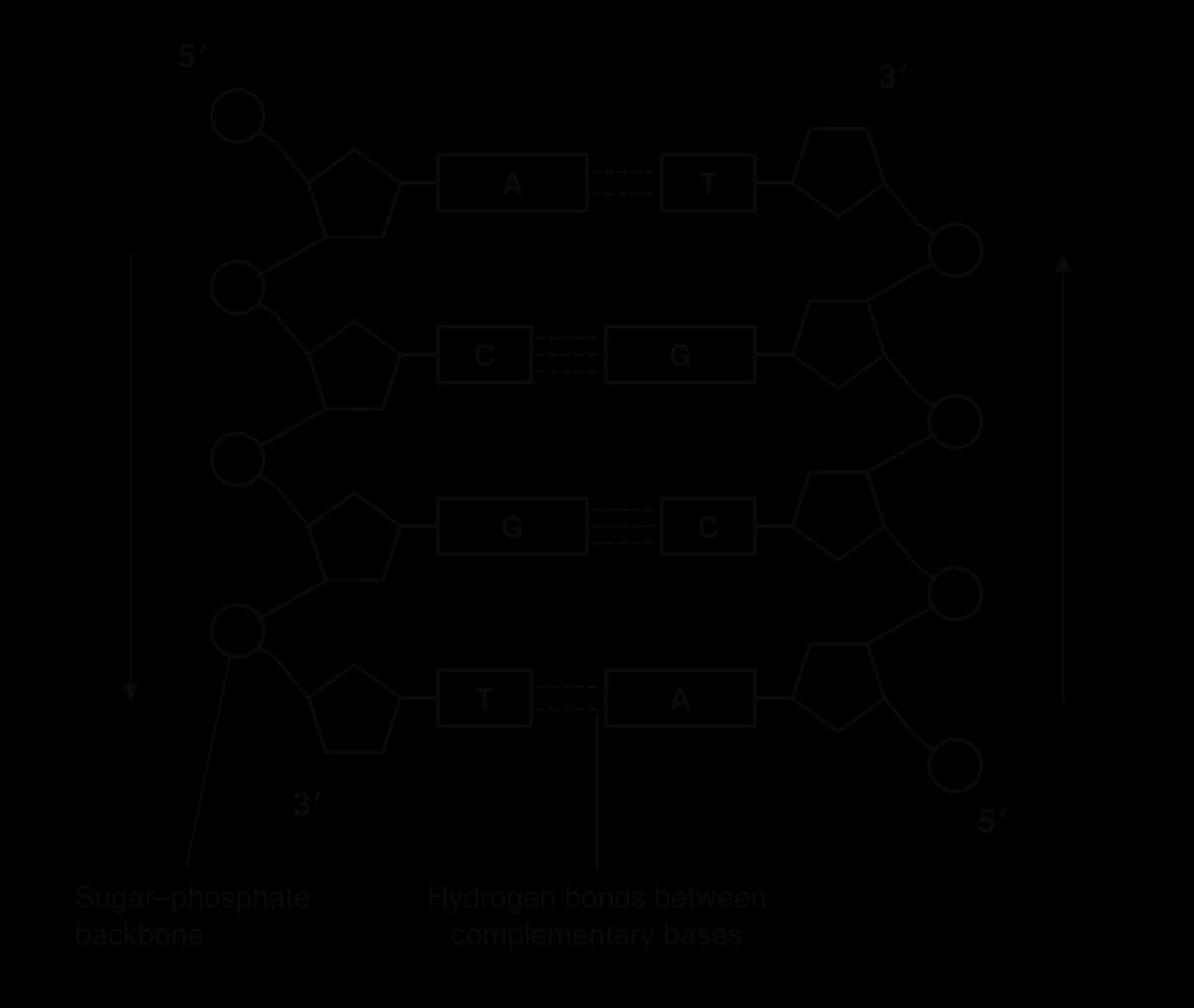
The enzymes that build nucleic acids can add a new nucleotide only to the –OH group on carbon 3′ at the end of a growing strand. The new nucleotide's phosphate (on its own 5′ carbon) bonds to that 3′ –OH by a condensation reaction. Strands therefore always grow in the 5′ → 3′ direction. This one rule has consequences for every process that uses nucleic acids:
Replication. DNA polymerase can only build a new strand 5′ → 3′. At a replication fork, the two template strands are antiparallel, so one new strand (the leading strand) can be built continuously towards the fork, while the other (the lagging strand) must be built in short fragments in the opposite direction and then joined (D1.1 DNA replication).
Transcription. RNA polymerase moves along the template strand of DNA inside a small region where the strands are separated (the transcription bubble), building the RNA 5′ → 3′. The template strand is read 3′ → 5′, so the RNA is antiparallel to it.
Translation. Ribosomes move along mRNA from its 5′ end towards its 3′ end, reading codons in that order. The start codon is near the 5′ end of the mRNA.
The four bases of DNA belong to two chemical groups. Adenine and guanine are purines, with a double-ring structure (a six-membered ring fused to a five-membered ring). Cytosine, thymine and uracil are pyrimidines, with a single six-membered ring, so purines are wider than pyrimidines.
Every complementary base pair contains one purine and one pyrimidine: A (purine) with T (pyrimidine), and G (purine) with C (pyrimidine). Each base pair is therefore the same width, about 2 nm across including the backbones, and the two sugar–phosphate backbones stay a constant distance apart along the whole molecule. A purine–purine pair would be too wide to fit and a pyrimidine–pyrimidine pair too narrow to reach across.
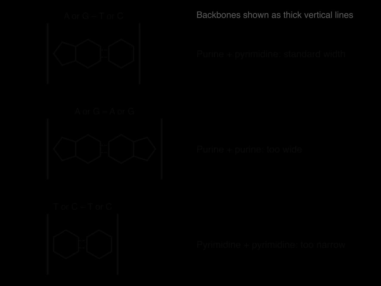
A constant diameter allows the base pairs to stack neatly on top of each other in the middle of the helix. Weak attractions between the flat, stacked bases add to the stability provided by hydrogen bonding. Together, the purine–pyrimidine rule and the specific hydrogen bonding explain why A pairs only with T and G only with C.
The DNA in a single human cell would measure about 2 m if stretched out end to end, yet it fits inside a nucleus only a few micrometres across. In eukaryotes, DNA is packed by winding it around proteins called histones. The basic unit of packing is the nucleosome.
The core of each nucleosome is a histone octamer: eight histone proteins, two each of the histones H2A, H2B, H3 and H4.
A length of DNA of about 147 base pairs is wrapped around the octamer about 1.65 times (often described as "twice").
A further histone, H1, binds to the DNA where it enters and leaves the nucleosome, holding the wrapped DNA in place and helping nucleosomes pack together.
A short stretch of linker DNA connects each nucleosome to the next, giving a "beads on a string" appearance under the electron microscope.
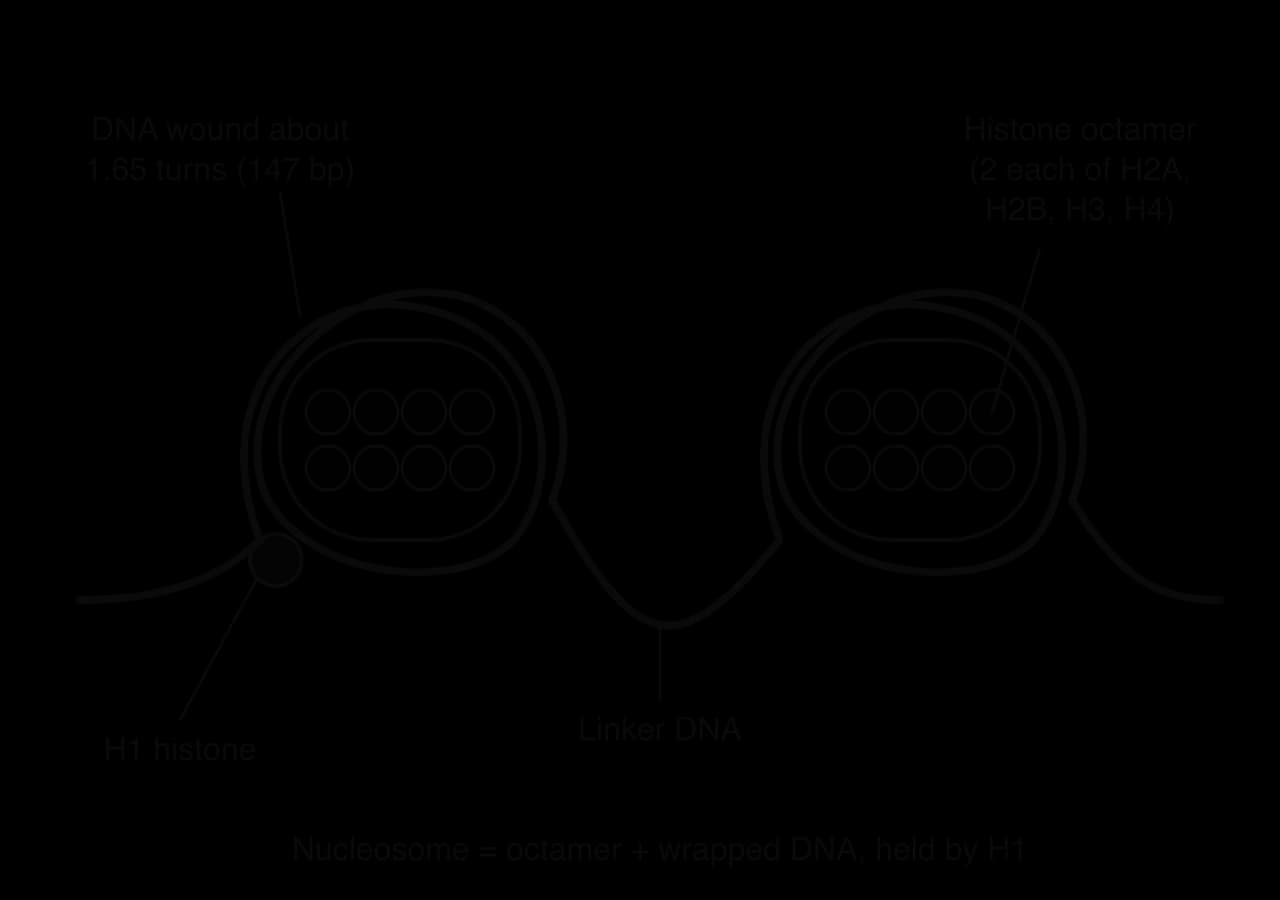
The binding between DNA and histones depends on charge. DNA is negatively charged along its whole length because of its phosphate groups. Histones contain many positively charged amino acids (lysine and arginine), which are attracted to the phosphates. This attraction holds the DNA tightly to the histone core without depending on the base sequence, so any DNA can be packed. Nucleosomes also help to control which genes are expressed, because tightly packed DNA is less accessible to the enzymes of transcription (D2.2 Gene expression, HL).
The detailed shape of the nucleosome was found by X-ray crystallography. In 1997, Karolin Luger and colleagues published the structure of the nucleosome core particle at a resolution of 2.8 Å, showing 146 base pairs of DNA wrapped about 1.65 turns around the histone octamer; later structures showed that the core particle holds 147 base pairs. Coordinates from structures like this are stored in the Protein Data Bank (the 1997 structure is entry 1AOI) and can be explored with free molecular visualisation software such as Jmol or Mol*.
With such software the molecule can be rotated and coloured by chain or by charge. Doing this shows how the DNA wraps around the outside of the octamer, how the tails of the histones extend between the DNA turns, and how the positively charged regions of the histones line up with the negatively charged phosphates of the DNA. Visualisation tools are an example of technology allowing scientists, and students, to see structures that are far too small to observe directly.
The DNA Learning Center animation shows how DNA is wound into nucleosomes and then coiled further into chromosomes.
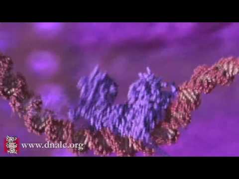 YouTube
By the 1940s, biologists knew that genes were carried on chromosomes, which contain both DNA and protein. Many thought protein was more likely to be the genetic material, because proteins are built from 20 different amino acids and seemed more able to carry complex information than DNA with only four bases.
In 1952, Alfred Hershey and Martha Chase tested this using a bacteriophage (a virus that infects bacteria) called T2. A T2 phage is made of only two kinds of molecule: a protein coat and DNA inside it. When it infects a bacterium, part of the phage enters the cell and directs it to make new phages. Whichever molecule enters the cell must be the genetic material.
The experiment depended on labelling each molecule with a different radioisotope: a radioactive form of an element that can be detected and tracked.
Proteins contain sulfur (in the amino acids cysteine and methionine) but DNA does not. Phages grown with radioactive sulfur, ³⁵S, therefore had radioactive protein coats.
DNA contains phosphorus (in its phosphate groups) but most proteins do not. Phages grown with radioactive phosphorus, ³²P, therefore had radioactive DNA.
Each batch of labelled phages was mixed with the bacterium Escherichia coli and allowed to infect it.
The mixture was agitated in a kitchen blender, which knocked the phage coats off the outside of the bacteria.
The mixture was centrifuged. The heavier bacteria formed a pellet at the bottom of the tube, while the lighter phage coats stayed in the liquid (the supernatant).
The radioactivity of the pellet and the supernatant was measured.
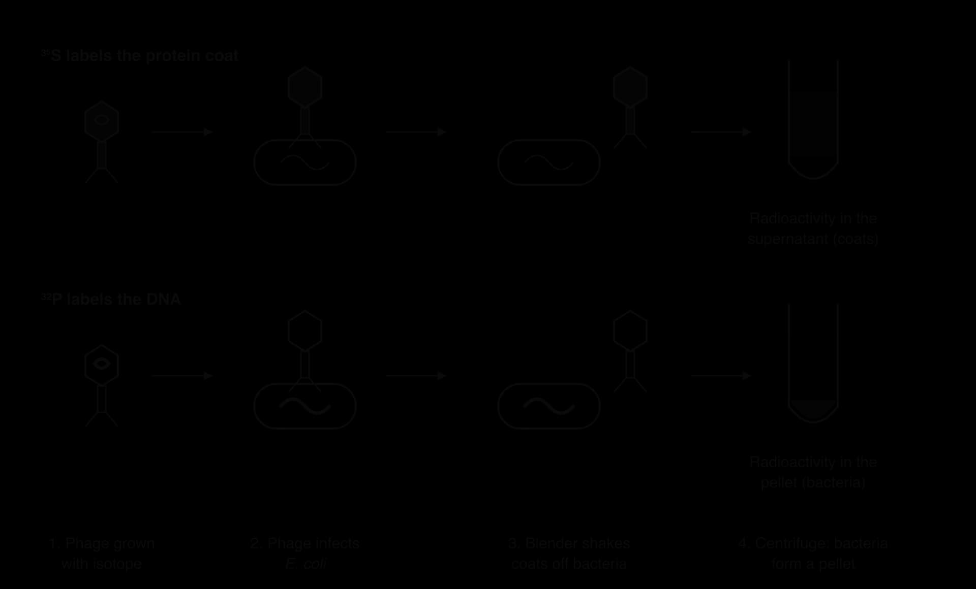
With ³⁵S-labelled phages, most of the radioactivity was in the supernatant, with the empty coats. With ³²P-labelled phages, most of the radioactivity was in the pellet, inside the bacteria. The bacteria infected with ³²P-labelled phages went on to produce new phages, and some of these contained ³²P. Hershey and Chase concluded that DNA, not protein, enters the bacterial cell and directs the production of new viruses, so DNA is the genetic material of T2.
The result was not perfectly clean: a little ³⁵S stayed with the pellet, because blending did not remove every coat. The conclusion was nevertheless clear, and it supported earlier evidence from Oswald Avery and colleagues in 1944 that DNA could transform the heritable characteristics of bacteria. Hershey shared the 1969 Nobel Prize in Physiology or Medicine.
In the 1920s and 1930s, many biochemists accepted the tetranucleotide hypothesis, proposed by Phoebus Levene. It stated that DNA was made of a simple repeating unit of the four bases in equal amounts, such as ACGT ACGT ACGT. If this were true, DNA would be a monotonous molecule with no variation between species, unable to carry genetic information. The hypothesis was one reason why protein was thought more likely to be the genetic material.
From the late 1940s, Erwin Chargaff measured the percentage of each base in DNA from many different organisms, using paper chromatography to separate the bases and ultraviolet absorption to measure them.
Organism | A / % | T / % | G / % | C / % |
|---|---|---|---|---|
Human (thymus) | 30.9 | 29.4 | 19.9 | 19.8 |
Sea urchin | 32.8 | 32.1 | 17.7 | 17.3 |
Grasshopper | 29.3 | 29.3 | 20.5 | 20.7 |
Wheat | 27.3 | 27.1 | 22.7 | 22.8 |
Yeast (Saccharomyces) | 31.3 | 32.9 | 18.7 | 17.1 |
Escherichia coli | 24.7 | 23.6 | 26.0 | 25.7 |
Bacteriophage φX174 (single-stranded DNA) | 24.0 | 31.2 | 23.3 | 21.5 |
Sources: Chargaff and Davidson, The Nucleic Acids, 1955 (human); other rows from Chargaff's data as tabulated by Bansal, Current Science, 2003.
Two patterns stand out in the table of base percentages, and they are now called Chargaff's rules:
In every species, the amount of adenine is about equal to the amount of thymine (A ≈ T), and the amount of guanine is about equal to the amount of cytosine (G ≈ C). It follows that the total of purines equals the total of pyrimidines (A + G ≈ T + C). The one row that breaks this pattern is the virus φX174, whose DNA is single-stranded, so its bases are not paired.
The proportions differ between species. Human DNA is about 60% A + T and sea urchin DNA about 65%, while the DNA of E. coli is only about 48% A + T. The amounts of A and G are not equal to each other.
The second pattern falsified the tetranucleotide hypothesis. If DNA were a repeating ACGT unit, every base would make up 25% of the DNA of every species, and the data show that this is not true. Rejecting a hypothesis because reliable measurements contradict its predictions is called falsification, and it applies even to a hypothesis that is widely accepted. The variation between species also showed that DNA was varied enough to carry genetic information.
The first pattern, A ≈ T and G ≈ C, was not explained until 1953. Watson and Crick's model, with A always paired with T and G with C, accounts for it exactly: if every A has a T opposite it, the two must be present in equal amounts. Chargaff's data are therefore also strong evidence for complementary base pairing. The small differences from exact equality in the table come from measurement error.
Nucleic acids appear in Paper 1A as questions on nucleotide structure, base pairing and the differences between DNA and RNA; in Paper 1B as data on base composition or experiments with labelled molecules; and in Paper 2 as drawings of DNA and RNA strands and explanations of how structure relates to replication and information storage. Common command terms are draw, state, distinguish, calculate, explain and, at HL, evaluate and deduce.
Part (b) shows why ratios are useful: the A + T to G + C ratio is 1.52 in humans but about 0.93 in E. coli, while the A to T ratio stays close to 1 in both.
Complementary base pairing is used again in DNA replication and in protein synthesis.
I can state that DNA is the genetic material of all living organisms and explain why RNA viruses do not contradict this.
I can draw a nucleotide with simple shapes and number the carbon atoms of the pentose.
I can draw a short RNA strand showing the sugar–phosphate backbone and condensation linkage.
I can draw a section of DNA showing antiparallel strands and complementary base pairs with the correct numbers of hydrogen bonds.
I can distinguish between DNA and RNA in three ways.
I can explain how complementary base pairing allows replication, transcription and translation.
I can calculate the number of possible sequences using 4ⁿ and explain the limitless capacity of DNA.
I can explain why the universal genetic code is evidence for common ancestry.
I can label the 5′ and 3′ ends of strands and explain why nucleotides are added only to the 3′ end. (HL)
I can explain how purine–pyrimidine pairing and nucleosomes contribute to DNA structure. (HL)
I can describe and evaluate the Hershey–Chase experiment. (HL)
I can use Chargaff's data to support base pairing and to falsify the tetranucleotide hypothesis. (HL)
[4 max]
Explain how the properties of water allow it to be transported to the top of tall trees.
7 marks
a. water molecules are polar and form hydrogen bonds with each other;
b. (so) water is cohesive / water molecules stick together;
c. water evaporates from the cell walls of leaf cells (and is lost by transpiration);
d. water is drawn from the xylem through the cell walls by capillary action;
e. (due to) adhesion of water to hydrophilic cellulose in the cell walls;
f. this creates tension / negative pressure in the xylem;
g. water is pulled up the xylem as a continuous column;
h. cohesion stops the column breaking under tension;
i. adhesion of water to the xylem walls also helps to support the column;
j. lignified xylem walls withstand the tension without collapsing;
k. the process is passive / needs no energy from the plant;
l. water also carries dissolved mineral ions to the leaves because it is a solvent;
[7 max]
One additional mark is available for the construction of the answer.
Which components are found in every nucleotide of DNA?
A purified nucleic acid sample contains adenine, cytosine, guanine and uracil but no thymine. What could the sample be?