 YouTube
Explain what organelles are, why cells are compartmentalised, and (HL) how mitochondria, chloroplasts, the nucleus, ribosomes, the Golgi apparatus and vesicles are adapted to their functions.
Each is in the Biology glossary, with every other term in the subject.
An 80S ribosome attached to the rough endoplasmic reticulum, synthesising a protein that enters the ER lumen for secretion, the plasma membrane or lysosomes.
The breaking open of cells and separation of their organelles into different fractions, usually by centrifugation at increasing speeds, so that each type can be studied.
One of the flattened, membrane-bound sacs that make up the Golgi apparatus or the endoplasmic reticulum.
A protein that assembles into a curved, cage-like coat on the cytoplasmic side of a membrane, bending it into a pit that buds off as a vesicle.
The division of a cell into separate membrane-bound compartments, each with its own conditions, enzymes and substrates, which concentrates reactants and keeps incompatible processes apart.
An 80S ribosome in the cytosol that is not attached to a membrane; it synthesises proteins for use within the cytoplasm.
The addition of carbohydrate chains to a protein (or lipid), which takes place in the rough ER and Golgi apparatus and produces glycoproteins.
The narrow space between the outer and inner membranes of a mitochondrion (or chloroplast) into which protons are pumped, building up a proton gradient.
A eukaryotic cell is divided into separate compartments, each surrounded by its own membrane and each holding a particular set of enzymes and molecules. These compartments and the other discrete structures in the cell are called organelles. Dividing the cell in this way allows processes that would interfere with each other to run side by side in the same cell.
The idea links the molecular level of organisation to the level of the cell. The structure of each organelle (the number of membranes, the folding of those membranes, the volume enclosed) matches the job it does, which makes organelles one of the clearest examples of form and function in biology.
39 exam-style questions on this subtopic, written in the style of the IB papers. The first has its markscheme open. Counted on
Which structure in a eukaryotic cell is classed as an organelle?
Organelles are discrete subunits of a cell adapted to a specific function; ribosomes (like nuclei, vesicles and the plasma membrane) count as organelles even though they have no membrane. The cell wall is an extracellular structure, the cytoskeleton is a network that extends throughout the cell rather than a discrete subunit, and the cytoplasm is the whole region in which the organelles are found.
The answer is C.
Before cell fractionation, liver tissue is homogenized in an ice-cold, buffered sucrose solution that is isotonic with the cells. Which statements give reasons for these conditions?
I. The low temperature slows the activity of enzymes that would digest the organelles.
II. The isotonic solution prevents organelles from bursting or shrinking by osmosis.
III. The high solute concentration makes the organelles denser so that they sediment at lower speeds.
The official deck for this part of the syllabus.
Why is a eukaryotic cell divided into separate membrane-bound compartments?
State three features of an organelle's structure that are matched to the job it does.
With a free account, Augment remembers where you stopped and what you have practiced.
A discrete subunit of a cell that has a distinct structure and is adapted to perform one or more specific functions, for example the nucleus, ribosomes, vesicles and the plasma membrane.
A membrane-bound vacuole formed around a particle, such as a bacterium, taken into a cell by phagocytosis; it fuses with lysosomes so that the particle is digested.
The release from a cell of useful substances that the cell has made, such as enzymes, hormones or mucus, usually by exocytosis.
A membrane-bound vesicle that carries substances made in the cell, usually proteins from the Golgi apparatus, to the plasma membrane and releases them by exocytosis.
A short sequence of amino acids at the start of a polypeptide that directs the ribosome making it to the rough ER, so the polypeptide enters the ER lumen.
A centrifuge that spins samples fast enough to produce forces of more than 100,000 times gravity, used to separate small organelles and large molecules.
Organelles are the working parts of a cell. Each one is a discrete unit with a recognisable structure, and each is adapted to carry out one or a few specific functions. Most organelles in a eukaryotic cell are bounded by one or two membranes, but some, such as ribosomes, have no membrane at all.
Using this definition, the nucleus, ribosomes, vesicles and the plasma membrane (also called the cell membrane) are all organelles. Each is a discrete structure with its own job: the nucleus holds the chromosomes, ribosomes synthesise polypeptides, vesicles carry materials around the cell, and the plasma membrane controls what enters and leaves.
Three parts of the cell are usually not counted as organelles, and the reasons follow from the definition:
Cell wall. The cell wall of a plant, fungus or bacterium lies outside the plasma membrane. It is made of material that the cell secretes (cellulose in plants), so it is an extracellular structure rather than a subunit of the cell itself.
Cytoskeleton. The cytoskeleton is a network of protein fibres (microtubules, microfilaments and intermediate filaments) that runs through the whole cytoplasm. It is not a discrete unit with a boundary, so it is treated as a system of the whole cell.
Cytoplasm. The cytoplasm is everything inside the plasma membrane apart from the nucleus. It contains the organelles, so it cannot also be one of them. The fluid part of it, without the organelles, is called the cytosol.
A useful way to sort organelles is by the number of membranes that surround them. The number of membranes is easy to see in an electron micrograph and is linked to the organelle's function and origin.
Number of membranes | Organelles | Main function |
|---|---|---|
None | Ribosomes; centrioles (animal cells) | Protein synthesis; organising microtubules |
Single membrane | Plasma membrane; rough and smooth endoplasmic reticulum; Golgi apparatus; lysosomes; vesicles; vacuoles | Controlling exchange with the surroundings; making, modifying, transporting and storing materials; digestion |
Double membrane | Nucleus; mitochondrion; chloroplast (plants and algae) | Holding the chromosomes; aerobic cell respiration; photosynthesis |
The double-membrane organelles are among the largest in the cell and have the most specialised internal structure. Mitochondria and chloroplasts also contain their own DNA and 70S ribosomes, evidence that they descend from bacteria taken in by an ancestral eukaryotic cell (endosymbiosis, A2.2 Cell structure).
Among the single-membrane organelles are secretory vesicles, small sacs that carry proteins made in the cell to the plasma membrane. There they fuse with the membrane and release their contents to the outside by exocytosis. This release of useful substances made by a cell is called secretion; digestive enzymes, hormones and mucus all leave cells this way.
YouTubeLight microscopes show only the largest organelles. Most of what is known about how organelles work comes from separating them from each other and studying each type on its own. The method is called cell fractionation, and it depended on a new piece of technology, the ultracentrifuge, which spins samples fast enough to produce forces of more than 100,000 times gravity (written 100,000 g).
Tissue such as liver is first cut up and broken open in a blender called a homogeniser. The resulting homogenate is spun in a centrifuge. The heaviest and densest organelles sink first and form a pellet at the bottom of the tube; lighter organelles stay in the liquid above, the supernatant. The supernatant is poured into a fresh tube and spun again at a higher speed, which brings down the next-heaviest organelles. Repeating this gives a series of pellets, each rich in one kind of organelle.
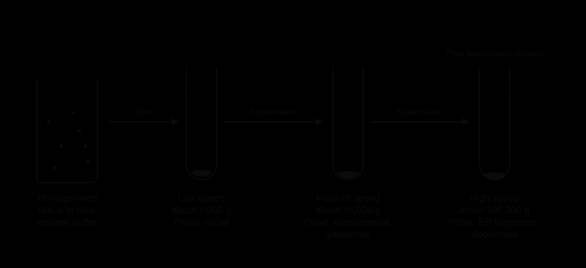
Three conditions of the homogenate protect the organelles. The buffer is kept ice-cold so that enzymes released from broken organelles (especially digestive enzymes from lysosomes) work too slowly to damage the other organelles. It is isotonic, with the same solute concentration as the cells, so that organelles neither gain water by osmosis and burst nor lose water and shrink. It is also buffered to keep the pH constant, so that proteins are not denatured.
The results of a fractionation experiment are usually shown as the share of an enzyme's activity found in each fraction. The table gives illustrative results of this kind for acid phosphatase in liver, set out as a practice question.
Fraction | Main contents | % of total acid phosphatase activity |
|---|---|---|
1 (1,000 g pellet) | Nuclei, unbroken cells | 6 |
2 (10,000 g pellet) | Mitochondria, lysosomes | 72 |
3 (100,000 g pellet) | ER fragments, ribosomes | 14 |
4 (final supernatant) | Cytosol | 8 |
The nucleus is the compartment that holds the chromosomes of a eukaryotic cell. Its envelope separates the chromosomes from the cytoplasm, and this separates two stages of gene expression in space. Transcription, in which a gene is copied into messenger RNA (mRNA), happens inside the nucleus. Translation, in which ribosomes use the mRNA to build a polypeptide, happens in the cytoplasm.
Because the two stages happen in different places, a newly made RNA transcript can be altered before any ribosome meets it. The first transcript of a eukaryotic gene is called pre-mRNA. Inside the nucleus it undergoes post-transcriptional modification: non-coding sections (introns) are cut out and the coding sections (exons) are joined together, and the two ends are protected by a cap and a tail. Only the finished mRNA is exported through the nuclear pores to the cytoplasm.
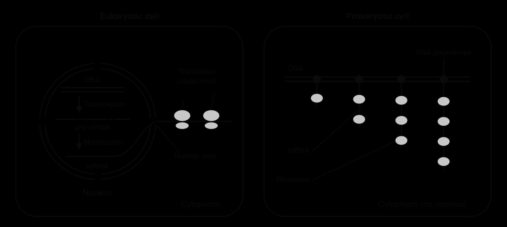
In a prokaryote there is no nucleus, so the DNA lies in the same compartment as the ribosomes. Ribosomes attach to the mRNA as soon as its first part has been made, while RNA polymerase is still transcribing the rest of the gene. Transcription and translation are therefore coupled, and the mRNA cannot be modified first. Prokaryotic genes almost never contain introns, so this is not a problem for them; eukaryotic genes do, and translating an unmodified transcript would produce a faulty polypeptide.
Separation also allows one gene to give more than one product. By removing different combinations of exons (alternative splicing), a single eukaryotic gene can be made into several different mRNAs and so several different proteins. Keeping the mRNA in the nucleus until it is finished also allows the cell to check transcripts and destroy faulty ones before they reach the ribosomes.
The same principle applies within the cytoplasm. Membranes divide it into many separate spaces, such as the lumen of the endoplasmic reticulum, the inside of a lysosome and the inside of a vesicle. This compartmentalisation has two main advantages.
An enzyme catalyses a reaction only when its substrate collides with its active site. The rate of collisions, and therefore the rate of reaction, rises with the concentration of both. Inside a small compartment, the enzymes of a metabolic pathway and their substrates are held together at high concentration instead of being diluted through the whole cytoplasm. The product of one reaction is also close to the enzyme for the next step, so a pathway with many steps runs quickly.
Each compartment can also keep its own conditions. The inside of a lysosome is kept at about pH 4.5–5, while the cytosol around it is close to pH 7.2. A proton pump in the lysosome membrane maintains this difference, using ATP to move H⁺ ions into the lysosome. The enzymes inside have their optimum pH in the acid range, so they work at full rate only in their own compartment.
Some processes would damage each other or the cell if they took place in the same space. Digestion is the clearest example. A cell contains many hydrolytic enzymes able to break down proteins, nucleic acids, lipids and carbohydrates. In the open cytoplasm these enzymes would digest the cell's own molecules and organelles. Enclosing them in lysosomes allows the cell to digest unwanted material safely.
A lysosome is a small vesicle, about 0.1–1 µm across, bounded by a single membrane and containing around 50 different hydrolytic enzymes. The enzymes are made on the rough endoplasmic reticulum and packaged into lysosomes by the Golgi apparatus. Lysosomes digest material taken into the cell, break down worn-out organelles (autophagy) and, in some cases, destroy the whole cell when it is no longer needed.
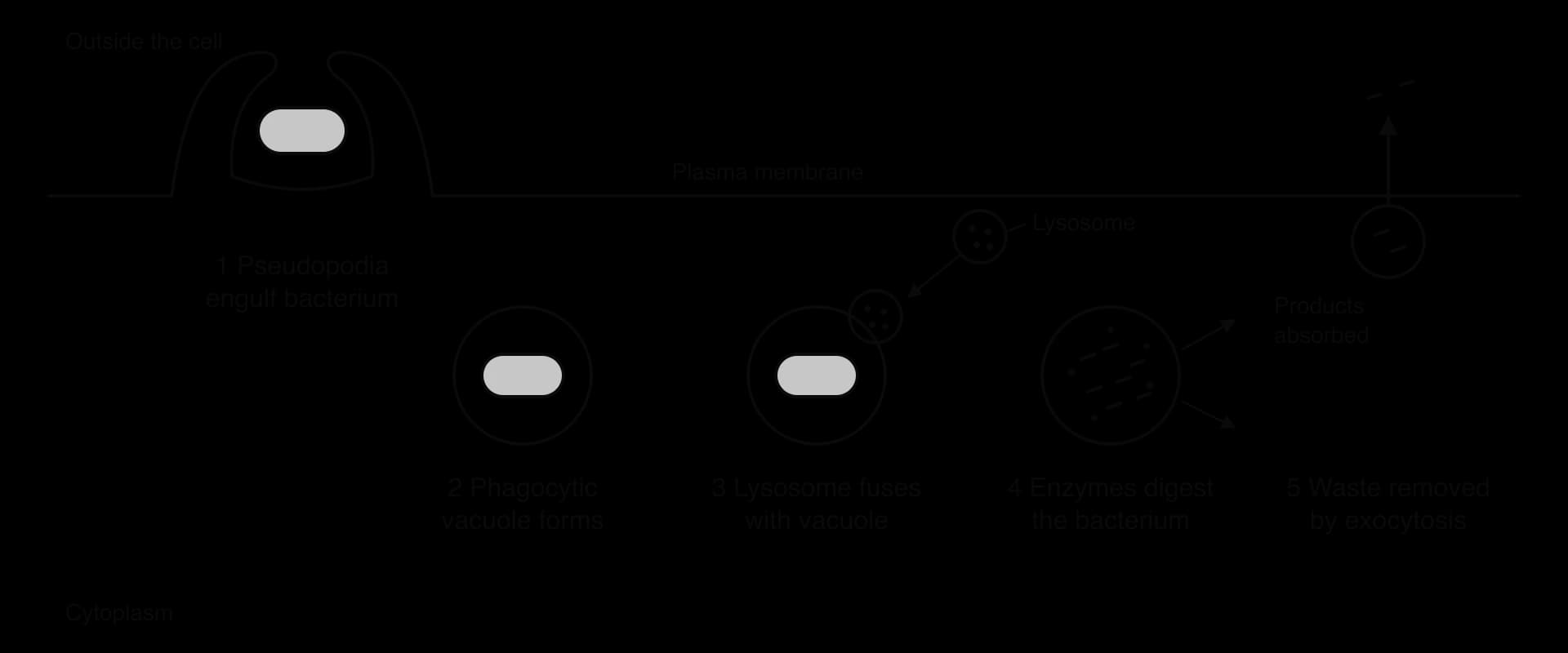
White blood cells called phagocytes, such as macrophages, use lysosomes to destroy bacteria. The phagocyte extends parts of its cytoplasm, called pseudopodia, around a bacterium. The pseudopodia meet and fuse, enclosing the bacterium in a membrane-bound sac called a phagocytic vacuole. This uptake of large particles is phagocytosis, a type of endocytosis (B2.1 Membranes and membrane transport).
Lysosomes then move to the vacuole and their membranes fuse with its membrane. The hydrolytic enzymes pour in and digest the bacterium, while the vacuole membrane keeps them away from the rest of the phagocyte. Small, useful products such as amino acids and sugars are absorbed into the cytoplasm, and the indigestible remains are removed from the cell by exocytosis. The role of phagocytes in defending the body is covered in C3.2 Defence against disease.
Tay–Sachs disease shows what happens when one compartment fails. Lysosomes in nerve cells normally break down a lipid called GM2 ganglioside, which is part of the membranes of neurons. The enzyme that does this is hexosaminidase A. In Tay–Sachs disease a mutation in the gene for this enzyme (HEXA) means that no working hexosaminidase A is made.
Without the enzyme, GM2 ganglioside is still taken into lysosomes but cannot be digested. It builds up inside them until the lysosomes swell and the neurons are destroyed. Affected babies seem healthy at birth, but from about three to six months they lose motor skills and then sight and hearing. Most die in early childhood. The disease is called a lysosomal storage disease, because the undigested material is stored in the lysosomes.
The disease is inherited as a recessive allele: a child must inherit the faulty allele from both parents to be affected. People with one copy are carriers and are healthy, because one working allele produces enough enzyme. The allele is unusually common in some populations, including Ashkenazi Jewish communities, where about 1 in 27–30 people is a carrier.
In 1971 a carrier-screening programme began in the Jewish community of the Baltimore–Washington area of the USA, led by Michael Kaback. Carriers are identified with a simple blood test that measures hexosaminidase A activity, which is about half the normal level in a carrier. Couples who are both carriers can then make informed decisions about having children. Screening spread through Jewish communities in North America and worldwide, and the number of affected babies fell sharply.
Measure | Value |
|---|---|
Carrier frequency among Ashkenazi Jews | About 1 in 27–30 |
Start of community carrier screening | 1971 |
Affected births a year among Jewish people in the USA and Canada before screening | About 50–60 |
Affected births a year in the same group after screening became widespread | About 2–5 |
Fall in affected births | More than 90% |
Sources: Kaback et al., JAMA 270:2307, 1993 (the screening programme); incidence figures as reported by JTA and The Times of Israel.
The case shows both advantages of compartmentalisation. The lysosome concentrates the substrate and the enzyme that digests it in one small space, and keeps the digestion away from the rest of the cell. When the enzyme is missing, the compartment fills with substrate that cannot be removed, and the cell fails. It also shows the interaction between levels of organisation: a change in one gene (molecular level) alters one organelle (cell level), which affects the whole nervous system (organism level).
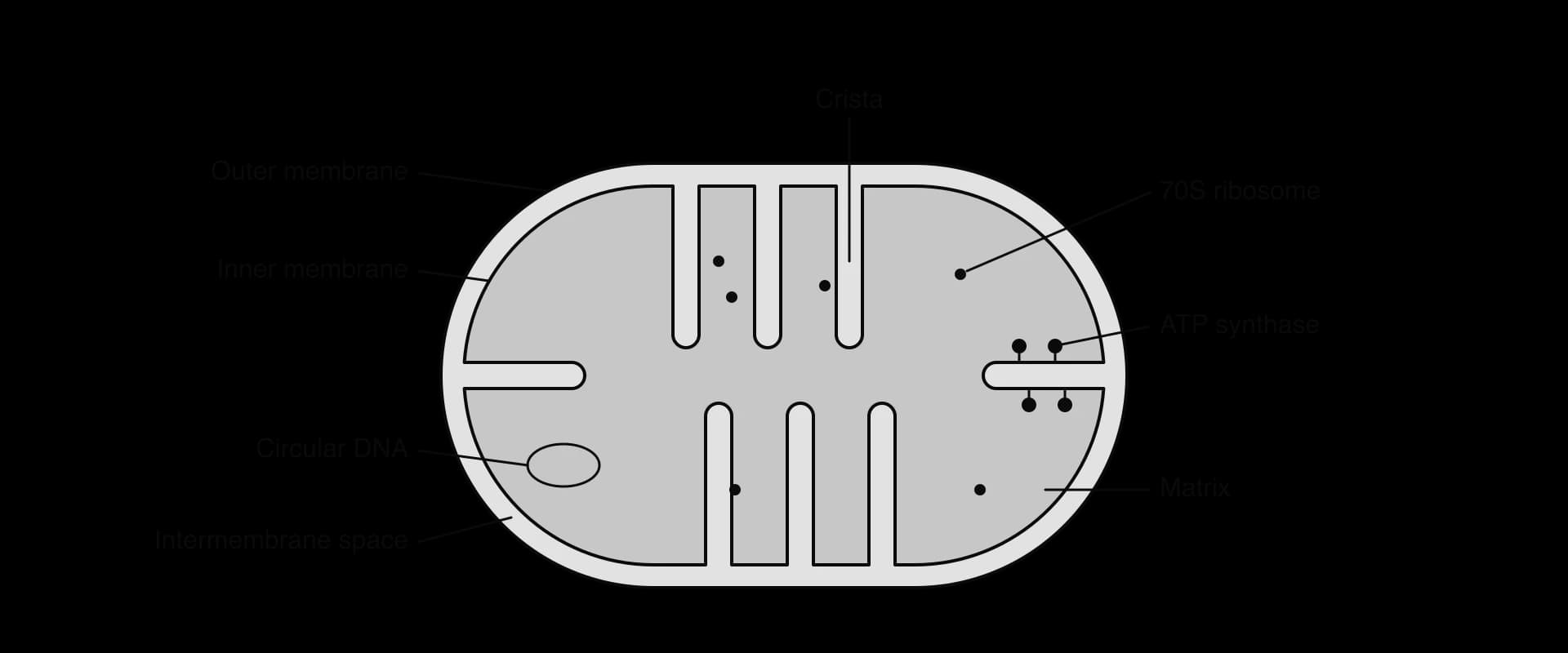
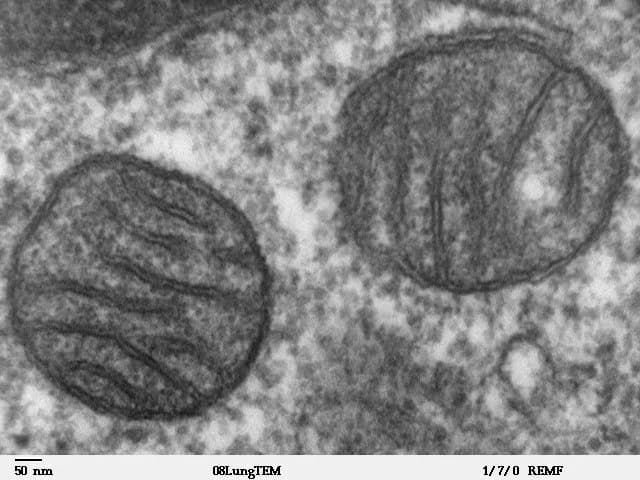
The mitochondrion is the site of aerobic cell respiration, which releases energy from organic molecules and uses it to make ATP. A typical mitochondrion is about 0.5–1 µm wide and a few micrometres long. It is bounded by two membranes, and the inner one is folded into shelf-like cristae.
Structure | Adaptation | How it helps ATP production |
|---|---|---|
Outer membrane | Contains transport proteins | Separates the mitochondrion from the cytoplasm; transports pyruvate and other substrates in |
Inner membrane and cristae | Folded into cristae, giving a large surface area | Holds many copies of the electron transport chain and ATP synthase, so more ATP can be made |
Intermembrane space | Very small volume | Protons pumped into it quickly reach a high concentration, so a steep proton gradient builds up fast |
Matrix | Contains the enzymes of the link reaction and Krebs cycle, plus DNA and 70S ribosomes | Enzymes and substrates are concentrated together; the mitochondrion can make some of its own proteins |
The functions of the two compartments are distinct. The link reaction and the Krebs cycle take place in the matrix, where the enzymes and their substrates are concentrated. These reactions release CO₂ and load the coenzymes NAD⁺ and FAD with electrons. The reduced coenzymes pass their electrons to the electron transport chain, a series of protein carriers in the inner membrane.
As electrons move along the chain, energy is used to pump protons (H⁺) from the matrix into the intermembrane space. Because the space is so narrow, the protons quickly reach a high concentration there, creating a steep proton gradient across the inner membrane. Protons flow back into the matrix through ATP synthase, which uses the energy of this flow to make ATP. Making ATP in this way is called chemiosmosis (C1.2 Cell respiration).
Cells with a high demand for ATP, such as heart muscle and the cells of kidney tubules, have many mitochondria with densely packed cristae. This correlation between ATP demand and the surface area of the inner membrane supports the idea that the cristae are an adaptation for ATP production.
The chloroplast is the site of photosynthesis in plants and algae. It is usually lens-shaped and about 5 µm long, bounded by a double membrane called the envelope. Inside is a system of flattened membrane sacs called thylakoids, surrounded by fluid called the stroma. Thylakoids are stacked into piles called grana (singular granum), and the grana are linked by single thylakoids called lamellae.
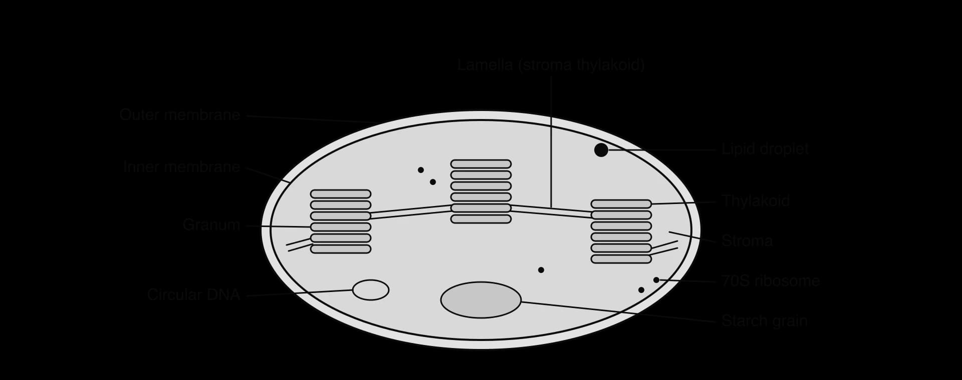
Photosynthesis takes place in two stages, in two different compartments. The light-dependent reactions take place on and across the thylakoid membranes. The Calvin cycle (the light-independent reactions) takes place in the stroma. The structure of the chloroplast is adapted to both.
Large surface area of thylakoid membranes. The thylakoid membranes hold the photosystems, which contain chlorophyll and absorb light, together with electron carriers and ATP synthase. Stacking thylakoids into grana packs a very large area of membrane into a small organelle, so more light can be absorbed and more ATP and NADPH made.
Small volume of fluid inside the thylakoids. Energy from excited electrons is used to pump protons into the thylakoid space. Because the space inside each thylakoid is so small, the proton concentration rises quickly and a steep gradient builds up. Protons flow back into the stroma through ATP synthase, producing ATP by chemiosmosis.
Compartmentalisation of the Calvin cycle in the stroma. The enzymes of the Calvin cycle, including Rubisco, and their substrates are concentrated in the stroma. The ATP and NADPH from the thylakoids are released directly into the stroma, close to the enzymes that use them.
The stroma also contains starch grains (stored products of photosynthesis), lipid droplets, circular DNA and 70S ribosomes. As in the mitochondrion, the inner compartment is enclosed by membranes that hold the light-capturing and proton-pumping machinery, while the fluid compartment holds the enzymes of a cycle.
The nucleus is bounded by the nuclear envelope, made of two membranes with a narrow space between them. The outer membrane is continuous with the membranes of the rough endoplasmic reticulum, and ribosomes are attached to it. The inner membrane is lined with a mesh of protein filaments, the nuclear lamina, which supports the envelope and anchors the chromosomes.
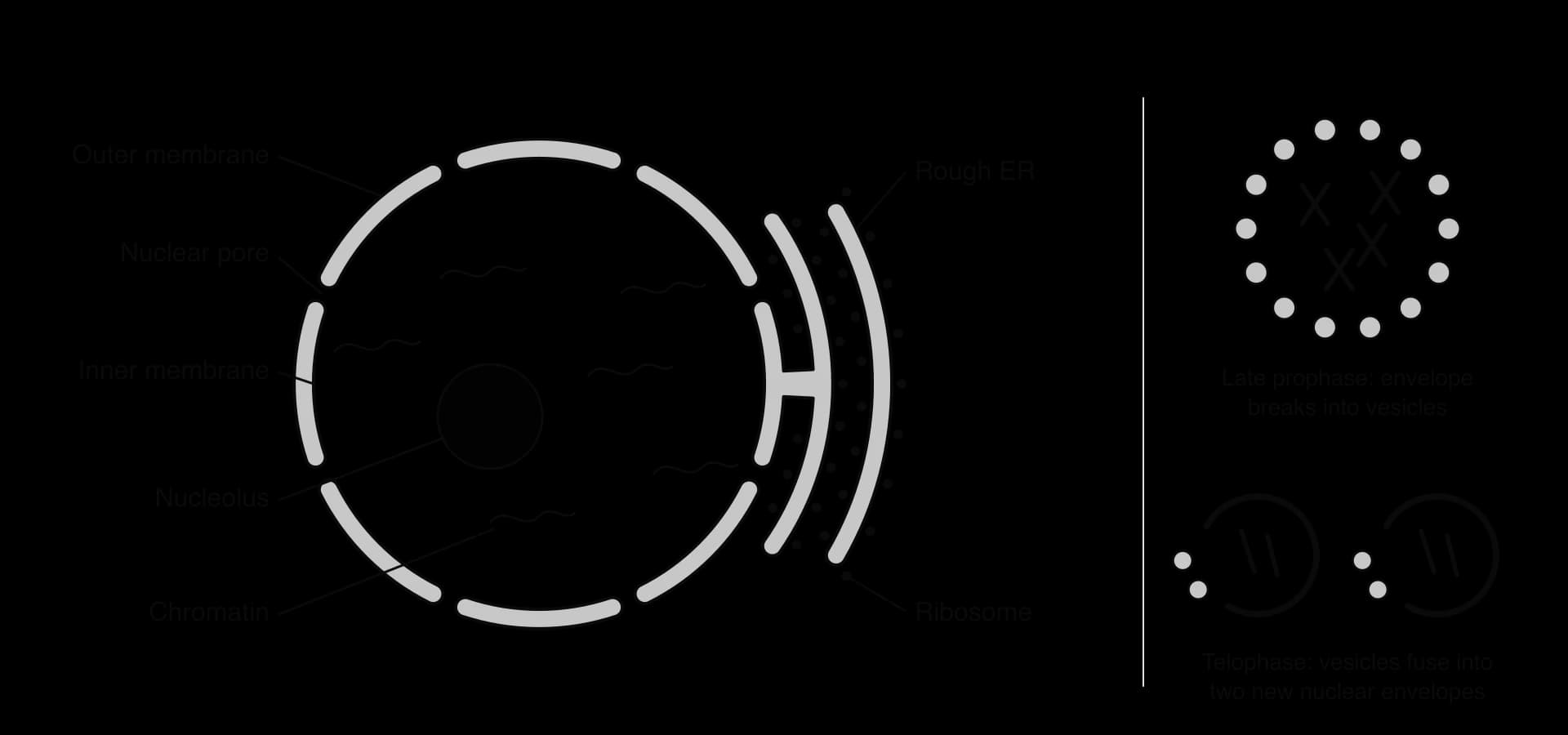
The two membranes fuse with each other at thousands of points to form nuclear pores, each lined by a large ring of proteins called the nuclear pore complex. Pores are needed because molecules made in one compartment are used in the other:
Out of the nucleus: mRNA (for translation in the cytoplasm) and ribosomal subunits (assembled in the nucleolus).
Into the nucleus: proteins made in the cytoplasm that are needed in the nucleus, such as histones, DNA polymerase, RNA polymerase and transcription factors; also nucleotides and ATP.
Small molecules and ions pass through the pores freely, but large molecules pass only if they carry the right signal, which is recognised by the proteins of the pore complex. The pores therefore let the nucleus exchange materials with the cytoplasm while still controlling which large molecules cross, so proteins such as transcription factors enter the nucleus only when they are needed.
During mitosis and meiosis the chromosomes must be moved by spindle microtubules, which form in the cytoplasm. In late prophase the nuclear lamina is taken apart and the nuclear envelope breaks up into many small vesicles, so the microtubules can reach and attach to the chromosomes. At telophase the vesicles gather around each group of chromosomes and fuse to form two new envelopes, and the pores reassemble. A double membrane can be divided into vesicles and rebuilt quickly in this way, which allows each new cell to inherit a complete nucleus (D2.1 Cell and nuclear division).
Ribosomes in the cytoplasm of eukaryotic cells are 80S ribosomes, built from a large and a small subunit. They exist in two places. Free ribosomes float in the cytosol. Bound ribosomes are attached to the outside of the membranes of the rough endoplasmic reticulum (rER), a network of flattened membrane sacs that extends from the nuclear envelope through the cytoplasm.
Free ribosomes | Bound ribosomes (on the rER) | |
|---|---|---|
Position | In the cytosol | Attached to the cytoplasmic face of the rER membrane |
Structure | 80S | 80S (identical) |
Where the polypeptide goes as it is made | Released into the cytosol | Threaded through the membrane into the lumen of the rER |
Destination of the protein | Used in the cytoplasm (for example, enzymes of glycolysis, haemoglobin in red blood cell precursors); some imported into the nucleus, mitochondria or chloroplasts | Secreted from the cell (for example, digestive enzymes, antibodies), placed in the plasma membrane, or sent to lysosomes |
Free and bound ribosomes are structurally identical, and a single ribosome can be free at one moment and bound the next. Its position depends on the polypeptide it is making. Proteins destined for the rER begin with a signal sequence, a short stretch of about 15–30 mostly hydrophobic amino acids at the start (N-terminus) of the chain.
Translation begins on a free ribosome in the cytosol.
As the signal sequence emerges from the ribosome, a signal recognition particle binds to it and pauses translation.
The signal recognition particle docks the ribosome onto a receptor on the rER membrane, next to a protein channel.
Translation resumes and the growing polypeptide is threaded through the channel into the lumen of the rER. The signal sequence is usually cut off.
In the lumen the polypeptide folds, often helped by other proteins, and carbohydrate chains may be added. It is then carried to the Golgi apparatus in transport vesicles.
Cells that secrete large amounts of protein have very extensive rER. Pancreatic acinar cells, which secrete digestive enzymes into the small intestine, are packed with rER, a large Golgi apparatus and many secretory vesicles. Plasma cells, which secrete antibodies, show the same pattern. The amount of each organelle in a cell matches the cell's function.
The Golgi apparatus is a stack of flattened, curved membrane sacs, each called a cisterna (plural cisternae). Unlike the rER, the cisternae are not connected to each other. The stack has two faces. The cis face, closest to the rER, receives transport vesicles; the trans face, on the opposite side, releases vesicles carrying finished proteins.
Proteins arrive from the rER in transport vesicles, which fuse with the cis face. They then move through the stack from cis to trans, and enzymes in the cisternae modify them on the way. The main modification is glycosylation: carbohydrate chains are added, removed or rebuilt, converting proteins into glycoproteins. Other modifications include adding phosphate groups and cutting some proteins into their active form.
At the trans face the proteins are sorted according to their destination and packed into different vesicles: secretory vesicles for the plasma membrane, vesicles for lysosomes, and vesicles carrying membrane proteins. Sorting depends on chemical tags added in the Golgi. Lysosomal enzymes, for example, receive a mannose-6-phosphate tag, which receptors in the trans cisternae recognise.
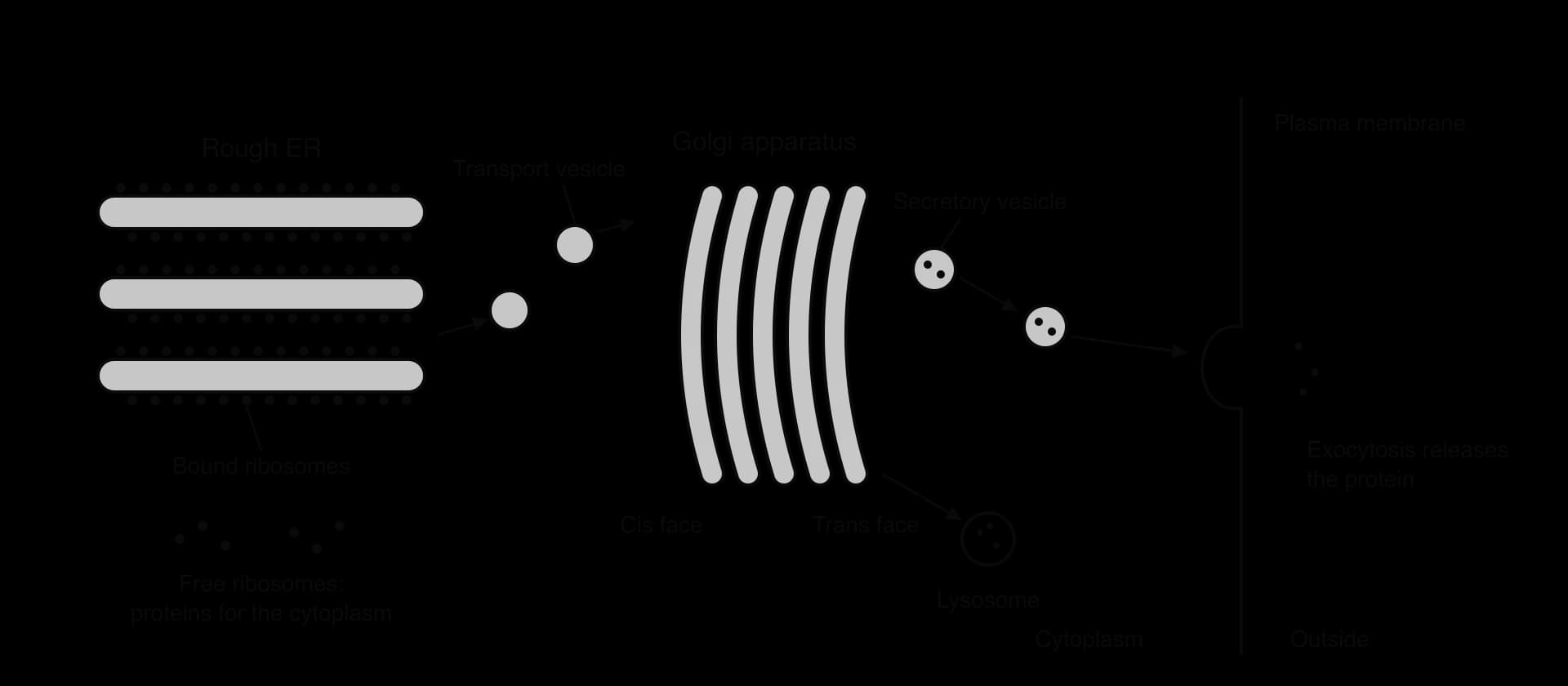
The importance of sorting is shown by I-cell disease (mucolipidosis II). Affected people lack the Golgi enzyme that adds the mannose-6-phosphate tag. Their lysosomal enzymes are made normally but, without the tag, are not sorted into lysosomes; they are packed into secretory vesicles and released from the cell instead. Lysosomes fill with undigested material, as in Tay–Sachs disease, but here many different enzymes are missing at once.
 YouTube
A vesicle is a small sac, usually well under 1 µm across, made of a single membrane that encloses a little fluid and its contents. Vesicles carry materials between the compartments of the endomembrane system (the nuclear envelope, ER, Golgi apparatus, lysosomes and plasma membrane). They form by budding from one membrane and deliver their cargo by fusing with another. Because the vesicle membrane joins the target membrane when it fuses, vesicles also move membrane itself, so new phospholipids and membrane proteins made in the ER are added to the plasma membrane.
Transport vesicles carry proteins from the rER to the Golgi apparatus.
Secretory vesicles carry proteins from the Golgi apparatus to the plasma membrane for exocytosis. Some release their contents continuously; others, such as those holding hormones or neurotransmitters, are stored until a signal triggers their release.
Lysosomes are vesicles from the trans face of the Golgi apparatus that hold hydrolytic enzymes.
Endocytic vesicles form when the plasma membrane folds inwards around material outside the cell.
A flat membrane does not bend into a sphere on its own. Many vesicles are shaped by coat proteins that assemble on the cytoplasmic side of the membrane. The best known is clathrin. Each clathrin unit has three legs joined at the centre (a triskelion). Triskelia link together into a curved cage of hexagons and pentagons, like the panels of a football, and as the cage grows it pulls the membrane into a pit and then a sphere.
Clathrin-coated vesicles are used in receptor-mediated endocytosis, the uptake of particular molecules that bind to receptor proteins on the cell surface. A well-studied example is the uptake of low-density lipoprotein (LDL), the particle that carries cholesterol in the blood:
LDL particles bind to LDL receptors in the plasma membrane.
Adaptor proteins link the receptors to clathrin, which assembles under the membrane and forms a coated pit.
The pit deepens until it is connected to the membrane only by a narrow neck; a protein called dynamin pinches the neck and the coated vesicle is released into the cytoplasm.
The clathrin coat is removed within seconds and recycled to form new pits. The uncoated vesicle fuses with other vesicles that deliver it to lysosomes, where the LDL is digested and cholesterol released; the receptors are returned to the plasma membrane.
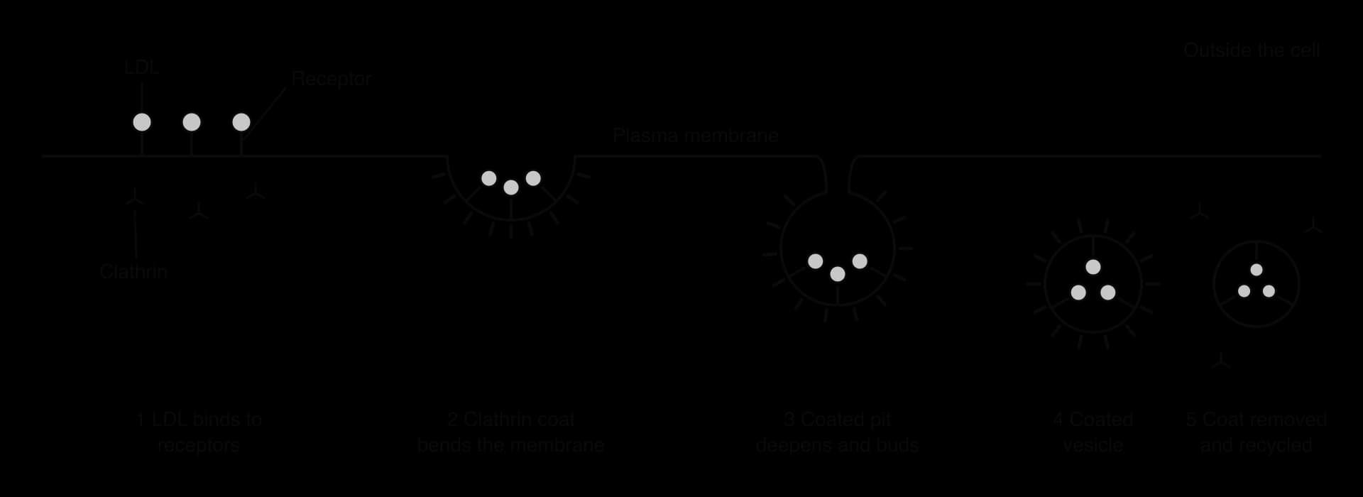
This pathway was worked out by Michael Brown and Joseph Goldstein, who studied people with familial hypercholesterolaemia, an inherited condition with very high blood cholesterol. They found that patients' cells either lacked LDL receptors or had receptors that could not gather in coated pits, so LDL was not taken up and stayed in the blood. Brown and Goldstein received the Nobel Prize in Physiology or Medicine in 1985. Clathrin coats are also used at the trans face of the Golgi apparatus, where they form the vesicles that carry tagged enzymes to lysosomes.
Questions on organelles appear in Paper 1A as identification of organelles from diagrams and electron micrographs, and as "which is an organelle" or "which has a double membrane" items. Paper 1B uses data from cell fractionation or from cells with different amounts of each organelle. Paper 2 asks for drawings of mitochondria and chloroplasts, explanations of the advantages of compartmentalisation, and outlines of the secretory pathway. Common command terms are state, label, draw, outline, explain and compare and contrast.
A good answer about compartmentalisation names the compartment, says what it concentrates or separates, and gives the benefit. "Lysosomes keep hydrolytic enzymes separate from the cytoplasm, so the cell's own molecules are not digested" is one complete marking point.
The multi-part question uses approximate values from electron micrographs of three types of mammalian cell, analysed to find the percentage of the cell volume occupied by two organelles.
Cell type | Rough ER / % of cell volume | Mitochondria / % of cell volume |
|---|---|---|
Pancreatic acinar cell | 22 | 7 |
Heart muscle cell | 1 | 35 |
Liver cell | 9 | 22 |
Approximate values from stereological studies of mammalian cells (pancreas: Bolender, 1974; liver: Weibel et al., 1969).
I can define an organelle and explain why the cell wall, cytoskeleton and cytoplasm are not organelles.
I can classify organelles as having no membrane, one membrane or two membranes.
I can outline how cell fractionation separates organelles and explain why the homogenate is cold, isotonic and buffered.
I can explain how separating the nucleus from the cytoplasm allows mRNA to be modified before translation.
I can explain two advantages of compartmentalisation, using lysosomes and phagocytic vacuoles.
I can describe how the loss of one lysosomal enzyme causes Tay–Sachs disease.
I can draw and label a mitochondrion and a chloroplast and explain their adaptations. (HL)
I can explain the benefits of a double nuclear membrane with pores, including its breakdown in mitosis and meiosis. (HL)
I can compare free ribosomes with ribosomes on the rough ER and explain the role of the signal sequence. (HL)
I can describe how the Golgi apparatus modifies, sorts and packages proteins. (HL)
I can explain how clathrin forms vesicles in receptor-mediated endocytosis. (HL)
I and II only
I and III only
II and III only
I, II and III
In the bacterium Escherichia coli, ribosomes can begin translating an mRNA molecule while the same molecule is still being transcribed. Why is this possible in prokaryotes but not in eukaryotes?