
Explain how phospholipids and proteins form membranes and how substances cross them by diffusion, osmosis, facilitated diffusion, active transport and (HL) vesicles, gated channels, pumps and cotransporters.
Each is in the Biology glossary, with every other term in the subject.
The movement of particles across a membrane against their concentration gradient, from a lower to a higher concentration, by pump proteins using energy from ATP.
An integral channel protein forming a narrow pore through which water molecules pass in single file, greatly increasing the permeability of a membrane to water.
A protein in the plasma membrane that binds to a matching molecule on a neighbouring cell, attaching cells to each other to form tissues; different CAMs form different junctions.
An integral membrane protein with a hydrophilic pore through which specific ions or polar molecules diffuse across the membrane; many channels are gated and can open or close.
A steroid lipid found among the phospholipids of animal cell membranes, which regulates membrane fluidity by reducing it at high temperatures and preventing stiffening at low temperatures.
A difference in the concentration of a substance between two regions; particles diffuse down it from higher to lower concentration, and moving them against it requires energy.
A change in the three-dimensional shape of a protein, for example when a substrate, signalling molecule or phosphate group binds to it.
Every cell is surrounded by a plasma membrane (also called the cell membrane), a film of lipid and protein only about 7–10 nm thick. It separates the cytoplasm from the surroundings and controls which substances enter and leave, so the chemistry inside a cell can differ from the chemistry outside it.
52 exam-style questions on this subtopic, written in the style of the IB papers. The first has its markscheme open. Counted on
Biological molecules and structures are held together both by covalent bonds and by weaker interactions.
Outline how condensation reactions produce polymers, using nucleic acids and one other type of macromolecule as examples.
4 marks
a. condensation links monomers by covalent bonds, releasing water;
b. one water molecule is released for each bond formed;
c. nucleotides are the monomers of nucleic acids / RNA / DNA;
d. the phosphate of one nucleotide bonds to the sugar of the next;
e. forming a sugar–phosphate backbone;
f. monosaccharides / glucose join to form polysaccharides / starch / cellulose / glycogen (by glycosidic bonds);
OR
f. amino acids join to form polypeptides (by peptide bonds);
g. polymers can be broken down into monomers by hydrolysis (using water);
[4 max]
Award f. for one correctly named other polymer with its monomer.
Explain how the structure of DNA is related to its functions.
7 marks
a. DNA is a double helix of two (antiparallel) strands of nucleotides;
b. nucleotides in each strand are linked by covalent bonds (between the sugar of one and the phosphate of the next);
One of 1 short multiple-choice questions on B2.1. Pick an answer to see why it's right or wrong.
What is osmosis?
The official deck for this part of the syllabus.
What is the plasma membrane made of, and about how thick is it?
State two roles of the plasma membrane.
With a free account, Augment remembers where you stopped and what you have practiced.
A membrane protein that moves two different particles across a membrane together in the same direction, using the movement of one down its concentration gradient to move the other against its gradient.
An early model of membrane structure, proposed in 1935, in which a phospholipid bilayer is sandwiched between two continuous layers of protein; falsified by later evidence.
The uptake of material into a cell by the plasma membrane folding inwards around it and pinching off to form a vesicle; requires ATP.
A membrane transport protein that moves one type of particle into the cell and another type out of the cell in the same cycle, such as the sodium–potassium pump.
The release of material from a cell by a vesicle fusing with the plasma membrane and emptying its contents outside the cell; requires ATP.
The passive movement of particles down a concentration gradient across a membrane through a channel protein (or with a carrier protein), without the use of energy from ATP.
The model of membrane structure in which proteins are embedded in or attached to a fluid phospholipid bilayer, with phospholipids and many proteins free to move sideways.
A lipid with a carbohydrate chain attached, found in the outer layer of the plasma membrane with the carbohydrate facing outside the cell; involved in cell recognition and adhesion.
Movement of a substance against its concentration gradient using energy from another substance moving down a concentration gradient that a pump has set up using ATP.
A membrane protein embedded in one or both layers of the phospholipid bilayer, held there by a hydrophobic region in the core; many span the whole membrane.
An ion channel that opens when a specific signalling molecule (ligand), such as a neurotransmitter, binds to it.
The ease with which phospholipids and proteins move within a membrane; it depends on temperature, the proportion of unsaturated fatty acids and, in animal cells, cholesterol.
A ligand-gated ion channel in postsynaptic membranes that opens when acetylcholine binds to it, allowing positive ions, mainly Na⁺, to diffuse into the cell.
The net movement of water across a partially permeable membrane from a region of lower solute concentration to a region of higher solute concentration.
A membrane that allows some particles to pass through it but not others; in cells, small non-polar molecules cross the bilayer while ions and large polar molecules need membrane proteins.
Movement of particles across a membrane down their concentration gradient using only their own kinetic energy, without energy from ATP; includes simple diffusion, facilitated diffusion and osmosis.
A membrane protein attached to one surface of the membrane, to phospholipid heads or to an integral protein, without entering the hydrophobic core of the bilayer.
A type of endocytosis in which a cell takes in large solid particles, such as bacteria, by enclosing them in a vesicle formed from the plasma membrane.
A type of endocytosis in which a cell takes in small droplets of the surrounding fluid, with the solutes dissolved in it, in small vesicles.
An integral membrane protein that uses energy from ATP to move specific particles across a membrane against their concentration gradient by changing shape.
A protein that binds a specific signalling molecule, such as a hormone or neurotransmitter, and responds by triggering a change in the cell.
The net movement of particles from a region of higher concentration to a region of lower concentration directly through the phospholipid bilayer, as a result of their random motion.
A cotransporter (SGLT) that moves glucose into a cell against its concentration gradient together with Na⁺ ions moving down their concentration gradient; found in the small intestine and nephron.
A pump protein in animal plasma membranes that uses one ATP per cycle to move three Na⁺ ions out of the cell and two K⁺ ions in, both against their concentration gradients.
An ion channel that opens or closes in response to a change in the potential difference (voltage) across the membrane, such as the Na⁺ and K⁺ channels of axons.
Eukaryotic cells also contain many internal membranes that divide the cytoplasm into compartments. The same structure, a double layer of lipid with proteins in it, is used in all of them. How molecules of lipid and protein assemble into membranes, and what determines whether a substance can cross one, links the molecular level of organisation directly to the level of the whole cell.
The main component of every biological membrane is the phospholipid. A phospholipid molecule has two parts with opposite properties. The head contains a negatively charged phosphate group joined to glycerol, so it is hydrophilic: it forms hydrogen bonds and other attractions with water. The two tails are long hydrocarbon chains of fatty acids, which are non-polar and therefore hydrophobic.
A molecule that has both a hydrophilic part and a hydrophobic part is described as amphipathic. Phospholipids are the most common amphipathic lipids in membranes; glycolipids and cholesterol are also amphipathic. The structure of phospholipids and the condensation reactions that build them from glycerol, fatty acids and phosphate are covered in B1.1 Carbohydrates and lipids.
When phospholipids are mixed with water they do not dissolve as single molecules. The hydrophilic heads are attracted to water, but the hydrophobic tails are not, and water molecules form hydrogen bonds with each other more strongly than with the tails. The arrangement with the lowest energy is one in which the tails are packed together away from the water and the heads face outwards into it. Both conditions are met by a sheet two molecules thick, with the heads on both surfaces and the tails in the middle, called a phospholipid bilayer.
No energy from the cell is needed for a bilayer to form. Phospholipids assemble into bilayers spontaneously, and a bilayer tends to close up on itself into a continuous sheet, because any free edge would leave hydrophobic tails exposed to water. For the same reason a small tear in a membrane reseals, and a bilayer that surrounds a volume of water forms a closed sac. These properties make the bilayer a suitable boundary for cells and for compartments within them.
A phospholipid bilayer is the basic structure of every membrane in a cell:
the plasma membrane around every cell, prokaryotic and eukaryotic;
the double membranes (two bilayers) around the nucleus, mitochondria and chloroplasts;
the single membranes of the rough and smooth endoplasmic reticulum, the Golgi apparatus, lysosomes, vesicles and vacuoles;
the thylakoid membranes inside chloroplasts.
The universality of the phospholipid bilayer is an example of unity in living things. All cells use the same basic membrane structure, while the proteins placed in each membrane differ and give each one its particular functions.
The core of a phospholipid bilayer is a layer of hydrocarbon tails about 3–4 nm thick. It is hydrophobic, so particles that are charged or polar cannot easily enter it. To cross the bilayer, an ion or polar molecule would have to leave the water, where it is surrounded by attracted water molecules, and pass through a non-polar region that does not attract it. Very few particles have enough energy to do this.
The permeability of the bilayer to a particle therefore depends mainly on two properties:
Size. Small molecules pass through the core more easily than large ones. Large molecules such as glucose, amino acids and proteins cannot cross the bilayer at a useful rate.
Polarity and charge. Non-polar molecules dissolve in the hydrocarbon core and cross it readily. Polar molecules cross slowly, and ions, which carry a full charge and are surrounded by a shell of water molecules, hardly cross at all.
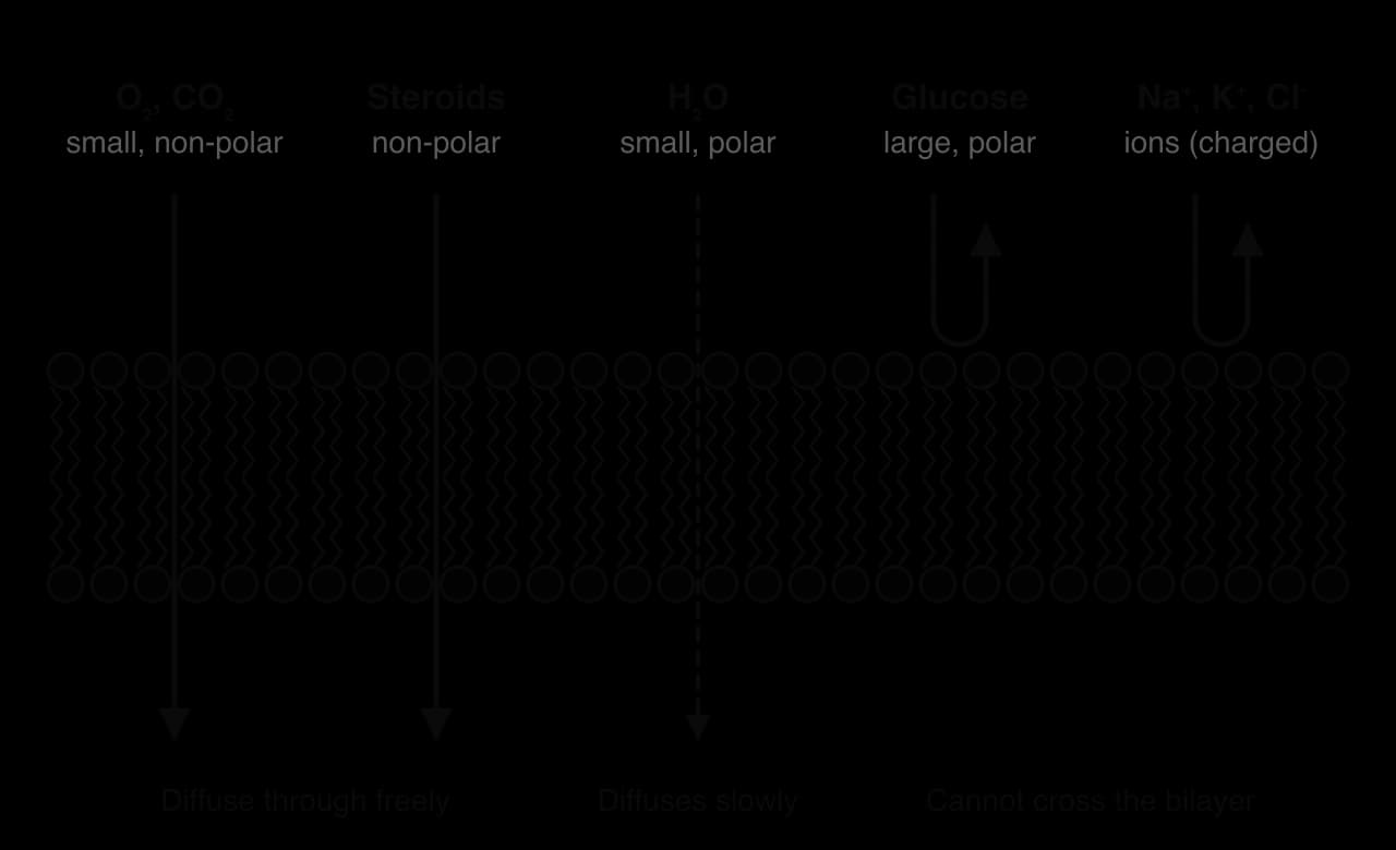
Because the bilayer blocks ions and most polar molecules, it is an effective barrier between two aqueous solutions, such as the cytoplasm and the tissue fluid around a cell. The cell can then control the movement of these substances using proteins in the membrane, and keep concentrations inside that are very different from those outside. The cytoplasm of a human cell, for example, has a much higher concentration of K⁺ and a much lower concentration of Na⁺ than the tissue fluid around it.
A partially permeable membrane does not have to be a living membrane. Visking tubing (dialysis tubing), made of cellulose, has tiny pores that let water and small solute molecules such as glucose through but not large molecules such as starch. Sucrose crosses it far more slowly than water, so a sucrose solution inside the tubing behaves almost as if the solute cannot escape. The tubing is often used as a model of a cell membrane in experiments.
Beetroot cells store a red pigment, betalain, in their large vacuoles. The pigment is a large polar molecule that cannot cross intact membranes, so pieces of beetroot placed in water release very little of it. If the membranes are damaged, the pigment leaks out and colours the water, and the intensity of the colour can be measured with a colorimeter. This simple practical is used to investigate how temperature or solvents such as ethanol affect membranes.
All particles in a liquid or gas are in constant random motion. Where particles are more concentrated, more of them move away by chance than move in from a less concentrated region. The result is a net movement from the region of higher concentration to the region of lower concentration, which continues until the particles are evenly spread. The difference in concentration between two regions is the driving force for this movement.
Diffusion along a concentration gradient needs no energy from the cell, because the kinetic energy of the particles themselves moves them. Any movement across a membrane that relies only on this kinetic energy is called passive transport. Diffusion of a substance through the phospholipid bilayer itself, without the help of any protein, is the simplest form of passive transport.
Oxygen (O₂) and carbon dioxide (CO₂) are small, non-polar molecules. They dissolve in the hydrophobic core of the bilayer and pass between the phospholipid molecules without any protein, so they cross membranes by simple diffusion.
In a respiring animal cell, cell respiration uses O₂ in the mitochondria, keeping its concentration in the cytoplasm low. The concentration of O₂ in the tissue fluid outside the cell is higher, so O₂ diffuses into the cell. At the same time, respiration produces CO₂, so its concentration inside the cell is higher than outside, and CO₂ diffuses out. The same gases move in the opposite directions in a photosynthesising leaf cell in the light. The cell keeps both gradients in place simply by using O₂ and producing CO₂, so the gases keep moving without the cell spending ATP on transport.
Concentration gradient. The steeper the gradient, the faster the net rate. Doubling the concentration difference roughly doubles the rate.
Size of the particle. Smaller particles diffuse faster.
Polarity and charge. Non-polar molecules cross the hydrophobic core faster than polar ones; ions hardly cross at all.
Temperature. Particles have more kinetic energy at higher temperatures, so they diffuse faster.
Surface area and distance. A larger membrane area allows more particles to cross at once, and a shorter distance means a shorter path. This is why gas exchange surfaces such as the alveoli are large and thin (see B3.1 Gas exchange).
The phospholipid bilayer sets the basic structure of a membrane, but proteins carry out most of its functions. Membranes differ greatly in their protein content. The inner membrane of a mitochondrion, which carries out many reactions of aerobic respiration, is about three-quarters protein by mass, whereas the myelin membranes that insulate some nerve fibres are mostly lipid. Membrane proteins vary in structure, in their position in the membrane and in their function.
An integral protein has a region of hydrophobic amino acids, often folded into α-helices, that sits in the hydrophobic core of the bilayer, and hydrophilic regions that project into the water on one or both sides. The hydrophobic region holds the protein firmly in the membrane, so integral proteins can only be removed by breaking up the bilayer, for example with detergents. Channel proteins and pumps are integral proteins that span the whole membrane.
A peripheral protein has no hydrophobic region embedded in the bilayer. It is held at the surface by weaker attractions to the phosphate heads or to integral proteins, and it can move away from the membrane more easily. Some peripheral proteins are anchored by a lipid chain that is attached to the protein and inserted into one layer of the bilayer. Peripheral proteins on the cytoplasmic side often link the membrane to the cytoskeleton, which holds the shape of the cell.
Function | What the protein does | Example |
|---|---|---|
Transport | Moves particles across the membrane, either passively (channels) or actively (pumps) | Aquaporins; the sodium–potassium pump |
Receptor | Binds a specific signalling molecule from outside the cell and passes on the signal | Insulin receptor in liver and muscle cells |
Enzyme | Catalyses a reaction at the membrane surface | Enzymes that digest disaccharides on the microvilli of the small intestine |
Cell adhesion | Binds to proteins on neighbouring cells or outside the cell, holding cells together | Cell-adhesion molecules in tissues (HL) |
Cell recognition | Acts as a marker that other cells identify, usually as a glycoprotein | Self markers recognised by white blood cells |
Electron carrier | Passes electrons along a chain in a membrane | Electron carriers in the inner mitochondrial membrane and thylakoids |
A receptor is a good example of how a membrane protein links a cell to its environment. Hormones such as insulin are too large and too polar to cross the bilayer. Instead, the hormone binds to a specific receptor on the outer surface, and the receptor changes shape inside the cell, which starts a response without the hormone entering. The details of these signalling pathways are covered in C2.1 Chemical signalling (HL).
Each function depends on the three-dimensional shape of the protein. A channel needs a pore of the right width and lining; a receptor needs a binding site that is complementary to its signalling molecule; an enzyme needs an active site. This is a clear case of form and function at the molecular level: the amino acid sequence determines how the protein folds, and the fold determines what the protein can do in the membrane.
Water molecules, like all particles, are in constant random motion and move across membranes in both directions. When the solutions on the two sides of a membrane contain different concentrations of solute, more water molecules cross in one direction than in the other. This net movement of water is osmosis, and it happens when the membrane is permeable to water but not to the solute.
Three conditions together explain the direction of osmosis:
Random movement of particles. Water molecules move at random and constantly collide with the membrane on both sides. Some of those that hit the membrane pass through it.
A membrane that is impermeable to the solute. The solute molecules (for example sucrose, or the many proteins and ions in cytoplasm) cannot cross, so the difference in solute concentration cannot even out by the solute moving. Only water can move to reduce the difference.
A difference in solute concentration. Solute molecules attract water molecules through hydrogen bonds and other interactions, and hold them in shells around themselves. In the solution with the higher solute concentration, a larger proportion of the water molecules is bound in this way and fewer are free to move through the membrane. More water molecules therefore cross from the side with the lower solute concentration than cross back, so there is a net movement of water towards the higher solute concentration.
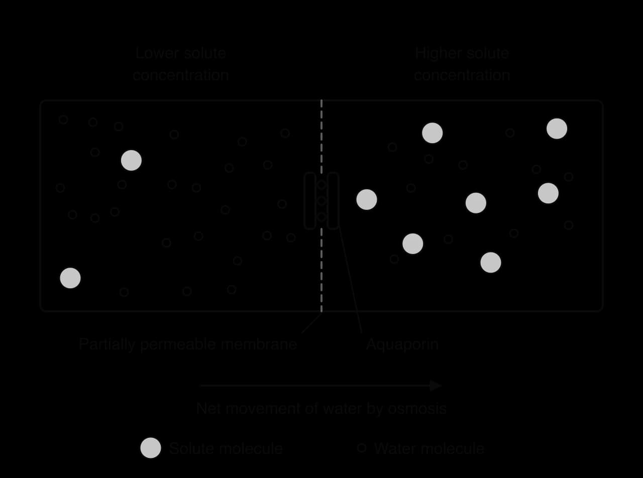
Osmosis stops when the solute concentrations on both sides are equal, or when another force, such as the pressure of a plant cell wall, balances it. Water molecules still cross in both directions at that point, but in equal numbers, so there is no net movement.
The plasma membrane is partially permeable, and the cytoplasm contains a high concentration of dissolved solutes, so cells take up or lose water by osmosis whenever the solution around them differs in solute concentration. A solution with a lower solute concentration than the cytoplasm is hypotonic, and a cell placed in it gains water. A solution with a higher solute concentration is hypertonic, and a cell placed in it loses water. In an isotonic solution there is no net movement. Animal cells such as red blood cells swell and can burst in hypotonic solutions, while plant cells become turgid because the cell wall resists expansion; these effects are studied in D2.3 Water potential.
Because water crosses the bilayer itself only slowly, many cells that need to move large volumes of water quickly have special channel proteins for water in their membranes, called aquaporins.
Each aquaporin is a transmembrane protein with a pore that is just wide enough for water molecules to pass one at a time. The pore is lined with amino acids that form hydrogen bonds with the water molecules as they pass. Positively charged amino acids near the narrowest point repel positively charged ions, including H⁺, so ions cannot pass even though they are small. The structure of the pore therefore makes the channel specific to water. Osmosis through aquaporins is still passive: the direction of movement depends only on the difference in solute concentration, and no ATP is used.
Aquaporins are especially common in cells that move a lot of water:
red blood cells, which must swell and shrink rapidly as they pass through regions of the kidney where the solute concentration of the tissue fluid changes sharply;
cells of the collecting ducts in the kidney, where the number of aquaporins in the membrane is adjusted to control how much water is reabsorbed from urine (see D3.3 Homeostasis);
root cells of plants, which take up water from the soil.
Aquaporins were identified by Peter Agre, who isolated a protein from red blood cell membranes and showed in 1992 that frog eggs became very permeable to water when they were made to produce it. Agre shared the 2003 Nobel Prize in Chemistry for this work. The discovery explained an old observation: some membranes let water through far faster than a pure phospholipid bilayer could.
Ions and polar molecules such as glucose cannot diffuse through the hydrophobic core of the bilayer at a useful rate. They can still move passively down their concentration gradients if the membrane contains proteins that give them a hydrophilic route across. This kind of passive transport is called facilitated diffusion, because a protein makes (facilitates) the movement, but the energy still comes from the random motion of the particles.
A channel protein is an integral protein that spans the membrane and folds around a central pore. The outer surface of the protein, which faces the hydrophobic core of the bilayer, is made of hydrophobic amino acids. The pore is lined with hydrophilic amino acids, so ions and polar molecules can pass through it without contact with the hydrophobic tails.
The diameter of the pore and the charges on the amino acids that line it determine which particles can pass. A potassium channel, for example, has a narrow region called the selectivity filter whose oxygen atoms are spaced to fit exactly around a K⁺ ion that has lost its shell of water molecules. A Na⁺ ion does not fit the filter as well and is rarely allowed through, even though it is smaller than K⁺. Each type of channel therefore lets through only one particle or a small group of similar particles, which makes the membrane selectively permeable. Roderick MacKinnon determined the structure of a potassium channel by X-ray crystallography in 1998 and shared the 2003 Nobel Prize in Chemistry with Peter Agre.
Many channel proteins can open and close. When the channel is open, specific ions diffuse through it rapidly; when it is closed, they cannot pass at all. These gated channels allow a cell to switch the permeability of its membrane to an ion on and off. Some channels open when a specific molecule binds to them, and others open when the voltage across the membrane changes. Both kinds of gated channel are needed for nerve impulses.
Facilitated diffusion has the same direction as simple diffusion: from higher to lower concentration. Some polar molecules, such as glucose, cross by a related kind of facilitated diffusion that uses carrier proteins instead of open channels. A carrier binds the molecule on one side of the membrane and changes shape to release it on the other side. Carrier-mediated facilitated diffusion is still passive, because the direction depends only on the concentration gradient.
Cells often need to take up substances that are already more concentrated inside the cell than outside, or to remove substances that are more concentrated outside. Moving particles against their concentration gradient, from lower to higher concentration, cannot happen by diffusion. It needs energy, which the cell supplies from ATP.
The proteins that carry out active transport are called pump proteins. A pump protein is an integral protein that spans the membrane. It has a binding site for a specific particle that is open to only one side of the membrane at a time. The cycle of a typical pump has four stages:
The particle to be moved binds to the specific binding site of the pump, on the side of the membrane where it is at a lower concentration.
ATP is hydrolysed to ADP and phosphate, releasing energy. In many pumps the phosphate group is attached to the pump protein itself.
The energy causes a conformational change in the pump: the protein changes shape so that the binding site now opens to the other side of the membrane.
The particle is released on the side where its concentration is higher. The pump returns to its original shape, ready to bind another particle.
Pumps are specific, because only particles that fit the binding site are moved. They also move particles in one direction only, so a pump that takes up a substance never carries it back out. Because the energy for each cycle comes from ATP, any factor that stops ATP production, such as a lack of oxygen or a poison such as cyanide that blocks aerobic respiration, soon stops active transport.
Mineral ion uptake by root hair cells. Nitrate, potassium and other mineral ions are usually much more concentrated in root cells than in the soil water. Pumps in the plasma membranes of root hair cells move these ions into the cells against the gradient.
The sodium–potassium pump. In almost all animal cells this pump moves Na⁺ out of the cell and K⁺ into it, both against their gradients, using energy from ATP.
Calcium pumps in muscle. Pumps in the membrane of the store of Ca²⁺ inside muscle fibres move Ca²⁺ back into the store after each contraction, so the muscle can relax.
Proton pumps. Pumps in the membranes of plant cells and in the lining of the stomach move H⁺ ions against steep concentration gradients.
 YouTube
A membrane is selectively permeable when it lets some particles through and holds others back, and when the cell can control which particles cross. The three ways of crossing a membrane differ in how selective they are.
Simple diffusion is not selective. Whether a particle can diffuse through the bilayer depends only on its size and on whether it is hydrophilic or hydrophobic. The cell cannot choose to admit O₂ but exclude another small non-polar molecule, and it cannot switch simple diffusion on or off.
Facilitated diffusion is selective. Each channel or carrier lets through only particles that fit its pore or binding site. By making or removing channels, or by opening and closing gated channels, a cell controls which particles cross and when.
Active transport is selective. Each pump has a binding site for specific particles, and the cell controls the direction of movement as well as which particles move.
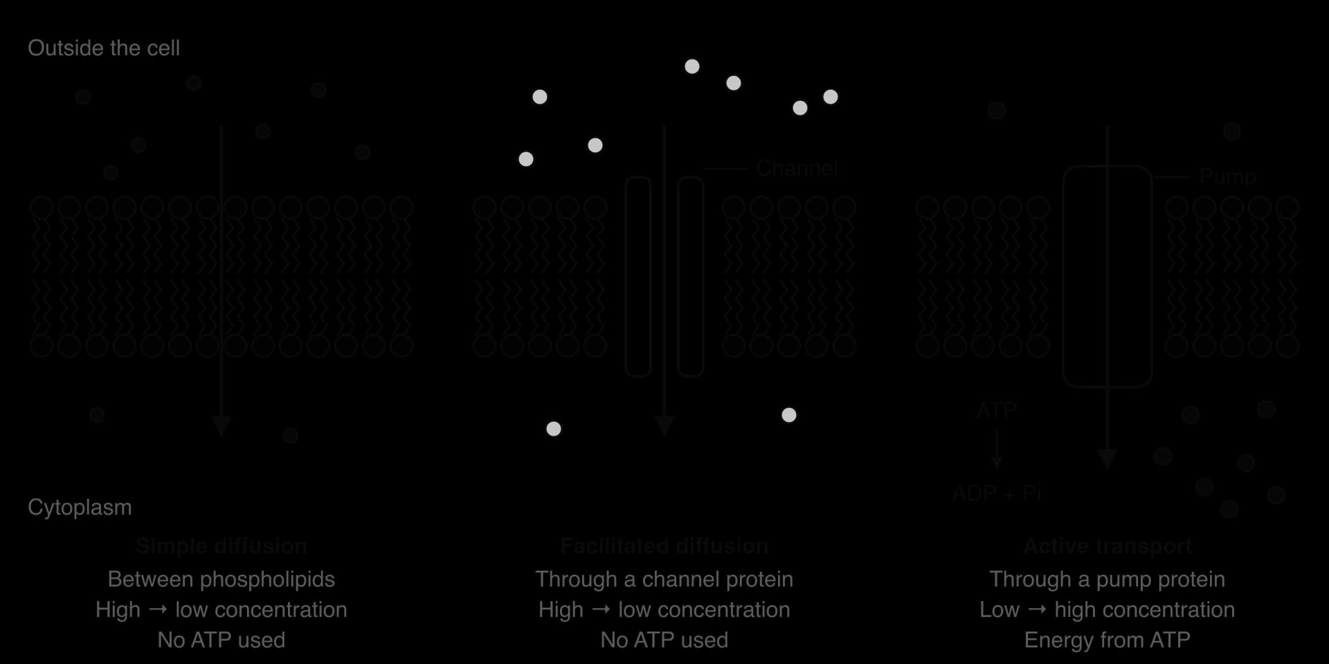
Because the bilayer itself lets almost no ions or large polar molecules through, the membrane proteins determine which of these substances a cell takes up or releases. This is why different cell types, with the same bilayer, can have very different permeabilities. A cell in the lining of the small intestine has many glucose transporters, while a nerve cell has large numbers of Na⁺ and K⁺ channels.
Feature | Simple diffusion | Facilitated diffusion | Active transport |
|---|---|---|---|
Route across the membrane | Between phospholipids | Through a channel (or carrier) protein | Through a pump protein |
Direction | Down the concentration gradient | Down the concentration gradient | Against the concentration gradient (can also move with it) |
Energy from ATP | No | No | Yes |
Selective | No: depends only on size and polarity | Yes: specific to particles that fit the channel | Yes: specific to particles that fit the binding site |
Can the cell control it? | No | Yes (number of channels; gating) | Yes (number of pumps; ATP supply) |
Examples | O₂, CO₂, steroid hormones | K⁺ through potassium channels; water through aquaporins | Na⁺ and K⁺ by the sodium–potassium pump; mineral ions into root hairs |
The rate at which a substance enters cells can show how it crosses the membrane. For simple diffusion, the rate is proportional to the concentration gradient, so a graph of rate against the concentration outside the cell is a straight line. For facilitated diffusion, the rate rises at first but then levels off, because there are a limited number of channel or carrier proteins in the membrane and at high concentrations all of them are in use.
Active transport has a similar levelling-off shape, because it also depends on a limited number of proteins. It can be told apart from facilitated diffusion by two tests: it continues when the concentration inside the cell is already higher than outside, and it stops when ATP production is stopped by a respiratory poison or by a lack of oxygen.
Many proteins and lipids in the plasma membrane have short chains of sugar units (oligosaccharides) attached to them. A protein with a carbohydrate chain attached is a glycoprotein, and a lipid with a carbohydrate chain attached is a glycolipid.
The carbohydrate chains of both glycolipids and glycoproteins are always on the extracellular side of the plasma membrane. They are added inside the endoplasmic reticulum and Golgi apparatus, on the side of the membrane that faces the inside of these organelles; when vesicles fuse with the plasma membrane, that side becomes the outer surface of the cell. Together, the carbohydrate chains form a sugar-rich layer on the outside of many animal cells called the glycocalyx.
Cell recognition. The carbohydrate chains are very varied in their sequence of sugars and in their branching, so they act as identification markers. The ABO blood groups are an example: the A and B antigens on red blood cells are carbohydrate chains on glycoproteins and glycolipids, differing by a single sugar at the end of the chain. The immune system recognises the cells of the body as "self" partly from glycoproteins on their surface, and attacks cells with unfamiliar markers.
Cell adhesion. Glycoproteins and glycolipids bind to molecules on neighbouring cells or in the material outside cells, helping cells to stick together in tissues. Hydrogen bonds between the sugar chains and water also help to keep the cell surface hydrated.
Receptors. Some glycoproteins are receptors, for example for hormones, and the carbohydrate part can be needed for binding.
The accepted model of membrane structure combines all of the components described so far. It is called the fluid mosaic model.
The name describes the two main ideas of the fluid mosaic model:
Fluid. The phospholipids are not bonded to each other. They are held together only by hydrophobic interactions between the tails and by attraction of the heads to water, so individual molecules move sideways within their own layer, millions of times per second. Many proteins also drift sideways. The membrane behaves like a two-dimensional liquid, which lets it bend, change shape and reseal.
Mosaic. Seen from above, the membrane is a mosaic of many different proteins scattered among the phospholipids, like tiles of different shapes in a pattern. The types and positions of proteins vary between membranes and change over time.
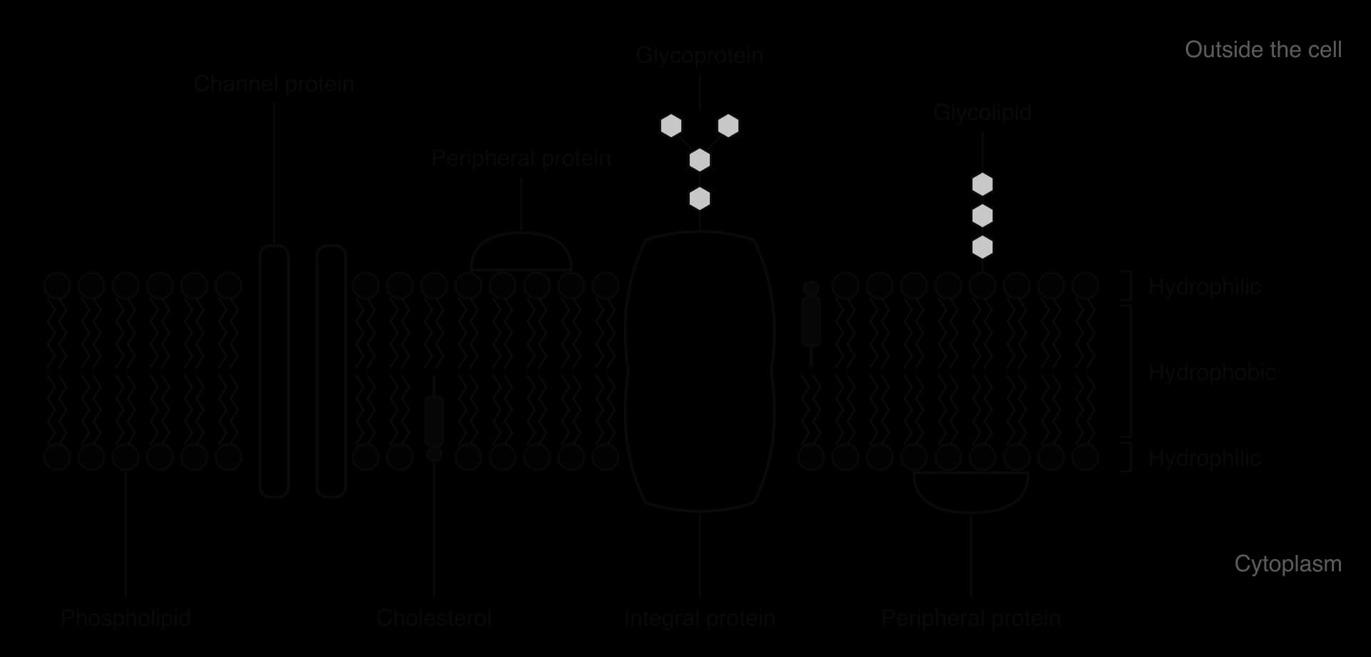
A two-dimensional drawing of the fluid mosaic model shows a section through the membrane. It includes phospholipids, at least one integral protein and one peripheral protein, a glycoprotein, cholesterol, and labels for the hydrophobic and hydrophilic regions. Glycolipids and the carbohydrate chains can be added, always on the outer surface.
 YouTube
Biologists could not see the structure of membranes directly, so they built models to explain the evidence available. In 1935 Hugh Davson and James Danielli proposed that a membrane is a phospholipid bilayer coated on both sides by continuous layers of protein, like a sandwich. The model explained why membranes are barriers to polar substances, and why they appeared in early electron micrographs as two dark lines (thought to be protein) separated by a paler band (thought to be lipid).
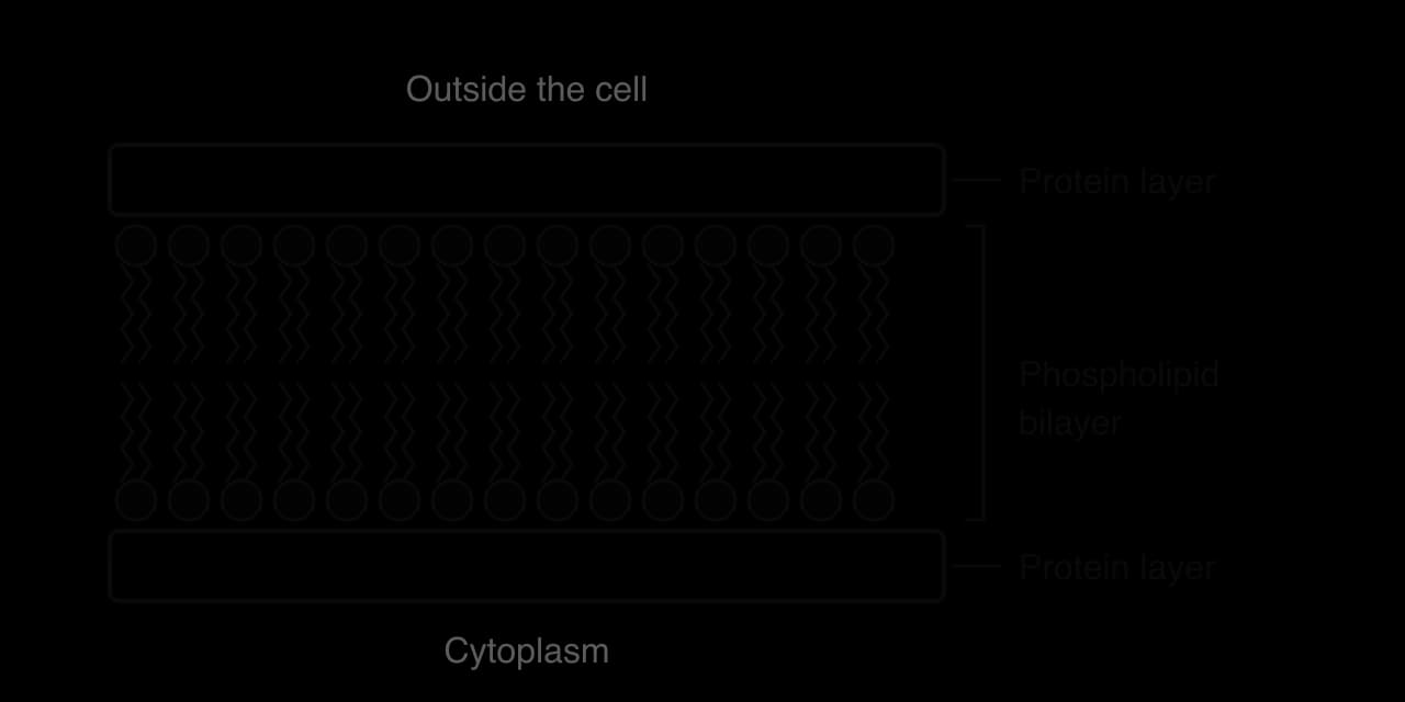
From the 1950s onwards, several kinds of evidence did not fit the Davson–Danielli model:
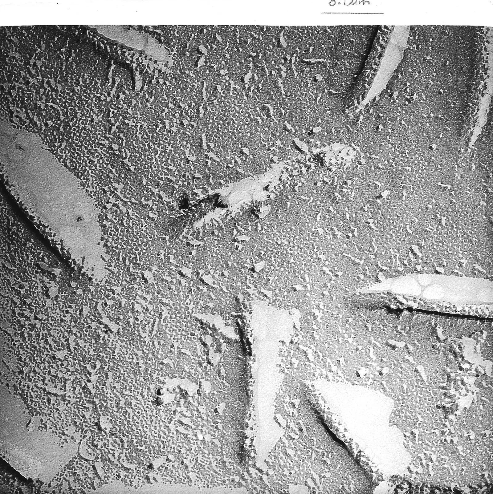
Freeze-fracture electron microscopy. Cells are frozen rapidly and then split with a blade. The fracture often runs along the middle of a membrane, between the two layers of phospholipids, and shows the inner faces. These faces are covered in globular particles, integral proteins that pass into or through the hydrophobic core.
Membrane proteins. When membrane proteins were extracted and studied, they turned out to vary greatly in size and to have hydrophobic regions. A protein with a hydrophobic surface would not sit stably in a layer exposed to water, but would fit inside the hydrophobic core.
Movement of proteins. In 1970 Larry Frye and Michael Edidin fused mouse cells with human cells and labelled the membrane proteins of each species with different fluorescent markers. At first the two sets of proteins stayed in separate halves of the hybrid cell, but within about 40 minutes at 37 °C they had mixed evenly. The proteins must be free to move in a fluid membrane, which a rigid protein coat would not allow.
In 1972 Jonathan Singer and Garth Nicolson proposed the fluid mosaic model, which explained all of this evidence. The history of membrane models shows how science progresses: a model is accepted while it explains the evidence, and it is tested by making predictions. When new techniques produce evidence that contradicts a model, the model is falsified and replaced by one that fits better. The fluid mosaic model itself has since been refined, for example by evidence that some lipids and proteins cluster into small regions rather than mixing freely.
Cystic fibrosis is a genetic disease that results from a fault in a single membrane protein. It is caused by mutations in the gene for CFTR (cystic fibrosis transmembrane conductance regulator), a channel protein in the plasma membrane of epithelial cells that line the airways, the pancreatic ducts, the gut and the sweat glands. CFTR is a chloride channel: when it opens, Cl⁻ ions move through it by facilitated diffusion, out of the epithelial cells into the fluid on their surface.
In the airways, Cl⁻ leaving the cells raises the solute concentration of the thin layer of liquid on the epithelium. Na⁺ ions follow the negative chloride ions, and water then moves out of the cells by osmosis, partly through aquaporins. This keeps the mucus thin enough for cilia to sweep it, with trapped bacteria and dust, out of the lungs. In cystic fibrosis too few working CFTR channels reach the membrane, so too little Cl⁻ and water leave the cells. The mucus becomes thick and sticky, clogging the airways and pancreatic ducts and allowing repeated lung infections. In the sweat glands, CFTR normally reabsorbs Cl⁻ from sweat, so people with cystic fibrosis have unusually salty sweat; measuring sweat chloride is the standard diagnostic test.
Nearly 90% of people with cystic fibrosis carry at least one copy of the F508del mutation, a deletion of one amino acid (phenylalanine) at position 508. The missing amino acid makes the protein misfold, and the cell destroys most of the misfolded protein before it reaches the plasma membrane. The few channels that do arrive open less often than normal.
Because the problem lies in the folding and opening of one protein, drugs have been designed to act on CFTR itself. The three-drug combination elexacaftor/tezacaftor/ivacaftor (sold as Trikafta) was approved by the US Food and Drug Administration on 21 October 2019. Two of the drugs, elexacaftor and tezacaftor, help the F508del protein fold correctly so that more of it reaches the membrane. The third, ivacaftor, keeps the channel open for longer once it is there. A trial of 403 patients with one F508del allele compared the combination with a placebo.
Outcome measured | Change with the drug compared with placebo |
|---|---|
Lung function (percentage of predicted FEV1) at week 4 | +13.8 percentage points |
Lung function through week 24 | +14.3 percentage points |
Sweat chloride concentration | −41.8 mmol dm⁻³ |
Rate of pulmonary exacerbations (flare-ups of lung disease) | 63% lower |
Source: Middleton et al., New England Journal of Medicine 381:1809, 2019.
The fall in sweat chloride shows that more working chloride channels were present in the membranes of sweat gland cells, and the improvement in lung function shows the effect on the airways. The case links several ideas from membrane biology: channel proteins make membranes selectively permeable, the conformation of a protein determines whether it can do its job, and the movement of ions through channels controls the movement of water by osmosis.
A membrane has to be fluid enough to bend, to let proteins move and to fuse with vesicles, but not so fluid that it becomes leaky or falls apart. How easily the phospholipids and proteins move within the bilayer is its fluidity, and it depends on temperature and on the lipids the membrane is made of.
The fatty acid tails of phospholipids can be saturated, with only single bonds between carbon atoms, or unsaturated, with one or more C=C double bonds (monounsaturated or polyunsaturated). In membranes most double bonds are in the cis form, which puts a kink in the chain.
Saturated tails are straight. They pack tightly side by side, and the many hydrophobic interactions between them hold them together. Lipids with saturated fatty acids have higher melting points, so a membrane rich in them is less fluid and stronger, and it stays stable at higher temperatures.
Unsaturated tails are kinked. The kinks stop the tails packing closely, so there are fewer interactions between neighbouring tails. Lipids with unsaturated fatty acids have lower melting points, so a membrane rich in them stays fluid and flexible at low temperatures.
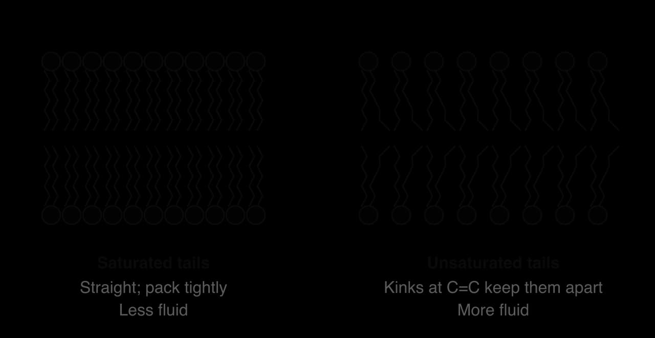
The ratio of saturated to unsaturated fatty acids therefore sets the temperature range over which a membrane has a suitable fluidity. Organisms that live at different temperatures, or whose temperature changes, adjust this ratio. Bacteria such as Escherichia coli grown at lower temperatures make membranes with a higher proportion of unsaturated fatty acids, which keeps their membranes about as fluid as those of cells grown at higher temperatures. This response, called homeoviscous adaptation, is an example of how organisms adapt to their habitat at the molecular level. Plants that survive frost, and fish that live in polar seas, also have membranes rich in unsaturated fatty acids, while organisms from hot springs have more saturated (and other heat-stable) lipids.
Animal plasma membranes contain large amounts of cholesterol, a steroid lipid, in some membranes as much as one molecule of cholesterol for each phospholipid. Cholesterol is amphipathic but very different in shape from a phospholipid.
A cholesterol molecule has a small hydrophilic hydroxyl (–OH) group at one end, four rigid hydrocarbon rings in the middle and a short hydrocarbon tail. It sits in one layer of the bilayer with the –OH group among the phosphate heads, where it forms hydrogen bonds with them, and with the rings and tail in the hydrophobic core alongside the fatty acid tails.
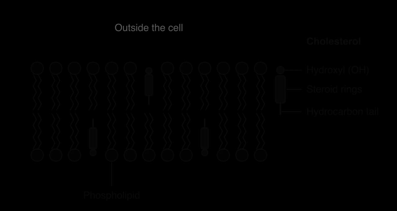
Cholesterol acts as a modulator, or adjustor, of membrane fluidity, with opposite effects at high and low temperatures:
At high temperatures the rigid rings restrict the movement of the fatty acid tails next to them, which reduces fluidity and makes the membrane less permeable to small water-soluble molecules and ions.
At low temperatures cholesterol molecules sit between the phospholipids and stop the tails packing closely together and crystallising, which prevents the membrane becoming stiff.
The overall effect is to keep the fluidity of the membrane within a narrower range over the temperatures the cell experiences. Cholesterol is found in animal membranes; plant membranes contain similar sterols such as stigmasterol instead, and bacterial membranes usually contain no sterols.
Because the bilayer is fluid, it can bend, pinch apart and join with other membranes without tearing. A small piece of membrane can bud off as a vesicle, and a vesicle can fuse with another membrane so that the two bilayers become one continuous sheet. In both processes the hydrophobic tails stay out of contact with water. Vesicles allow cells to move large molecules, particles and large volumes of fluid that could not pass through channels or pumps.
In endocytosis a region of the plasma membrane folds inwards (invaginates) around the material to be taken in. The edges of the fold meet and fuse, and a vesicle containing the material separates from the membrane into the cytoplasm. The process uses energy from ATP, and proteins of the cytoskeleton pull the membrane into shape.
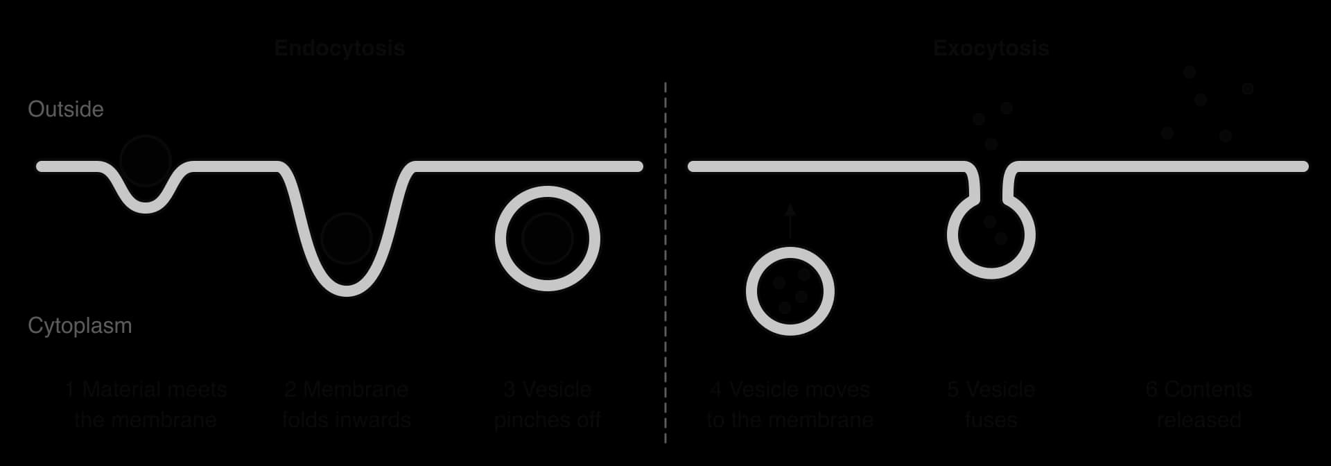
There are two main kinds of endocytosis:
Phagocytosis ("cell eating") takes in large solid particles. White blood cells called neutrophils and macrophages extend parts of their cytoplasm around bacteria, enclose them in a large vesicle and then digest them with enzymes from lysosomes; amoebas feed in the same way.
Pinocytosis ("cell drinking") takes in small droplets of the surrounding fluid, with the solutes dissolved in it, in small vesicles.
A more specific form of endocytosis uses receptors on the cell surface to bind particular molecules before the vesicle forms. Liver cells, for example, take up cholesterol carried in the blood in particles called LDL in this way; the coated vesicles involved are described in B2.2 Organelles and compartmentalisation.
In exocytosis a vesicle moves through the cytoplasm, carried along the cytoskeleton, to the plasma membrane. The vesicle membrane fuses with the plasma membrane and opens, and the contents are released outside the cell. Examples include:
secretion of digestive enzymes by gland cells of the pancreas, which make the enzymes on the rough endoplasmic reticulum, package them in the Golgi apparatus and release them into the pancreatic duct;
release of neurotransmitters such as acetylcholine from the end of a neuron at a synapse, triggered by the entry of Ca²⁺ (see C2.2 Neural signalling);
removal of indigestible waste after phagocytosis.
Endocytosis and exocytosis also change the area of the plasma membrane. Every vesicle that forms by endocytosis removes a piece of membrane, and every vesicle that fuses adds one. A cell that is secreting rapidly would keep growing in surface area if it did not also recycle membrane by endocytosis.
Neurons depend on channels that open only at particular moments. A channel that is closed most of the time but opens in response to a signal allows ions to rush through it down their concentration gradients for a short time, which changes the voltage across the membrane. There are two main ways of opening a gated channel.
A ligand-gated channel has a binding site on its outer surface for a specific chemical, its ligand. When the ligand binds, the channel undergoes a conformational change that opens the pore. When the ligand leaves the binding site, the channel closes again.
The nicotinic acetylcholine receptor is a ligand-gated channel in the postsynaptic membrane at many synapses, including the neuromuscular junctions between motor neurons and muscle fibres. It is made of five protein subunits arranged in a ring around a central pore. Two of the subunits have binding sites for the neurotransmitter acetylcholine. When acetylcholine binds to both sites, the pore opens and positively charged ions pass through, mainly Na⁺ diffusing into the cell. The entry of positive charge depolarises the postsynaptic membrane. The receptor is called nicotinic because nicotine also binds to it and opens it.
A voltage-gated channel opens and closes in response to changes in the voltage across the membrane, the membrane potential. Parts of the channel protein carry charged amino acids, and when the voltage changes these parts move, changing the shape of the protein. Voltage-gated channels for Na⁺ and K⁺ in the membranes of neuron axons produce action potentials:
At the resting potential (about −70 mV, inside negative) both types of channel are closed.
When the membrane is depolarised to a threshold, voltage-gated Na⁺ channels open. Na⁺ diffuses into the axon down its concentration gradient, making the inside positive.
Within about a millisecond the Na⁺ channels close (they become inactivated) and voltage-gated K⁺ channels open. K⁺ diffuses out, so the inside becomes negative again (repolarisation).
The K⁺ channels close, and the sodium–potassium pump restores the original concentrations of the ions over time.
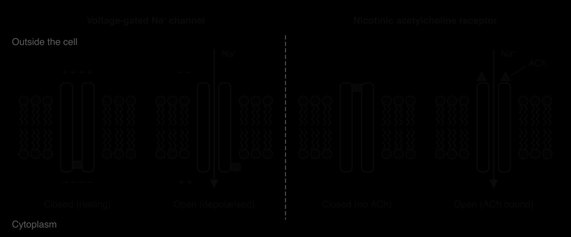
Gated channels are a clear example of interaction between molecules and cells: a neurotransmitter released by one cell changes the permeability of another cell's membrane within a fraction of a millisecond. How action potentials travel along axons and how synapses pass on signals are covered in C2.2 Neural signalling.
The sodium–potassium pump is an integral protein in the plasma membrane of almost every animal cell. It uses ATP to pump three Na⁺ ions out of the cell and two K⁺ ions into the cell in each cycle, both against their concentration gradients. Because it moves two different ions in opposite directions, it is an exchange transporter (antiporter).
The pump is open to the cytoplasm. Three Na⁺ ions from the cytoplasm bind to sites inside the pump.
Binding of Na⁺ allows ATP to transfer a phosphate group to the pump (the pump is phosphorylated), and ADP is released.
Phosphorylation causes a conformational change: the pump closes on the inside and opens to the outside of the cell. Its shape no longer fits Na⁺, so the three Na⁺ ions are released outside.
Two K⁺ ions from outside the cell bind to the pump.
Binding of K⁺ causes the phosphate group to be released from the pump.
Loss of the phosphate makes the pump return to its original shape, open to the cytoplasm. The two K⁺ ions are released inside the cell, and the cycle can begin again.
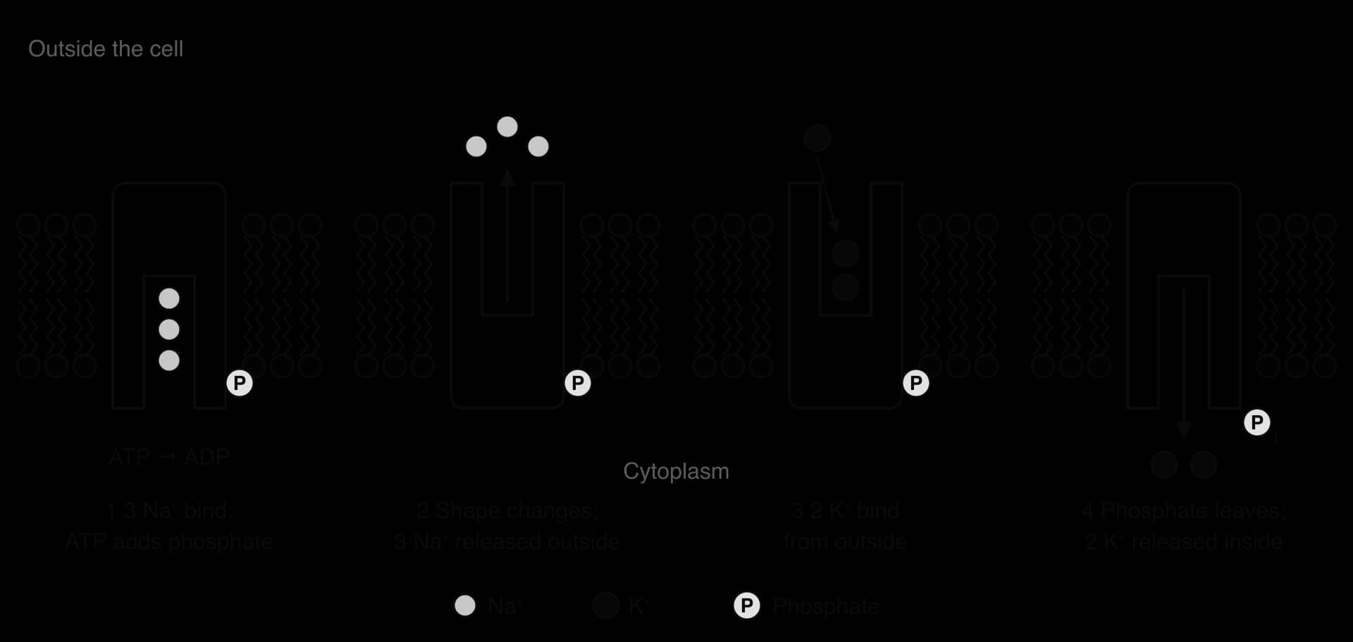
The pump builds up a high concentration of Na⁺ outside the cell and a high concentration of K⁺ inside. It also moves more positive charges out (three) than in (two) in each cycle, so it directly makes the inside of the cell slightly negative. Most of the resting potential comes from K⁺: the membrane at rest contains open K⁺ channels, so some K⁺ diffuses out down the gradient that the pump created, leaving the inside negative. In neurons the result is a resting potential of about −70 mV.
The concentration gradients set up by the pump are a store of potential energy. They drive the movement of Na⁺ into neurons through voltage-gated channels during action potentials, and the uptake of glucose and other substances by cotransporters. The pump also helps cells control their volume, by keeping the concentration of ions in the cytoplasm low enough to prevent too much water entering by osmosis. Running the pump continuously uses a large share of the ATP produced by a resting human body, and an even larger share in the brain. Jens Skou discovered the pump in nerve membranes from crabs in 1957 and shared the 1997 Nobel Prize in Chemistry for the discovery.
Some transport proteins move two different substances across a membrane together, in the same direction. These proteins are cotransporters (symporters). A cotransporter does not use ATP itself. Instead, one substance moves down its concentration gradient and the energy released drives the other substance against its gradient.
The sodium-dependent glucose cotransporter (SGLT) binds Na⁺ and glucose on the outside of the cell. Na⁺ is much more concentrated outside the cell than inside, because the sodium–potassium pump keeps removing it. As Na⁺ moves into the cell down its concentration gradient, the cotransporter changes shape and carries glucose in as well, even when the glucose concentration inside the cell is already higher than outside.
This is indirect active transport (also called secondary active transport). The movement of glucose against its gradient needs energy, but the energy comes from the Na⁺ gradient, not directly from ATP. ATP is used by the sodium–potassium pump to make the Na⁺ gradient in the first place, so if the pump stops, glucose uptake by SGLT stops too.
Epithelial cells lining the small intestine absorb glucose from digested food in three steps, each through a different membrane protein:
The sodium–potassium pump in the basal membrane (the side next to the blood) pumps Na⁺ out of the cell, using ATP. This keeps the Na⁺ concentration inside the cell low.
SGLT1 in the membrane of the microvilli (the side facing the lumen) moves two Na⁺ and one glucose molecule into the cell together. Na⁺ moves down its gradient, and glucose is carried in against its gradient, so glucose builds up inside the cell.
Glucose leaves the cell through glucose carrier proteins in the basal membrane by facilitated diffusion, down its concentration gradient, into the tissue fluid and then the blood.
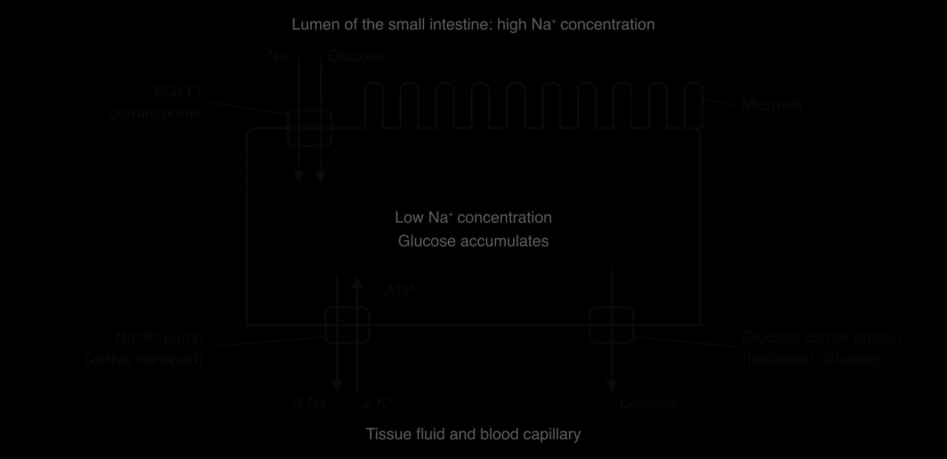
Oral rehydration solution, used to treat the severe diarrhoea of cholera, depends on this mechanism. The solution contains both glucose and sodium salts. SGLT1 in the intestine absorbs Na⁺ and glucose together, and water follows by osmosis, so a patient can be rehydrated by drinking even while losing large volumes of fluid. Because it is cheap and simple, oral rehydration therapy has saved millions of lives, especially of young children.
The kidneys filter blood, and the filtrate initially contains glucose at the same concentration as blood plasma. Cells lining the first part of the nephron, the proximal convoluted tubule, reabsorb the glucose so that it is not lost in urine. They use the same mechanism as the intestine, with a sodium–potassium pump on the blood side and sodium-dependent glucose cotransporters (mainly SGLT2) on the side facing the filtrate. Normally this reabsorbs all of the filtered glucose. Drugs called SGLT2 inhibitors, such as dapagliflozin and empagliflozin, block this cotransporter, so glucose is lost in urine and the blood glucose concentration falls; they are used to treat type 2 diabetes.
In multicellular organisms, most cells are attached to neighbouring cells. These attachments hold the cells of a tissue together, keep them in the right arrangement and allow some tissues to withstand stretching. The attachments are made by proteins in the plasma membrane called cell-adhesion molecules.
Cell-adhesion molecules (CAMs) are integral proteins that span the membrane. The part outside the cell binds to a CAM on the neighbouring cell, and the part inside is linked to the cytoskeleton, so the attachment is strong and the force is spread through the cells. Different CAMs are used to make different types of junction between cells: some make tight seals that stop fluid leaking between the cells of an epithelium, and others make strong anchoring points in tissues that are stretched, such as skin and heart muscle. One well-known family, the cadherins, only binds when Ca²⁺ ions are present.
CAMs also allow cells to recognise one another, because a CAM usually binds only to a matching CAM on another cell. During development this helps cells of the same type to sort themselves into the same tissue. When cancer cells lose some of their CAMs, they become detached from their neighbours and can spread to other parts of the body (metastasis), which links membrane structure to the behaviour of whole tissues.
Membranes appear throughout the course, so questions on B2.1 are common in every paper. Paper 1A multiple-choice questions often test the position of components in the fluid mosaic model, the differences between the types of transport, and the direction of osmosis. Paper 1B uses data from practical work, such as beetroot pigment leakage or the change in mass of tissue in different solutions. Paper 2 includes drawing the fluid mosaic model, short explanations of transport, and extended responses on membrane structure and transport. The command terms most often used are draw, outline, explain, distinguish and compare and contrast.
Extended responses on membranes reward precise terms used correctly: partially permeable, concentration gradient, hydrophilic, hydrophobic, conformational change, specific binding site and ATP. Annotated diagrams are accepted in extended answers when they are clearly labelled.
A good set of notes connects membranes to other parts of the course. Membrane proteins appear in B2.2, C1.2, C2.1 and C2.2, and the principles of osmosis are developed further in D2.3.
I can explain why phospholipids form continuous bilayers in water and why the bilayer is a barrier to ions and large polar molecules.
I can describe simple diffusion of O₂ and CO₂ and explain the factors that affect its rate.
I can distinguish integral from peripheral proteins and list six functions of membrane proteins.
I can define osmosis in terms of solute concentration and explain the role of aquaporins.
I can compare simple diffusion, facilitated diffusion and active transport, and explain how channels and pumps make membranes selectively permeable.
I can outline the structure and functions of glycoproteins and glycolipids.
I can draw and label the fluid mosaic model and explain the evidence that falsified the Davson–Danielli model.
I can explain how fatty acid composition and cholesterol affect membrane fluidity. (HL)
I can describe endocytosis and exocytosis with an example of each. (HL)
I can explain how ligand-gated and voltage-gated ion channels work in neurons. (HL)
I can describe the sodium–potassium pump cycle and its role in the resting potential. (HL)
I can explain glucose uptake by sodium-dependent glucose cotransporters and the role of CAMs in tissues. (HL)
[7 max]
Marks may be awarded for information in a clearly annotated diagram.
Outline how hydrophobic and hydrophilic interactions lead to the formation of lipid bilayers in cell membranes.
4 marks
a. phospholipids are amphipathic / have hydrophilic and hydrophobic parts;
b. the phosphate heads are hydrophilic / attracted to water;
c. the fatty acid / hydrocarbon tails are hydrophobic / not attracted to water;
d. in water the tails are kept together / away from water, and the heads face the water;
e. forming a continuous double layer / bilayer spontaneously;
f. the hydrophobic core is a barrier to ions / polar molecules;
[4 max]
One additional mark is available for the construction of the answer.
Cultured human cells were treated with a drug that prevents clathrin from binding to membranes. The rate of uptake of three substances was then measured and expressed as a percentage of the rate in untreated cells.
Substance taken up | Uptake rate / % of untreated cells |
|---|---|
Transferrin (an iron-carrying protein) | 9 |
Oxygen | 101 |
Glucose | 97 |
Which conclusion is best supported by these results?
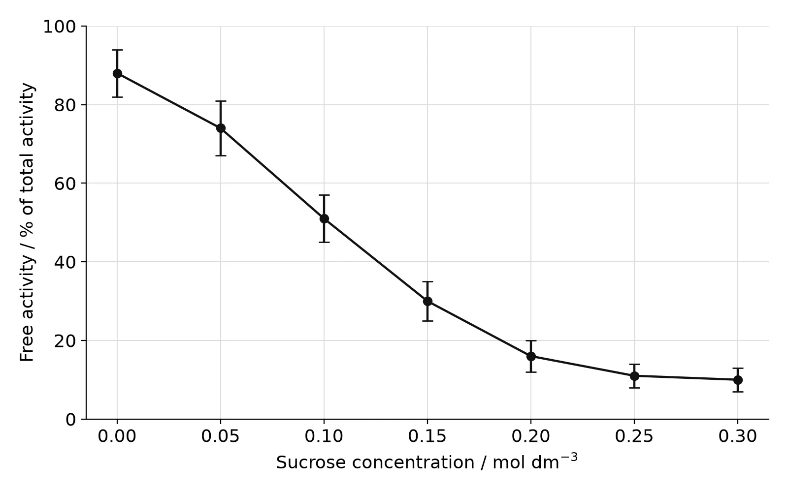
A fraction rich in lysosomes was prepared from liver cells. Equal samples were suspended in sucrose solutions of different concentrations for 15 minutes at 25 °C. The activity of acid phosphatase, a lysosomal enzyme, was then measured by adding its substrate to the medium. The substrate cannot cross the lysosome membrane, so only enzyme that has escaped from lysosomes is active (free activity). Total activity was measured in a separate sample after adding a detergent that dissolves all membranes. Five replicates were done at each concentration.
The graph shows the mean free activity as a percentage of total activity. Error bars show ± one standard deviation.
State the independent variable in this investigation.
1 mark
Suggest why free activity was expressed as a percentage of total activity.
1 mark
Describe the effect of sucrose concentration on free activity.
2 marks
Deduce whether there is a significant difference between the free activity at 0.25 mol dm-3 and at 0.30 mol dm-3.
1 mark
Explain the high free activity in low sucrose concentrations.
2 marks
Using the results, explain the advantage to a living cell of keeping hydrolytic enzymes inside lysosomes.
2 marks