Explain how cells differentiate, what stem cells are and how potency differs, how size and SA:V limit cells, and (HL) how pneumocytes, muscle cells and gametes are adapted.
Each is in the Biology glossary, with every other term in the subject.
A vesicle over the front of a sperm head containing hydrolytic enzymes, released when the sperm meets the egg so that it can digest a path through the zona pellucida.
Striated muscle of the heart, made of short, branched cells with usually one nucleus each, joined end to end by intercalated discs.
A vesicle just beneath the plasma membrane of an egg whose contents, released after fertilisation, harden the zona pellucida and prevent other sperm entering.
The process by which an unspecialised cell becomes specialised for a particular function, by expressing some of its genes and not others.
An organism in the early stages of development after the zygote begins to divide; in humans, up to about eight weeks after fertilisation.
A haploid sex cell (a sperm or an egg) that fuses with another gamete at fertilisation to form a zygote.
A specialised junction joining cardiac muscle cells end to end, containing gap junctions that allow electrical impulses to pass between cells and strong junctions that hold them together.
An inward folding of a membrane or layer of cells, which increases surface area; in some cells the plasma membrane folds deeply into the cytoplasm.
A human body contains more than 200 types of cell, from red blood cells that carry oxygen to neurons that carry electrical signals over a metre. All of them come from a single fertilised egg. Cell specialisation explains how one cell gives rise to so many different kinds, and how each kind is shaped for its function.
Two principles underlie cell specialisation. Every cell in a body has the same genes but uses only some of them, so specialisation is a question of gene expression. Each specialised cell also shows form and function at the level of the cell: its size, shape and contents match the job it does, within the limits set by its surface area and volume.
39 exam-style questions on this subtopic, written in the style of the IB papers. The first has its markscheme open. Counted on
The structure of specialized cells and tissues is closely related to their function.
Compare and contrast the structure of xylem vessels and phloem sieve tubes in relation to their functions.
7 marks
Similarities:
a. both are tubes made of cells joined end to end;
b. both are found together in vascular bundles;
c. both have no nucleus when mature / both have reduced or no cell contents, allowing flow;
d. both have cellulose cell walls;
e. both carry fluid by mass flow;
Differences:
f. xylem vessels are dead / phloem sieve tube elements are living;
g. xylem walls are lignified / phloem walls are not;
h. xylem has no / incomplete end walls / phloem has sieve plates with pores;
i. xylem has pits in the side walls / phloem has plasmodesmata linking to companion cells;
j. sieve tube elements have companion cells with many mitochondria / xylem has no companion cells;
k. xylem carries water and mineral ions / phloem carries sucrose and other organic compounds;
l. xylem flow is one way, from roots to leaves / phloem flow is from source to sink, in either direction;
m. xylem contents are under tension / phloem contents are under positive pressure;
[7 max]
At least one similarity and one difference are needed for full marks.
Describe the adaptations of type I and type II pneumocytes in the alveoli.
The official deck for this part of the syllabus.
How many types of cell are there in the human body, and from which single cell do they all come?
Do the different specialised cells of one body contain different genes? Explain.
With a free account, Augment remembers where you stopped and what you have practiced.
A signalling chemical that forms a concentration gradient across an embryo and causes cells to develop differently according to the concentration they receive.
Describes a stem cell that can differentiate into a limited range of cell types, usually within one tissue, such as blood stem cells in bone marrow.
The large, non-motile female gamete, with a haploid nucleus and cytoplasm containing nutrient stores and organelles for the early embryo.
Describes a stem cell that can differentiate into any type of cell in the body, but not extra-embryonic cells, so cannot form a whole organism alone.
The ability of a stem cell to divide repeatedly by mitosis while keeping a population of unspecialised stem cells.
A cell whose structure and contents are adapted to carry out a particular function, as a result of differentiation.
The small, motile male gamete, with a haploid nucleus in its head, mitochondria in its midpiece and a flagellum for swimming.
An unspecialised cell that can divide repeatedly to make more stem cells and can differentiate into one or more types of specialised cell.
A microenvironment in a tissue that maintains stem cells and controls whether they remain unspecialised or divide and differentiate, for example in bone marrow or hair follicles.
The surface area of a cell or organism divided by its volume; it decreases as size increases, because volume increases faster than surface area.
Describes a stem cell that can differentiate into any type of cell, including extra-embryonic cells such as the placenta, and so can form a whole organism.
An extremely thin, flattened cell that forms most of the alveolar wall, keeping the diffusion distance for gas exchange very short.
A rounded cell in the alveolar epithelium that secretes surfactant from lamellar bodies, reducing surface tension, and can divide to replace type I pneumocytes.
A thick layer of glycoproteins surrounding the plasma membrane of a mammalian egg, to which sperm bind before fertilisation.
The diploid cell formed when the nuclei of a male and a female gamete fuse at fertilisation; the first cell of a new organism.
Sexual reproduction begins with fertilisation, when a sperm and an egg fuse. The cell produced is a zygote, with a full diploid set of chromosomes. The zygote divides repeatedly by mitosis, producing an embryo of genetically identical cells. In the first few divisions the cells are unspecialised: they look alike and none has yet taken on a particular role.
As the embryo grows, its cells start to become different from each other. This process is differentiation. A differentiating cell switches on some genes and switches off others, so it makes a particular set of proteins. Those proteins give it its structure and function. A cell that has completed this process is a specialised cell, such as a muscle cell full of contractile proteins or a red blood cell full of haemoglobin.
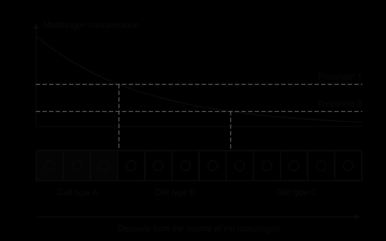
Cells in different parts of an embryo must become different types: the cells at one end form the head, those at the other end the tail. They do this by responding to signalling chemicals called morphogens. A morphogen is released by cells in one region and diffuses away through the embryo, so its concentration is high near the source and falls with distance. A concentration gradient is set up across the tissue.
Each cell detects the concentration of morphogen where it sits. Morphogens act on gene expression: above one threshold concentration a certain set of genes is switched on, between two thresholds a different set, and below the lower threshold another. Cells at different distances from the source therefore express different genes and differentiate into different types. In this way the position of a cell in the embryo determines which type of cell it becomes.
A well-studied example is the fruit fly Drosophila melanogaster. The mother places mRNA for a protein called Bicoid at the front end of the egg. After fertilisation, Bicoid protein spreads backwards and forms a gradient. Bicoid is a transcription factor: high concentrations switch on the genes for head structures, and lower concentrations switch on genes for the thorax. Embryos without Bicoid develop no head.
Some cells remain unspecialised and keep the ability to produce new specialised cells. These are stem cells. They are found in large numbers in the early embryo, and in smaller numbers in many adult tissues, where they replace cells that wear out or are damaged.
Stem cells have two properties that together set them apart from other cells:
Self-renewal: they can divide by mitosis endlessly, producing more stem cells. When a stem cell divides, at least one of the two daughter cells usually stays a stem cell, so the population is never used up.
Differentiation along different pathways: their daughter cells can differentiate into specialised cells. Depending on the stem cell, the range of possible cell types may be large or small.
Most specialised cells have lost one or both properties. A neuron or a red blood cell does not divide, and a skin cell cannot become a muscle cell. The combination of the two properties is what makes stem cells useful in development, in repair and in medicine.
Adult stem cells are found in particular microenvironments called stem cell niches. The cells around a stem cell, the molecules they secrete and the materials between them control its behaviour. A niche can keep stem cells in a resting, unspecialised state, or signal them to divide and differentiate when new cells are needed.
Niche | Stem cells | What they produce | Function |
|---|---|---|---|
Bone marrow (inside large bones such as the femur, pelvis and sternum) | Haematopoietic (blood-forming) stem cells | All types of blood cell: red blood cells, all types of white blood cell, and platelets | Replace blood cells, which have short lives; human red blood cells last about 120 days |
Hair follicles (the bulge region of each follicle in the skin) | Follicle stem cells | Cells of the hair follicle and the growing hair; also epidermis cells after injury | Regrow hair in each hair growth cycle; help repair the skin when it is wounded |
The bone marrow niche is the basis of bone marrow transplants. E. Donnall Thomas pioneered transplantation of bone marrow to treat leukaemia, a cancer of white blood cells, and shared the Nobel Prize in Physiology or Medicine in 1990. The patient's diseased marrow is destroyed by chemotherapy or radiation, and haematopoietic stem cells from a matched donor are infused into the blood. The donor stem cells find their way to the marrow niches, settle there and rebuild the whole blood system.
Adult stem cells from other niches are used in medicine too. In 2015 Holoclar became the first therapy containing stem cells to be approved in the European Union. It uses stem cells taken from the edge of the cornea (the limbus) of a patient's healthy eye, grown in the laboratory and transplanted to repair a cornea damaged by burns.
Stem cells differ in their potency, the range of cell types they can produce. Potency falls as development goes on.
Type | What it can become | Where it is found |
|---|---|---|
Totipotent | Any type of cell, including the cells of the placenta and other extra-embryonic membranes; a single cell can form a whole organism | The zygote and the cells of the very early embryo (the first few divisions) |
Pluripotent | Any type of cell in the body, but not the placenta; cannot form a whole organism alone | The inner cell mass of the blastocyst (about 5 days after fertilisation in humans); induced pluripotent stem cells |
Multipotent | A limited range of cell types, usually within one tissue | Adult stem cells, for example haematopoietic stem cells in bone marrow |
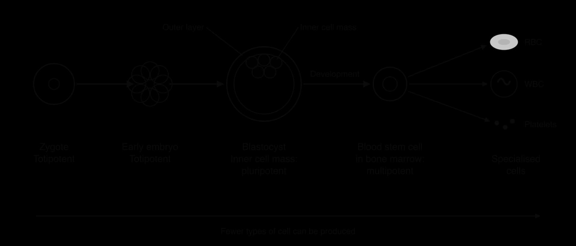
Cells of the early embryo are totipotent, but soon become pluripotent. By about five days after fertilisation the human embryo is a hollow ball called a blastocyst. Its outer layer will form the placenta, and the cluster of cells inside, the inner cell mass, will form the whole body of the fetus. Inner cell mass cells are pluripotent: they can produce every cell type of the body, but no longer the placenta. Embryonic stem cells used in research come from the inner cell mass.
 YouTube
Before 2006 the only source of pluripotent human cells was the early embryo, and obtaining embryonic stem cells destroyed the embryo, which raised ethical objections. Specialised mammalian cells were thought to be fixed in their state. In 2006 Kazutoshi Takahashi and Shinya Yamanaka in Kyoto showed that specialised cells could be returned to a pluripotent state by changing the expression of a few genes.
They took fibroblasts, ordinary specialised cells from the connective tissue of mice, and added just four genes: Oct3/4, Sox2, Klf4 and c-Myc. Each codes for a transcription factor that is active in embryonic stem cells. With these four genes switched on, the fibroblasts changed back into cells that looked and behaved like embryonic stem cells. They were pluripotent: they could form cells of all three germ layers of the embryo. The new cells were called induced pluripotent stem cells (iPSCs). In 2007 the same was achieved with human skin cells.
The result showed that a specialised cell still contains all the genes needed to make any cell type, and that changing the expression of a few regulatory genes is enough to reset it. Yamanaka shared the 2012 Nobel Prize in Physiology or Medicine with John Gurdon, who had shown in the 1960s that the nucleus of a specialised frog cell could support the development of a whole tadpole.
The first medical use followed quickly. In wet age-related macular degeneration, the layer of cells that supports the light-sensitive cells of the retina (the retinal pigment epithelium) is damaged, and central vision is lost. In 2014 Masayo Takahashi's team at RIKEN in Kobe took skin cells from a woman in her 70s with the disease, reprogrammed them into iPSCs, and differentiated those into retinal pigment epithelium. A small sheet of these cells was transplanted under her retina.
Stage | Date | Detail |
|---|---|---|
Mouse iPSCs from fibroblasts | 25 August 2006 (published) | Four transcription factor genes: Oct3/4, Sox2, Klf4, c-Myc |
Human iPSCs | 2007 | The same approach with human skin cells |
Nobel Prize in Physiology or Medicine | 2012 | Shinya Yamanaka and John Gurdon |
First transplant of iPSC-derived cells | 12 September 2014, Kobe | A 1.3 mm × 3.0 mm sheet of retinal pigment epithelium made from the patient's own skin cells |
Result after one year | 2015 (reported 2017) | The sheet was intact; vision had neither improved nor worsened |
Sources: Takahashi and Yamanaka, Cell 126:663, 2006; Nobel Assembly at Karolinska Institutet, 2012; Mandai et al., New England Journal of Medicine 376:1038, 2017.
Because the cells came from the patient herself, her immune system did not reject them, so no immunosuppressive drugs were needed. The main aim of this first trial was safety, and the result showed that the transplanted cells survived for at least a year and caused no harm. iPSCs also avoid the ethical problem of destroying embryos, although they carry their own risks: one of the four original genes, c-Myc, can cause cancer, and reprogrammed cells must be checked carefully for mutations before use.
Most human cells are between about 5 µm and 20 µm across, too small to see without a microscope. Specialisation can push cells far outside this range in either direction, and the size of a cell is itself an adaptation to its function.
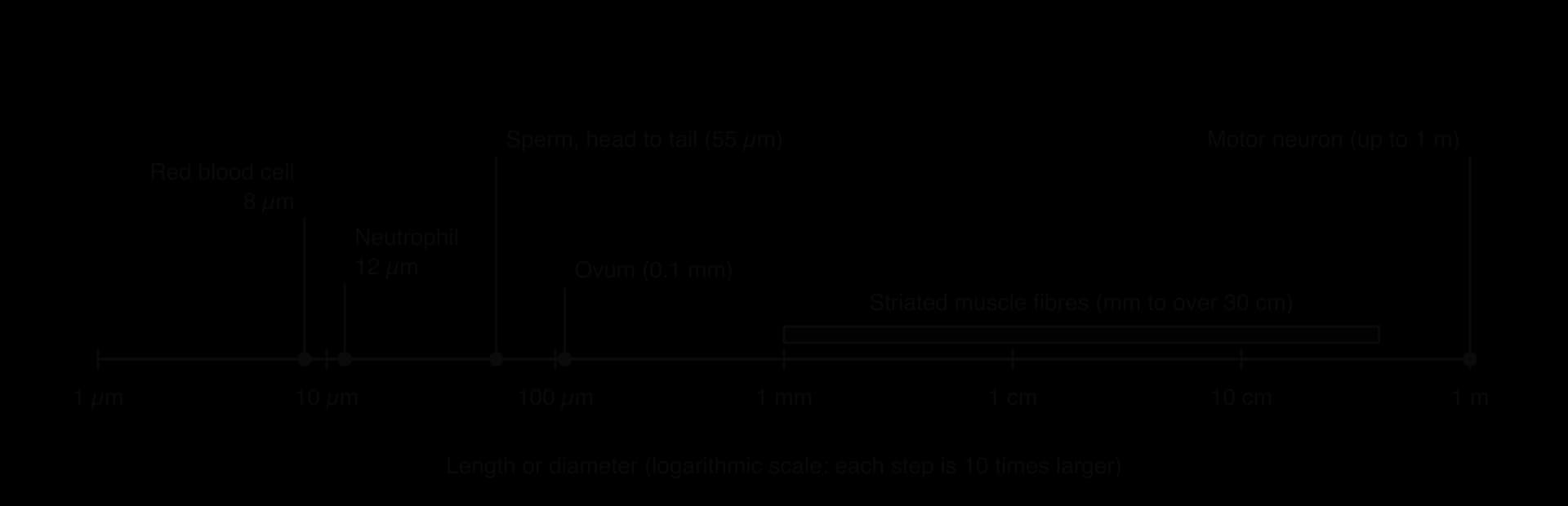
Cell | Typical size | How size relates to function |
|---|---|---|
Ovum (egg cell) | About 0.1 mm diameter; just visible to the naked eye | Large volume of cytoplasm stores nutrients and organelles for the early embryo |
Sperm | Head about 5 µm long; about 55 µm including the tail | Small and streamlined, with little cytoplasm, so it can swim fast; it contributes only a haploid nucleus and a centriole |
Red blood cell | 7–8 µm diameter, about 2 µm thick | Small enough to squeeze through capillaries about the same width; large surface area relative to volume for oxygen uptake |
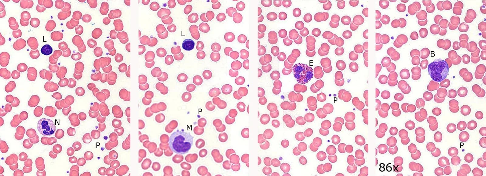
White blood cells | About 7–20 µm, depending on type (a neutrophil about 12 µm) | Larger than red blood cells, with a nucleus and organelles; phagocytes need room to engulf bacteria |
Motor neuron | Cell body about 0.1 mm; axon up to about 1 m (spinal cord to foot) | A single long cell carries a signal over the whole distance without delays at junctions |
Striated muscle fibre | Diameter about 10–100 µm; length from millimetres to more than 30 cm | One fibre contracts along its whole length as a unit |
The two gametes show the contrast most clearly. The ovum is one of the largest cells in the body and the sperm one of the smallest. Both are haploid, and both deliver one set of chromosomes, but the egg also provides the cytoplasm, mitochondria and food reserves for the first days of development. The sperm is reduced to what is needed to reach and enter the egg.
 YouTube
Every cell exchanges materials with its surroundings. Oxygen and nutrients enter, and carbon dioxide and other wastes leave, all across the plasma membrane. Heat produced by metabolism is also lost through the surface. The rate at which a cell can exchange materials depends on its surface area. The rate at which it needs to exchange materials depends on its volume, because the volume holds the cytoplasm where metabolism takes place.
The relationship between the two is described by the surface area-to-volume ratio (SA:V). As a cell grows, its volume increases faster than its surface area. Doubling the side of a cube multiplies its surface area by 4 (2²) but its volume by 8 (2³), so its SA:V ratio halves.
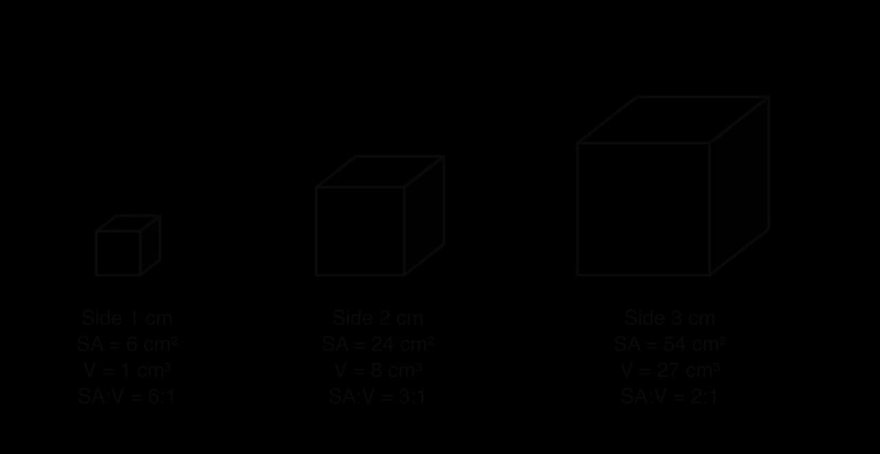
For a cube of side length :
A large cell therefore has relatively little surface for its volume. If a cell grew too large, materials could not enter or leave fast enough to supply its metabolism, and the centre of the cell would be far from the surface, so diffusion would take too long. Waste products and heat would also build up. This is why most cells are small, and why a cell that has grown large usually divides. Cells that are very large, such as the egg, have a low rate of metabolism, or have a shape that keeps every part of the cytoplasm close to the surface, such as long, thin neurons and muscle fibres.
The effect of SA:V can be shown with cubes of agar containing an indicator, such as phenolphthalein, which is pink in alkaline conditions. When cubes of different sizes are placed in dilute acid, the acid diffuses in and the pink colour disappears from the outside inwards. Small cubes lose their colour completely in a short time, while the centre of a large cube stays pink much longer.
Cells that must exchange materials quickly, without simply being small, use three main adaptations to increase their SA:V ratio:
Flattening. A flat cell has a much larger surface for its volume than a rounded cell, and no part of its cytoplasm is far from the surface.
Microvilli. These are finger-like projections of the plasma membrane, about 1 µm long, which greatly increase the area of membrane on one surface of the cell.
Invagination. The plasma membrane folds inwards into the cell, increasing the area of membrane without increasing the volume much.
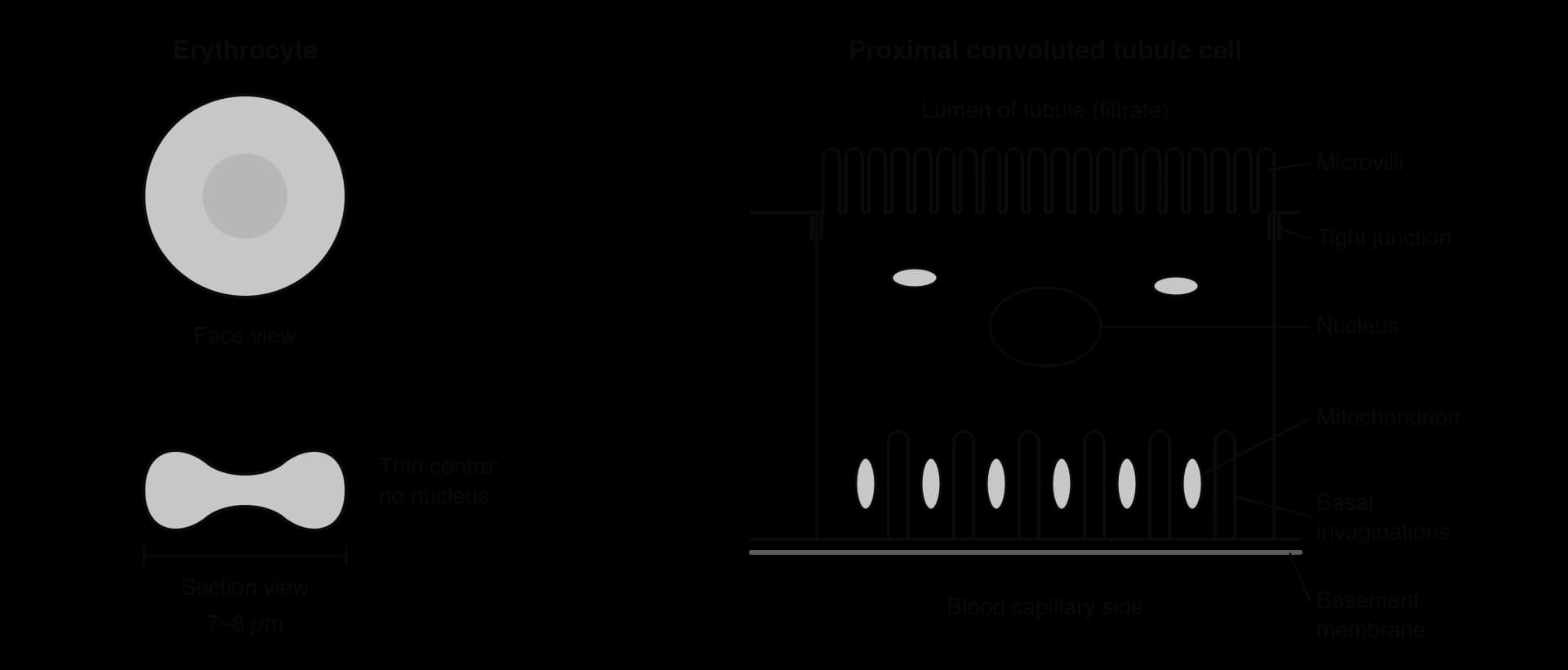
Red blood cells (erythrocytes) are biconcave discs: flattened, and thinner in the centre than at the rim. Each is about 7–8 µm across but only about 2 µm thick. The shape gives a large surface area for the volume of the cell, so oxygen diffuses in and out quickly, and no haemoglobin molecule is more than about 1 µm from the plasma membrane. Mature mammalian red blood cells also have no nucleus and no mitochondria, which leaves more space for haemoglobin. The flattened shape is flexible, so the cells can fold to pass through narrow capillaries.
In the kidney, the proximal convoluted tubule of each nephron reabsorbs useful substances from the filtrate back into the blood. Its walls are made of a single layer of cuboidal cells, which reabsorb about two-thirds of the water and sodium in the filtrate and all of the glucose. These cells show both microvilli and invaginations:
On the side facing the lumen (the apical side), dense microvilli form a brush border that greatly increases the area of membrane for absorbing glucose, amino acids and ions from the filtrate.
On the side facing the blood (the basal side), the plasma membrane is deeply invaginated. The folds increase the area for pumping substances out of the cell towards the blood.
Many mitochondria are packed between the basal invaginations, close to the membrane. They supply ATP for the Na⁺/K⁺ pumps there, which drive the active transport of reabsorption.
The walls of the alveoli in the lungs (B3.1 Gas exchange) are lined by an epithelium made of two kinds of cell, called pneumocytes. The two types have very different structures because they do very different jobs.
Type I pneumocytes are extremely thin, flattened cells. Over most of their area the cytoplasm is only a fraction of a micrometre thick, bulging only where the nucleus sits. They cover about 95% of the alveolar surface. Their thinness keeps the diffusion distance between air and blood as short as possible: together with the capillary wall, the barrier is less than 1 µm across over much of the alveolus.
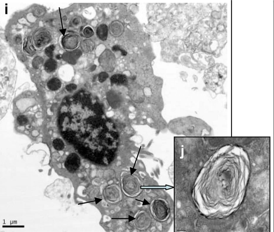
Type II pneumocytes are rounded (cuboidal) cells scattered among the type I cells. Their cytoplasm contains many lamellar bodies, secretory vesicles full of surfactant in tightly packed layers. They release surfactant by exocytosis onto the moist inner surface of the alveolus, and have microvilli on their exposed surface. They can also divide and differentiate to replace damaged type I cells.
Surfactant is a mixture of phospholipids and proteins that forms a film on the water lining the alveoli. It reduces surface tension, so the alveoli do not collapse when air is breathed out and are easier to inflate on the next breath. The importance of type II cells was shown in 1959 when Mary Ellen Avery found that the lungs of premature babies who died of respiratory distress syndrome lacked surfactant. Their type II pneumocytes had not yet started producing enough of it. Treatment with surfactant given into the airways became standard care for very premature babies in the 1990s.
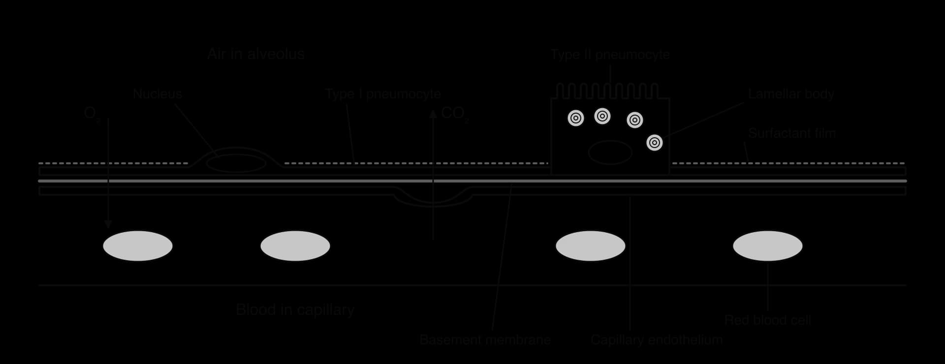
The alveolar epithelium shows that a tissue may need more than one type of cell. A thin cell cannot also be packed with secretory vesicles, and a thick secretory cell would slow diffusion. Dividing the work between two cell types allows both functions at once.
Skeletal muscle (the muscle attached to bones) and cardiac muscle (the muscle of the heart) are both striated: under the microscope they show regular light and dark bands. The bands come from myofibrils, long bundles of the contractile proteins actin and myosin arranged in repeating units, which fill most of the cytoplasm (B3.3 Muscle and motility). The way these myofibrils are packaged into cells is very different in the two tissues.
Feature | Cardiac muscle cell | Striated (skeletal) muscle fibre |
|---|---|---|
Shape | Short, roughly cylindrical, with branches | Very long, cylindrical, unbranched |
Length | About 0.1 mm | Millimetres to more than 30 cm |
Nuclei | Usually one, near the centre | Many, close to the plasma membrane |
Junctions between cells | Intercalated discs join cells end to end | No junctions along the fibre |
Contraction | Rhythmic and involuntary; the cells contract together | Voluntary, under the control of motor neurons |
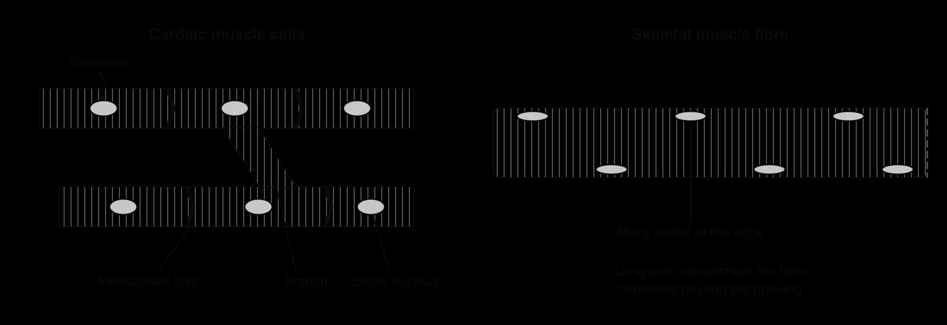
Several hypotheses link these structural differences to function:
Branching and intercalated discs in cardiac muscle. Intercalated discs contain gap junctions, which let ions and electrical impulses pass directly from cell to cell, and strong junctions that hold the cells together when they contract. Branches connect the cells into a three-dimensional network, so a wave of excitation spreads quickly through the whole wall of a heart chamber and the cells contract together, squeezing the blood out.
Length and many nuclei in skeletal muscle. Each skeletal muscle fibre forms when many precursor cells (myoblasts) fuse together during development, which explains its many nuclei. A single long fibre can contract along its whole length at once when a motor neuron stimulates it, without signals having to cross from cell to cell. The many nuclei, spread along the fibre, can make the large amounts of protein needed to build and repair such a large volume of cytoplasm.
A single nucleus in cardiac muscle. Cardiac cells are small enough for one nucleus to control. Cardiac muscle cells are not made by fusion, and almost all of them stop dividing after birth, which is why damage from a heart attack is not repaired.
Cell theory states that living things are made of cells, and a typical cell has one nucleus surrounded by cytoplasm and a single plasma membrane. A skeletal muscle fibre fits only part of this description. It is surrounded by one continuous plasma membrane, and its cytoplasm is continuous along its length, which suggests it is one cell. It also has hundreds of nuclei and was formed by the fusion of many cells, which suggests it is several cells joined together, or a structure that does not fit the usual idea of a cell. Some biologists therefore describe it as a syncytium, a mass of cytoplasm with many nuclei.
Atypical examples such as muscle fibres, aseptate fungal hyphae (also multinucleate) and red blood cells (no nucleus) test the generality of cell theory. Most biologists keep the theory and treat these as exceptions that can be explained by how they develop, rather than as evidence against it.
Human gametes are adapted to opposite roles. The sperm must swim to the egg and deliver its nucleus. The egg must attract and accept one sperm only, and then support the embryo until it implants in the uterus.

Structure | Adaptation and function |
|---|---|
Sperm: acrosome | A large vesicle over the front of the head, containing hydrolytic enzymes that digest a path through the zona pellucida |
Sperm: haploid nucleus | Holds 23 chromosomes, tightly condensed so that the head is small and streamlined |
Sperm: centriole | Forms the base of the flagellum and, after fertilisation, helps form the zygote's first mitotic spindle |
Sperm: midpiece with mitochondria | Mitochondria wound in a helix around the start of the flagellum produce ATP for swimming |
Sperm: flagellum (tail) | Microtubules slide past each other, using ATP, so the tail beats and drives the sperm forwards |
Egg: zona pellucida | A thick layer of glycoproteins around the egg; sperm bind to it and it triggers the acrosome reaction |
Egg: plasma membrane | Carries proteins that bind sperm, so the sperm membrane can fuse with the egg membrane |
Egg: cortical granules | Vesicles just beneath the plasma membrane; after one sperm enters, they release enzymes that harden the zona pellucida and stop other sperm entering |
Egg: haploid nucleus | Holds 23 chromosomes; with the sperm nucleus it restores the diploid number |
Egg: lipid droplets and other stores | Provide nutrients and energy for the embryo in its first days, before it implants |
Egg: mitochondria | Supply ATP for the early embryo; all the mitochondria of the new individual come from the egg |
The large difference in size between the two gametes reflects the division of labour. Producing millions of small, mobile sperm raises the chance that one reaches the egg. Producing a few large, well-stocked eggs gives each embryo a good start. The events of fertilisation (the acrosome reaction and the cortical reaction) are described in D3.1 Reproduction.
Cell specialisation is assessed in Paper 1A through potency, niches and SA:V questions, often with a calculation. Paper 1B uses data such as cube or agar-block experiments and cell size measurements. Paper 2 asks for SA:V calculations with units, explanations of how size limits cells, and at HL drawings of sperm and egg cells and comparisons of muscle types. Common command terms are calculate, distinguish, explain, outline, draw and discuss.
Answers about stem cell niches score when they name the niche (bone marrow or hair follicle), the stem cells, and what the niche does (maintains the stem cells or triggers division and differentiation). "Stem cells are found in many places" earns nothing.
I can outline how a zygote develops into specialised cells by differentiation.
I can explain how morphogen gradients cause cells in different positions to express different genes.
I can state the two properties of stem cells.
I can describe the stem cell niches in bone marrow and hair follicles.
I can distinguish between totipotent, pluripotent and multipotent stem cells, with examples.
I can describe how induced pluripotent stem cells are made and why they show that differentiation does not remove genes.
I can relate the sizes of human cells to their functions.
I can calculate SA:V for a cube and explain why it limits cell size.
I can explain how flattening, microvilli and invagination increase SA:V. (HL)
I can explain the adaptations of type I and type II pneumocytes. (HL)
I can compare cardiac muscle cells with striated muscle fibres and discuss whether a fibre is a cell. (HL)
I can draw and annotate human sperm and egg cells. (HL)
a. type I pneumocytes are extremely thin / flattened;
b. giving a short diffusion distance for gas exchange;
c. type I pneumocytes cover most of the alveolar surface;
d. type II pneumocytes have many secretory vesicles / lamellar bodies;
e. which release surfactant into the alveolar lumen;
f. surfactant reduces surface tension, preventing the alveoli from collapsing / sticking together;
g. the alveolar wall is a tissue with more than one cell type;
[4 max]
Outline how valves maintain the one-way flow of blood and lymph in mammals.
4 marks
a. valves open when pressure behind them is higher than in front, and close when the pressure difference reverses;
b. atrioventricular valves close as the ventricles contract, preventing backflow into the atria;
c. semilunar valves close when ventricular pressure falls below arterial pressure, preventing backflow into the ventricles;
d. pocket valves in veins prevent backflow where blood pressure is low;
e. so contraction of skeletal muscle squeezing the veins moves blood only towards the heart;
f. valves in lymph ducts prevent backflow of lymph;
[4 max]
One additional mark is available for the construction of the answer.
Multicellular organisms depend on cooperation between specialized cells.
Outline the evolution of multicellularity and its advantages.
4 marks
Explain how unspecialized cells develop into specialized cells.
7 marks
Describe how phloem sieve tube elements and companion cells are adapted for translocation.
4 marks
What is meant by cell differentiation?