Notes on B.1.3 Muscular function
Learning objectives
Outline the structure of skeletal muscle from the whole muscle to the sarcomere, and identify the major muscles of the body.
Describe a motor unit and explain how the all-or-none principle, recruitment and firing frequency grade the force of a contraction.
Outline why muscle contraction depends on a continuous supply of ATP.
Distinguish between type I, type IIa and type IIx fibres and explain how their recruitment depends on the intensity of an activity.
Explain how hypertrophy and atrophy change the size and recruitment of motor units.
Distinguish between isometric, isotonic concentric, isotonic eccentric and isokinetic contractions, using sporting examples.
Analyse a movement joint by joint, naming the agonist, antagonist and type of contraction, and explain reciprocal inhibition.
Explain the sliding filament theory, including the roles of calcium, ATP, actin, myosin, troponin and tropomyosin (HL).
Describe how the bands and zones of a sarcomere change during contraction (HL).
Introduction
Muscular function is the study of how skeletal muscles produce force. Every movement, from holding a plank to sprinting, depends on muscle fibres contracting in a controlled way, with the right muscles working, the right number of fibres switched on and the right type of contraction for the task.
The same knowledge serves both lenses of the course. For performance, it explains why sprinters and marathon runners differ in muscle make-up, and how strength training builds force. For health, it explains why muscles waste during bed rest, injury and ageing, and why resistance exercise helps older adults keep their independence. It also links the course’s themes: muscles are the effectors of the nervous system, their fuel comes from the energy systems, and they pull on bones to create the levers that move the body.
Three types of muscle tissue
The body contains three kinds of muscle tissue. All of them contract, but they differ in structure, in where they are found and in how they are controlled. Skeletal muscle is attached to bones by tendons and produces movement and posture. Smooth muscle lines hollow organs such as blood vessels, the gut and the airways. Cardiac muscle forms the wall of the heart, the myocardium.
Feature | Skeletal muscle | Cardiac muscle | Smooth muscle |
|---|---|---|---|
Location | Attached to bones by tendons | Wall of the heart only | Walls of blood vessels, gut, bladder, airways |
Control | Voluntary (somatic nervous system) | Involuntary; the heart generates its own impulses | Involuntary (autonomic nervous system and hormones) |
Appearance | Striated (striped); long, cylindrical fibres with many nuclei | Striated; branched cells joined end to end | Not striated; spindle-shaped cells with one nucleus |
Fatigue | Fatigues, at a rate that depends on fibre type | Highly fatigue resistant | Fatigue resistant; slow, sustained contractions |
Role in exercise | Produces movement, posture and heat | Pumps blood; output rises with exercise intensity | Redistributes blood by vasoconstriction and vasodilation |
The rest of this topic deals with skeletal muscle. There are more than 600 skeletal muscles in the body, and together they make up roughly 40% of body mass in an adult man and slightly less in an adult woman. The stripes seen under a microscope, called striations, are the repeating pattern of protein filaments that produce contraction.
The structure of skeletal muscle
A skeletal muscle is built like a cable made of smaller and smaller strands. Each level is wrapped in connective tissue, and all the wrappings join at each end of the muscle to form the tendon that attaches the muscle to bone. When the fibres contract, the force passes through these wrappings and the tendon to the bone.
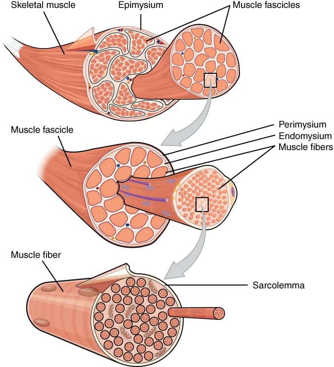
Whole muscle: surrounded by the epimysium, a tough outer layer of connective tissue.
Fascicle: a bundle of up to a few hundred muscle fibres, surrounded by the perimysium. Fascicles can be seen as the grain of a piece of meat.
Muscle fibre: a single muscle cell, surrounded by the endomysium. Fibres are long and thin; in some muscles a single fibre runs for several centimetres. Each fibre has many nuclei and is bounded by a cell membrane called the sarcolemma.
Myofibril: one of hundreds to thousands of thread-like structures packed inside each fibre, running its full length.
Sarcomere: the repeating unit of a myofibril, joined end to end like the links of a chain. It contains the protein filaments actin (thin) and myosin (thick) that slide past each other to shorten the sarcomere.
Inside the fibre, the cytoplasm is called the sarcoplasm. It holds the myofibrils, many mitochondria (especially in endurance fibres), stores of glycogen and fat droplets, and a network of membranes called the sarcoplasmic reticulum that stores calcium ions. Folds of the sarcolemma, called transverse tubules (T-tubules), carry the electrical signal deep into the fibre so that all its myofibrils contract together.

The arrangement explains how a tiny change becomes a large movement. Each sarcomere is only a few micrometres long and shortens by a fraction of that, but thousands of sarcomeres lie end to end along each myofibril. When every one of them shortens at the same moment, the whole fibre shortens by a useful distance, and the forces of all the fibres add together through the tendon.
The major skeletal muscles
Movement analysis uses the names of the major muscles and the joint actions they produce. A muscle usually crosses at least one joint and is attached at two ends: the origin, on the bone that stays more fixed, and the insertion, on the bone that moves. When the muscle shortens, it pulls the insertion towards the origin. The biceps brachii, for example, originates on the scapula and inserts on the radius, so its shortening flexes the elbow.
Muscle | Location | Main action(s) | Sporting example |
|---|---|---|---|
Deltoid | Over the shoulder | Abduction, flexion and extension of the shoulder | Lifting the arms in a lateral raise; the arm action in a front crawl recovery |
Pectoralis major | Front of the chest | Horizontal flexion (adduction) and medial rotation of the shoulder | Pushing phase of a bench press or a chest pass |
Latissimus dorsi | Back, below the shoulder blades | Extension and adduction of the shoulder | The pull phase of a swimming stroke or a pull-up |
Trapezius | Upper back and neck | Elevation, retraction and depression of the scapula | Shrugging the shoulders in a clean |
Biceps brachii | Front of the upper arm | Flexion of the elbow | Upward phase of an arm curl |
Triceps brachii | Back of the upper arm | Extension of the elbow | Push-up; releasing a shot put |
Rectus abdominis | Front of the abdomen | Flexion of the trunk (spine) | Sit-up; stabilising the trunk in a throw |
Obliques (internal and external) | Sides of the abdomen | Rotation and lateral flexion of the trunk | Trunk rotation in a golf swing or a javelin throw |
Hip flexors (iliopsoas) | Deep, from lumbar spine to femur |
The table uses the movement terms flexion, extension, plantarflexion and dorsiflexion for joint actions. These muscle names matter because almost every analysis of a movement starts by naming the muscle that produces it, and the same muscle can act in different ways at different moments of a skill.
Motor units and the all-or-none principle
Skeletal muscle contracts only when it is stimulated by the nervous system. Each motor neuron runs from the spinal cord (or the brainstem) to a muscle, where its axon divides into branches. Each branch ends on a single muscle fibre at a neuromuscular junction. One motor neuron therefore controls a group of fibres, and that group always contracts together.
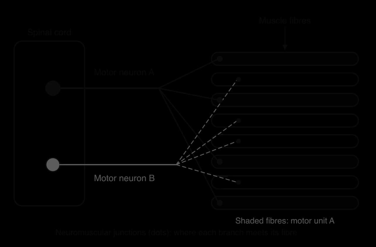
All the fibres in one motor unit are of the same fibre type, because the motor neuron determines the properties of the fibres it supplies. A muscle contains many motor units of different sizes and types. The number of fibres in a unit varies widely. Muscles that control fine movements, such as those that move the eye or the fingers, have small motor units of only a few fibres each. Large muscles that produce gross movements, such as the gastrocnemius or the quadriceps, have motor units with hundreds or more than a thousand fibres.
Small motor units give fine control because the force can be increased in small steps. A darts player or a snooker player relies on the small units of the forearm and hand. Large motor units produce large forces but less precise control, which suits the leg drive of a sprinter or a weightlifter.
The all-or-none principle applies to each motor unit, not to the whole muscle. A single unit cannot make a weak contraction or a half contraction. If the impulse reaches the threshold, every fibre in the unit contracts with its maximum force for that moment. The same muscles can still lift a pen gently or a heavy suitcase, because the nervous system controls how many units are active and how often they fire.
Grading the force of a contraction
The nervous system grades muscle force in two main ways: by changing the number of motor units that are active, and by changing how often each active unit is stimulated. Both are controlled by the motor cortex and the spinal cord, without conscious thought about individual units.
Recruiting more motor units
Motor unit recruitment is the activation of additional motor units to increase the force of a contraction. To pick up a pen, only a few small motor units are active. To lift a heavy suitcase, many more are recruited, including the largest. A trained weightlifter can recruit a higher proportion of the motor units in a muscle than an untrained person, which is one reason strength rises in the first weeks of training before the muscle grows.
Firing the units more often
A single nerve impulse produces a brief twitch of a motor unit. If a second impulse arrives before the fibres have fully relaxed, the second twitch adds to the first and the force rises. This adding together is called summation. When impulses arrive so frequently that the fibres have no time to relax at all, the twitches fuse into one smooth, sustained contraction of maximum force, called tetanus (not to be confused with the bacterial disease). Most voluntary movements use contractions close to this smooth, fused state.
Motor units also take turns. During a long, submaximal effort such as holding a posture or jogging, different motor units are switched on and off in rotation so that each one can partly recover. This asynchronous firing helps delay fatigue, while the force produced by the whole muscle stays steady.
The size principle of recruitment
The size principle was first described by the neurophysiologist Elwood Henneman in the 1950s and is often called Henneman’s size principle. Small motor neurons have a low threshold, so they are excited by a weak signal from the spinal cord. Large motor neurons need a stronger signal. As the effort increases, the signal strengthens, and progressively larger units join in. When the effort falls, the units switch off in reverse order, largest first.
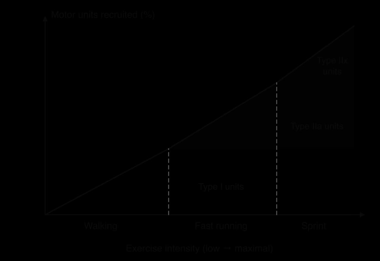
The order makes sense for the body. Most daily activity, such as standing, walking and slow running, needs little force and can be sustained for hours by small, fatigue-resistant type I units. The large, powerful but quickly fatiguing type IIx units are kept in reserve for short, maximal efforts such as a sprint, a jump or lifting a heavy load. Because recruitment depends on the force required, not on the speed of the movement, heavy resistance training at slow speeds still recruits the largest units.
Muscle contraction requires ATP
Muscle contraction is powered by the breakdown of adenosine triphosphate (ATP). The energy released when ATP is split into ADP and phosphate is used directly by the myosin filaments to pull on the actin filaments, and also by the pumps that return calcium ions to their store so that the muscle can relax.
A muscle fibre holds only enough ATP for a few seconds of maximal work, so ATP has to be resynthesised as fast as it is used. This is the job of the three energy systems: the phosphagen system (using phosphocreatine), the glycolytic system and the oxidative system. The fibre types described next differ mainly in which of these systems they rely on, which is why their speed and fatigue resistance differ so much.
When ATP resynthesis cannot keep up with demand, for example during repeated sprints or at the end of a long race, the force each fibre can produce falls. This is one part of peripheral fatigue. ATP in a working muscle never falls to zero: the fibre slows down and loses force before that point, which protects it from damage.
Muscle fibre types
Muscle fibres are not all alike. Motor units differ in the type of fibre they contain and in the diameter of their motor neuron, and these differences decide how fast, how forceful and how fatigue resistant a unit is. Human muscle contains three main fibre types, named by the type of myosin they contain: type I, type IIa and type IIx. They are also called slow-twitch (type I) and fast-twitch (type II) fibres.
Type I fibres are built for endurance. They have many large mitochondria, a high concentration of myoglobin (an oxygen-storing protein that makes them red) and many capillaries around each fibre, so they can supply ATP through the oxidative system for long periods. Their motor neurons are small, with thin axons that conduct impulses relatively slowly, and their motor units contain relatively few fibres. They are recruited first and are active in posture, walking and endurance events such as marathon running, road cycling and cross-country skiing.
Type IIa fibres are intermediate. They contract quickly and forcefully but also have a fair number of mitochondria and capillaries, so they resist fatigue better than type IIx fibres. They are heavily used in sustained high-intensity events such as the 400 m, the 800 m, rowing over 2,000 m and the repeated sprints of team sports. Their properties change readily with training.
Type IIx fibres are built for power. Their large motor neurons conduct impulses rapidly, their motor units contain many large fibres, and they store plenty of phosphocreatine and glycogen for the phosphagen and glycolytic systems. They have few mitochondria and little myoglobin, so they appear pale (white). They are recruited only for maximal or near-maximal efforts, such as a 100 m sprint, a shot put or a maximal lift.
Characteristic | Type I (slow oxidative) | Type IIa (fast oxidative glycolytic) | Type IIx (fast glycolytic) |
|---|---|---|---|
Speed of contraction | Slow | Fast | Fastest |
Force produced | Low | High | Highest |
Resistance to fatigue | High | Moderate | Low |
Motor neuron size and conduction velocity | Small, slow | Large, fast | Largest, fastest |
Fibres per motor unit | Few | Many | Most |
Fibre diameter | Small | Medium to large | Large |
Mitochondrial density | High | Moderate | Low |
Myoglobin content and colour | High; red | Moderate; red to pink | Low; white |
Capillary density | High | Moderate | Low |
Phosphocreatine and glycogen stores | Low | High | High |
Triglyceride (fat) stores | High | Moderate |
 YouTube
Recruitment patterns in different activities
Because of the size principle, the fibre types used in an activity depend on its intensity rather than on the sport itself. In a marathon run at a steady pace, type I fibres do most of the work and some type IIa fibres are recruited, especially on hills or as type I fibres fatigue late in the race. In a 100 m sprint, almost all motor units are recruited, including the type IIx units. In a football match, the pattern changes from moment to moment: type I units support walking and jogging, while sprints, jumps and tackles recruit type IIa and IIx units.
Fibre-type proportions vary between people and between muscles. An untrained adult has roughly half type I and half type II fibres in the leg muscles, but individuals range widely. Elite endurance athletes tend to have a high proportion of type I fibres in the muscles they use most, and elite sprinters a high proportion of type II fibres. Studies of track athletes in the 1970s concluded that an athlete’s preference for strength, speed or endurance events is partly a matter of genetic endowment, and that shot-putters, unlike sprinters and distance runners, had a wide variety of fibre compositions.
Sources: Costill et al., Journal of Applied Physiology, 1976.
Hypertrophy, atrophy and motor unit recruitment
Muscle is a living tissue that adapts to the demands placed on it. Regular, heavy loading makes fibres larger; disuse makes them smaller. Both changes alter how many motor units are needed for a given task and which fibre types are recruited.
Hypertrophy
Hypertrophy is the main long-term adaptation to resistance training with progressively heavier loads. The mechanical tension of heavy lifting, together with the metabolic stress of repeated sets, stimulates the fibres to build more actin and myosin. New myofibrils are added inside each fibre, so the fibre becomes thicker and can produce more force. Type II fibres usually grow more than type I fibres, because heavy loads recruit them and they respond strongly to tension. Hypertrophy depends on adequate protein and energy intake and on recovery between sessions.
Strength rises before the muscle grows. In the first weeks of a strength programme, most of the gain comes from neural adaptations: the nervous system learns to recruit more motor units, to fire them at higher frequencies, to synchronise them better and to relax the antagonist more fully. A classic eight-week training study of young men and women found that neural factors accounted for most of the early strength gain, and that hypertrophy became the dominant factor only after three to five weeks. Strength also increased in the untrained arm, an effect called cross education that is thought to depend entirely on changes in the nervous system.
Sources: Moritani and deVries, American Journal of Physical Medicine, 1979.
Hypertrophy changes recruitment in a useful way. A larger fibre produces more force, so fewer motor units are needed to lift a given load. A load that once demanded the recruitment of type IIx units may now be lifted with type I and IIa units alone, so it feels easier and can be lifted more times before fatigue. Training also shifts the fibre-type profile. Endurance and resistance training both convert some type IIx fibres into type IIa fibres, which are almost as forceful but more resistant to fatigue. There is little reliable evidence that type I fibres become type II fibres, or the reverse, in trained adults.
Atrophy
Atrophy is the reverse of hypertrophy. When a muscle is not loaded, protein breakdown exceeds protein synthesis and the fibres shrink. It begins within days of immobilisation, which is why a footballer whose leg is in a cast after a fracture loses visible thigh muscle within a few weeks. Bed rest, spinal cord injury, long illness, low energy availability and spaceflight all cause atrophy. In space, the lack of gravity removes the loading of postural muscles such as the soleus, so astronauts on long missions follow daily exercise programmes, including resistance exercise, to limit the loss.
Atrophy changes recruitment in two ways. First, smaller fibres produce less force, so a greater proportion of motor units must be recruited to do the same everyday task, and the task becomes tiring sooner. Second, severe disuse shifts the fibre profile towards fast, fatigable fibres. Studies have found that after 90 days of strict bed rest the IIx form of myosin made up about 39% of the muscle, mostly in hybrid fibres, and that people with long-standing spinal cord injury have about 40% pure type IIx fibres, far more than active people.
Sources: Trappe et al., Journal of Applied Physiology, 2015 (summarising earlier bed rest and spinal cord injury studies).
Ageing combines both effects. From middle age onwards, inactive adults gradually lose muscle mass and strength, a condition called sarcopenia when it becomes severe. Type II fibres shrink most, and some motor neurons are lost, so the remaining motor units become larger and less precise. The result is slower, weaker movements and a higher risk of falls. Resistance training slows these losses at any age, which is why health guidelines for older adults include muscle-strengthening activity.
Change | Health lens | Performance lens |
|---|---|---|
Hypertrophy | Maintains strength for daily tasks, supports bone health and blood glucose control, reduces falls in older adults | Increases force and power for sprinting, jumping, throwing and contact sports |
Neural adaptation | Quick strength gains in the first weeks of rehabilitation or a new programme | Better recruitment and firing of motor units; improved rate of force development |
Atrophy | Disuse after injury, illness or bed rest reduces independence; sarcopenia in older adults | Detraining in the off-season or after injury; reversibility of training gains |
Fibre-type shift | Disuse shifts fibres towards fast, fatigable types | Training converts type IIx fibres to more fatigue-resistant type IIa fibres |
Types of muscle contraction
A muscle produces tension whenever its motor units are active, but the muscle does not always shorten. Whether it shortens, stays the same length or lengthens depends on the balance between the force the muscle produces and the external force (the load) acting on it. Contractions are described in four ways: isometric, isotonic concentric, isotonic eccentric and isokinetic.
In an isometric contraction, the muscle force exactly matches the load, so the joint does not move. Examples include holding a plank, the abdominal muscles stabilising the trunk during a throw, a gymnast holding the crucifix position on the rings, and the quadriceps holding a skier in a tuck. Isometric contractions are important for posture and stability, because they fix one part of the body so that another can move.
The word isotonic means "same tension", although in practice the tension changes during a movement as the joint angle and the muscle’s pulling angle change. An isotonic contraction is divided into two types.
In a concentric contraction, the muscle force exceeds the load and the muscle shortens, pulling its insertion towards its origin. Examples are the biceps brachii during the upward phase of an arm curl, the quadriceps during the upward phase of a squat and the gastrocnemius at take-off in a jump.
In an eccentric contraction, the muscle is still active but is being stretched, so it acts as a brake. The biceps contracts eccentrically while lowering the dumbbell under control, and the quadriceps contracts eccentrically on the way down in a squat, when landing from a jump and when running downhill.
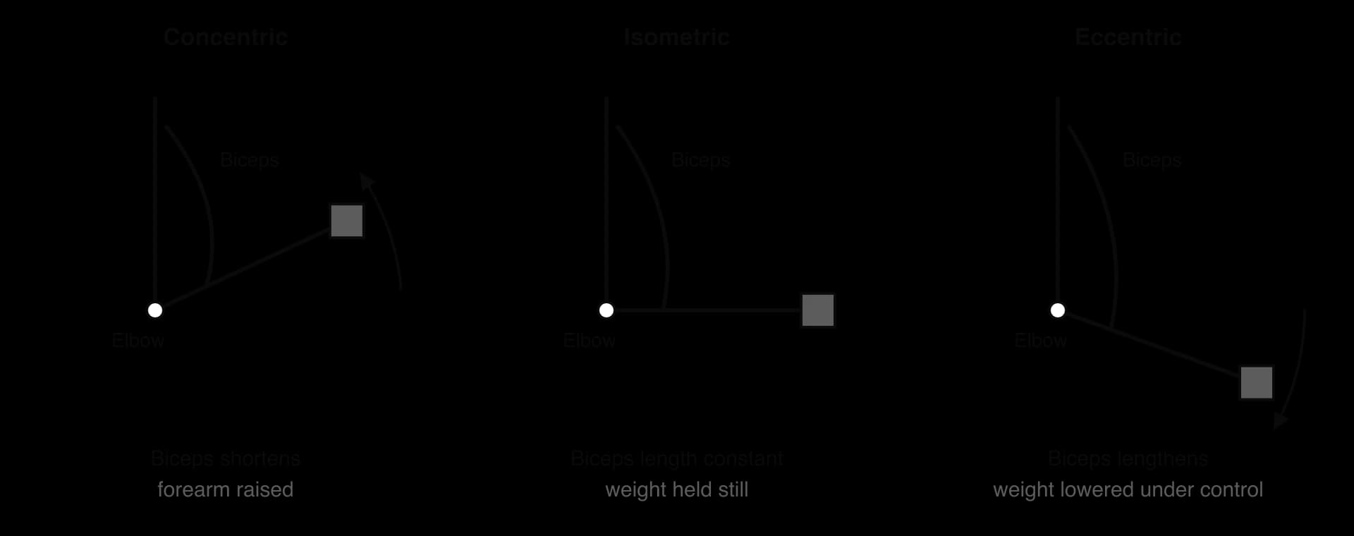
Eccentric contractions produce more force than concentric contractions at the same speed, and they use less energy, because some of the force comes from the stretched structures of the muscle. They also cause more microscopic damage to the fibres, which is why unaccustomed downhill running or heavy lowering leads to delayed-onset muscle soreness (DOMS) a day or two later. Eccentric training is used deliberately in rehabilitation and injury prevention, for example the Nordic hamstring exercise to reduce hamstring strains in footballers.
In an isokinetic contraction, the speed of movement stays the same throughout the range of motion. This rarely happens naturally, so it needs an isokinetic dynamometer, a machine that increases its resistance whenever the person pushes harder, keeping the speed constant. The muscle can therefore work maximally at every joint angle.
Isokinetic machines are used in rehabilitation, for example to rebuild quadriceps and hamstring strength after an anterior cruciate ligament (ACL) reconstruction, and in testing, where they give reliable measures of strength at set speeds and allow the injured and uninjured legs to be compared. In biomechanics research, the same machines measure maximal joint torques, as in studies of speed skaters’ ankle and knee strength.
Swimming is often given as the nearest natural example of an isokinetic action, because the resistance of the water rises as the arm moves faster, so the arm tends to move at a fairly constant speed through the pull. This is an approximation; true isokinetic work needs a machine.
Type | Muscle length | Muscle force vs load | Joint movement | Examples |
|---|---|---|---|---|
Isometric | No change | Equal | None | Plank; holding a handstand; trunk muscles fixing the spine during a tackle |
Isotonic concentric | Shortens | Muscle force greater | Yes; muscle produces the movement | Biceps lifting in an arm curl; quadriceps rising from a squat; gastrocnemius at take-off |
Isotonic eccentric | Lengthens | Load greater | Yes; muscle controls (brakes) the movement | Biceps lowering a dumbbell; quadriceps landing from a jump or running downhill |
Isokinetic | Shortens or lengthens at constant speed | Resistance matched by a machine | Yes, at constant velocity | Knee extension on an isokinetic dynamometer in rehabilitation after ACL surgery |
Muscles work in pairs
Muscles can only pull; they cannot push. To move a joint in one direction and back again, at least two muscles are needed on opposite sides of the joint. Each muscle plays a role in a given movement, and the role changes when the movement changes.
Every movement at a joint therefore involves a muscle that produces it and a muscle on the opposite side that must give way.
The agonist (prime mover) is the muscle mainly responsible for the movement. The antagonist lies on the opposite side of the joint and relaxes as the agonist contracts. Together they form an antagonistic muscle pair. In elbow flexion, the biceps brachii is the agonist and the triceps brachii is the antagonist. In elbow extension, the roles swap: the triceps is the agonist and the biceps the antagonist. A muscle is therefore not "an agonist" in general; it is the agonist for a particular movement.
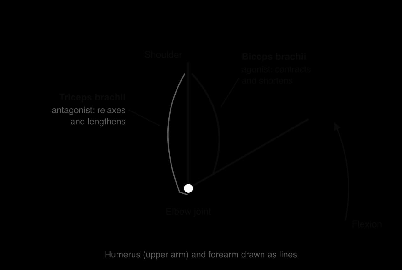
Two further roles help the agonist. A synergist assists the agonist or reduces unwanted movement: the brachialis and brachioradialis help the biceps to flex the elbow. A fixator stabilises the bone to which the agonist is attached, so that the agonist’s pull moves only the intended bone. During an arm curl, muscles around the shoulder, such as the trapezius and the rotator cuff, contract isometrically as fixators to stop the shoulder moving.
Reciprocal inhibition is a reflex at the level of the spinal cord. When the motor neurons of the agonist are excited, a branch of the same signal excites small inhibitory neurons that switch off the motor neurons of the antagonist. When the agonist is stretched suddenly, its muscle spindles (stretch receptors inside the muscle) trigger a reflex contraction of the agonist and, at the same time, the inhibition of its antagonist. The knee-jerk reflex shows this: a tap on the patellar tendon stretches the quadriceps, which contract, while the hamstrings relax.
Without reciprocal inhibition, both muscles of a pair would pull against each other, wasting energy and slowing the movement. A small amount of co-contraction of the antagonist does occur, especially near the end of a fast movement, to brake the limb and protect the joint. Beginners often show too much co-contraction, which makes their movements stiff and tiring; as a skill is learned, the timing of agonist and antagonist activity improves. This is one way in which motor learning changes the activity of muscles.
Joint | Movement | Agonist | Antagonist |
|---|---|---|---|
Elbow | Flexion | Biceps brachii | Triceps brachii |
Elbow | Extension | Triceps brachii | Biceps brachii |
Knee | Extension | Quadriceps group | Hamstring group |
Knee | Flexion | Hamstring group | Quadriceps group |
Hip | Flexion | Hip flexors (iliopsoas) | Gluteus maximus |
Hip | Extension | Gluteus maximus (with hamstrings) | Hip flexors (iliopsoas) |
Ankle | Plantarflexion | Gastrocnemius and soleus | Tibialis anterior |
Ankle | Dorsiflexion | Tibialis anterior | Gastrocnemius and soleus |
Shoulder | Flexion | Anterior deltoid | Latissimus dorsi |
Trunk (spine) | Flexion | Rectus abdominis | Erector spinae |
Analysing a movement joint by joint
A full joint-action analysis records, for each major joint in each phase of a skill, the movement, the agonist, the antagonist and the type of contraction. The following table analyses the upward (concentric) and downward (eccentric) phases of a barbell back squat.
Phase | Joint | Movement | Agonist | Antagonist | Contraction of agonist |
|---|---|---|---|---|---|
Downward | Hip | Flexion | Gluteus maximus (controlling) | Hip flexors | Eccentric |
Downward | Knee | Flexion | Quadriceps (controlling) | Hamstrings | Eccentric |
Downward | Ankle | Dorsiflexion | Gastrocnemius and soleus (controlling) | Tibialis anterior | Eccentric |
Upward | Hip | Extension | Gluteus maximus | Hip flexors | Concentric |
Upward | Knee | Extension | Quadriceps | Hamstrings | Concentric |
Upward | Ankle | Plantarflexion | Gastrocnemius and soleus | Tibialis anterior | Concentric |
The downward phase is the one most often analysed wrongly. The hips and knees are flexing, but the hip flexors and hamstrings are not pulling the body down: gravity is. The gluteus maximus and the quadriceps, which straighten the hips and knees, are working eccentrically to lower the body under control. Some textbooks list the "agonist" in a lowering phase as the muscle that would produce that movement if there were no gravity; the clearest approach is to name the muscle that is actually active and give its contraction type.
The sliding filament theory (HL only)
The sliding filament theory explains how a muscle fibre shortens. The filaments themselves do not change length. Instead, the thin actin filaments slide over the thick myosin filaments towards the centre of each sarcomere, pulled by the heads of the myosin molecules. Because thousands of sarcomeres shorten at the same time, the whole fibre shortens.
Structure of the sarcomere
Each sarcomere runs from one Z line to the next. The proteins that matter for contraction are arranged in a precise, repeating pattern, which produces the light and dark bands seen under a microscope.
Z line (also called the Z disc): the boundary at each end of the sarcomere, to which the actin filaments are anchored.
Actin: the thin filament, attached to the Z line, carrying binding sites for myosin.
Myosin: the thick filament in the centre of the sarcomere, with many protruding heads that can bind to actin and use ATP.
Tropomyosin: a long, thin protein that winds along the actin filament and, at rest, covers the binding sites for myosin.
Troponin: a protein complex attached to tropomyosin that binds calcium ions; when it does, it changes shape and pulls tropomyosin off the binding sites.
M line: the line at the centre of the sarcomere that holds the myosin filaments in place.
A band: the dark band, equal to the full length of the myosin filaments (including the parts that overlap with actin).
I band: the light band, containing only actin; it spans a Z line and the ends of two neighbouring sarcomeres.
H zone: the lighter region in the centre of the A band, containing only myosin, where actin does not overlap.
The regular repeat of Z lines and bands along every myofibril, lined up across the fibre, gives skeletal muscle its striped appearance. The thin filament is actually a combination of three proteins: actin, which forms the strand, and tropomyosin and troponin, which act as its on–off switch.
Each myosin head is also an enzyme, an ATPase, which splits ATP into ADP and inorganic phosphate (Pi) and stores the energy released by changing its shape, like setting a spring.
In a resting muscle, calcium is low and troponin holds tropomyosin in the blocking position, so myosin cannot attach and the muscle is relaxed even though ATP is present.
From nerve impulse to calcium release
Contraction begins with a signal from a motor neuron. The process that links the electrical signal to the mechanical action is called excitation–contraction coupling.
A nerve impulse (action potential) travels down the motor neuron to the neuromuscular junction.
The axon terminal releases the neurotransmitter acetylcholine (ACh), which diffuses across the small gap (the synaptic cleft) and binds to receptors on the sarcolemma.
This depolarises the sarcolemma and starts a muscle action potential, which spreads along the sarcolemma and down the T-tubules into the fibre.
The action potential causes the sarcoplasmic reticulum to release calcium ions (Ca²⁺) into the sarcoplasm around the myofibrils.
Ca²⁺ binds to troponin. Troponin changes shape and moves tropomyosin away from the binding sites on actin.
The binding sites on actin are now exposed, and the myosin heads can attach.
Acetylcholine acts for only a moment: an enzyme in the synaptic cleft, acetylcholinesterase, breaks it down almost immediately, so each impulse produces only a brief stimulus. This allows the nervous system to control contraction precisely through the frequency of impulses.
The cross-bridge cycle
Once the binding sites are exposed, each myosin head goes through a repeating cycle of attaching, pulling, detaching and re-cocking. Each cycle moves the actin filament a tiny distance, so a single contraction involves many cycles by each head.
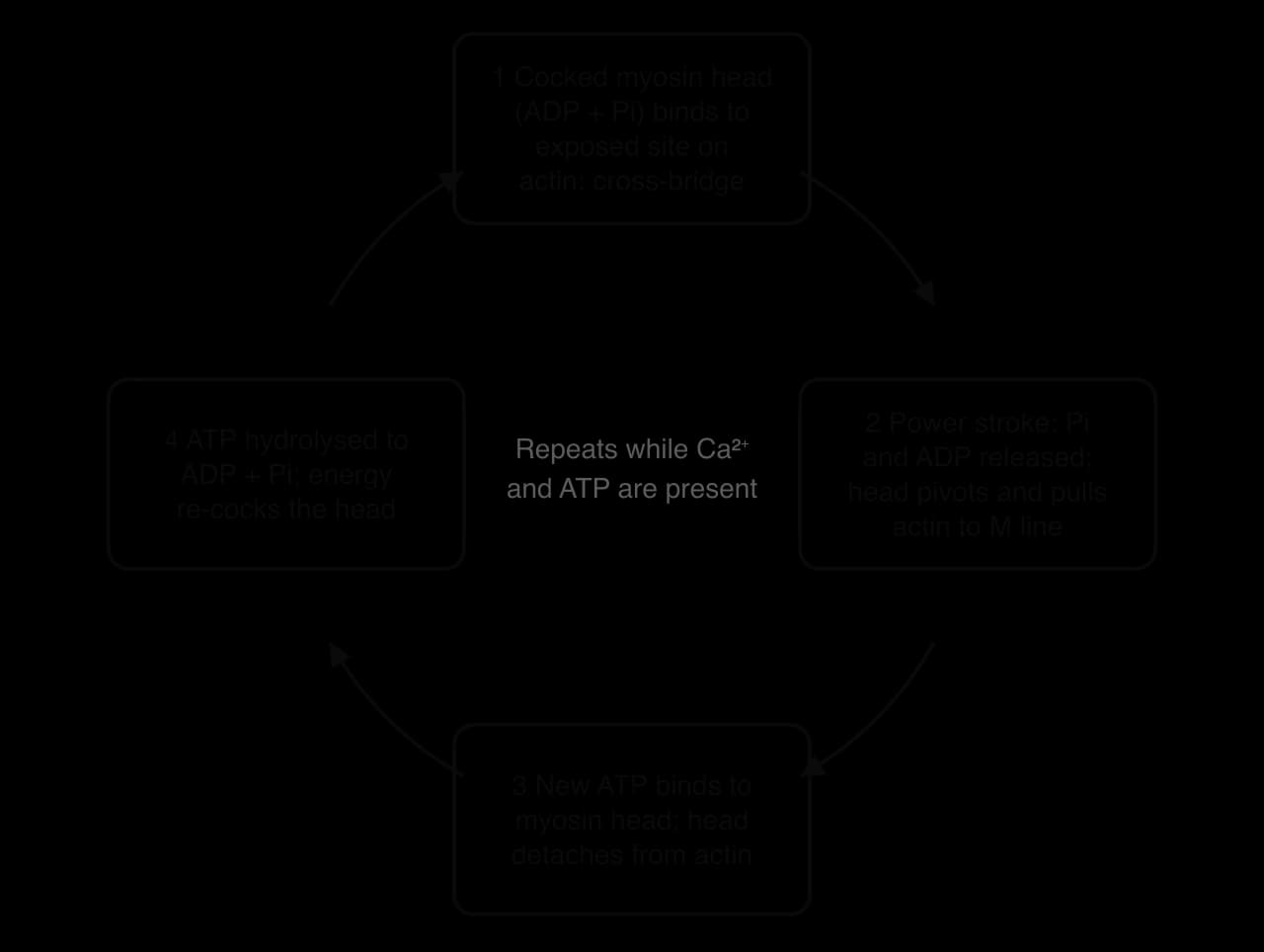
Attachment. A myosin head that has already split ATP (holding ADP and Pi, in its high-energy "cocked" position) binds to an exposed site on actin, forming a cross-bridge.
Power stroke. The head releases Pi and pivots, pulling the actin filament towards the M line. ADP is then released.
Detachment. A new molecule of ATP binds to the myosin head, which causes the head to let go of actin.
Re-cocking. The myosin head splits the ATP into ADP and Pi. The energy released returns the head to its cocked position, ready to bind further along the actin filament.
The cross-bridge cycle continues as long as Ca²⁺ keeps the binding sites exposed and ATP is available. The heads do not all move together: at any moment some are attached and pulling while others are detaching and re-cocking, so the pull on the actin is smooth, in the same way that a team of people hauling a rope hand over hand keeps the rope moving.
The power stroke is the step that produces force. The energy for it came from the ATP split in the previous cycle, and the release of Pi triggers the stroke.
Relaxation
Nerve impulses stop, and acetylcholine is broken down by acetylcholinesterase, so no new muscle action potentials are produced.
Ca²⁺ is actively pumped back into the sarcoplasmic reticulum. This pumping uses ATP.
With calcium removed from troponin, tropomyosin moves back over the binding sites on actin.
Myosin can no longer attach. The cross-bridges detach, and the sarcomere returns to its resting length as the muscle is stretched by its antagonist or by gravity.
Rigor mortis, the stiffness of muscles a few hours after death, illustrates the role of ATP. After death, calcium leaks from the sarcoplasmic reticulum, so cross-bridges form, but no new ATP is made, so the myosin heads cannot detach. The muscles stay locked until the proteins begin to break down.
How the bands change during contraction
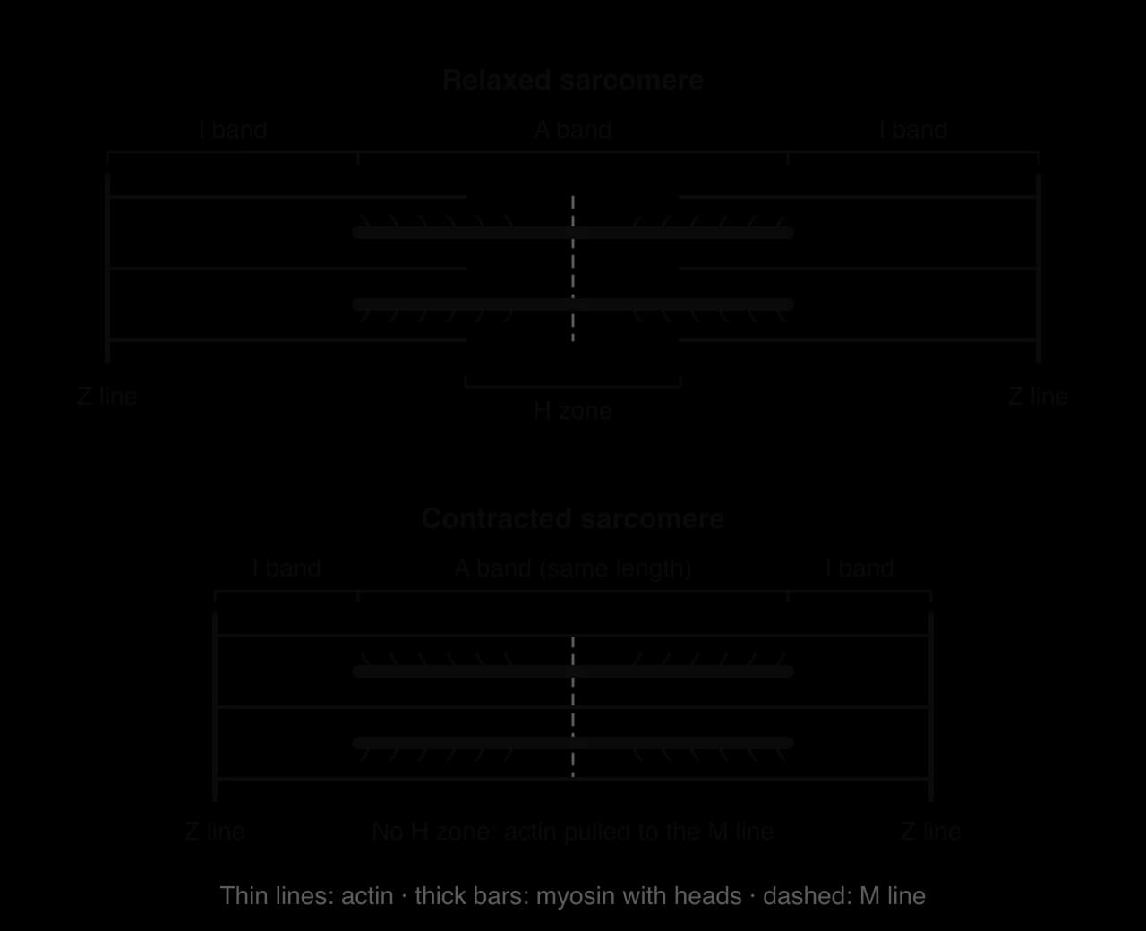
Part of the sarcomere | Change during contraction | Reason |
|---|---|---|
Sarcomere (Z line to Z line) | Shortens | Actin filaments, anchored to the Z lines, are pulled towards the M line |
A band | Stays the same length | It equals the length of the myosin filaments, which do not change length |
I band | Shortens | Actin slides further into the A band, so less of each actin filament lies outside the myosin |
H zone | Shortens and may disappear | Actin filaments from both ends move into the centre and overlap more with myosin |
Actin and myosin filaments | Stay the same length | They slide past each other; neither filament shortens |
Molecule or ion | Role in contraction |
|---|---|
Acetylcholine | Neurotransmitter released at the neuromuscular junction; starts the muscle action potential |
Calcium ions (Ca²⁺) | Released from the sarcoplasmic reticulum; bind to troponin to expose the binding sites on actin |
Troponin | Binds Ca²⁺ and changes shape, moving tropomyosin |
Tropomyosin | Blocks the binding sites on actin at rest; moves aside when troponin binds Ca²⁺ |
Actin | Thin filament with binding sites; pulled towards the M line |
Myosin | Thick filament; its heads form cross-bridges, perform the power stroke and split ATP |
ATP | Detaches myosin from actin, re-cocks the myosin head (when split) and powers the Ca²⁺ pump during relaxation |
 YouTube
The theory also links to nutrition. Calcium for contraction comes from the fibre’s own store, so a short-term shortage in the diet does not stop muscles working, because blood calcium is tightly regulated using calcium from bone. ATP, however, must be resynthesised continuously from food fuels, which is why carbohydrate and protein intake and their timing affect how long muscles can keep contracting and how well they recover.
Case study: muscle fibre types in elite runners
Fibre type is one reason some runners excel over 100 m and others over 10,000 m. The physiologist David Costill and colleagues at Ball State University in the United States took needle biopsies from the gastrocnemius of track athletes in the 1970s. In one study of 14 elite distance runners, 18 middle-distance runners and 19 untrained men, the elite distance runners’ muscles were characterised by a high percentage of slow-twitch (type I) fibres: 79% on average.
Their type I fibres were also about 22% larger in cross-sectional area than their fast-twitch fibres, and the activity of an oxidative enzyme (succinate dehydrogenase) in their muscle was 3.4 times that of the untrained men.
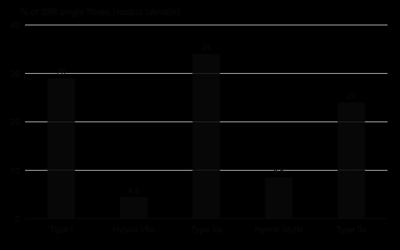
Almost 40 years later, researchers from the same laboratory studied the opposite extreme: a retired world champion sprint hurdler who had held the world record for the 110 m hurdles (12.91 s) and the indoor 60 m hurdles (7.30 s). They used a more precise method, analysing the type of myosin in 329 individual fibres from a biopsy of the vastus lateralis (part of the quadriceps), and measured the force, speed and power of single fibres.
Measure | Elite distance runners (gastrocnemius) | Champion sprint hurdler (vastus lateralis) |
|---|---|---|
Participants | 14 men | 1 man (retired) |
Method | Histochemical staining of muscle sections | Myosin type of 329 single fibres |
Type I (slow-twitch) fibres | 79% | 29% (plus 4.5% I/IIa hybrids) |
Fast-twitch fibres | About 21% | About 71%, including 24% pure IIx |
Single-fibre power, normalised (W L⁻¹) | Not measured | Type I 2.5; type IIa 17.1; type IIx 35.1 |
Sources: Costill, Fink and Pollock, Medicine and Science in Sports, 1976; Trappe et al., Journal of Applied Physiology, 2015.
The single-fibre data explain why the fast fibres matter so much for sprinting. The sprinter’s type IIx fibres were about twice as powerful as his type IIa fibres and about 14 times as powerful as his type I fibres. Speed and power followed the order IIx > IIa > I, exactly as the fibre-type table predicts. In a 100 m race or a hurdle clearance, where force must be produced in a fraction of a second, a high proportion of large, fast IIx motor units recruited at maximal effort is a clear advantage.
The distance runners show the opposite design. A high proportion of type I fibres, with many mitochondria and capillaries and high oxidative enzyme activity, allows a high rate of ATP resynthesis by the oxidative system for a long time without fatigue. Their type I fibres were even larger than their fast fibres, the reverse of the usual pattern, suggesting that years of running had made the most-used fibres grow.
Whether these athletes were born with their fibre types or created them by training cannot be settled by these studies alone. The original track athlete studies concluded that event preference is partly a matter of genetic endowment, and the researchers in the sprinter study pointed to genetic, evolutionary and environmental factors. Because training turns type IIx fibres into type IIa fibres and rarely turns type I into type II fibres, a sprinter with a very high IIx content is likely to have inherited much of it.
Fibre type sets limits on performance, but running economy, technique, tendon stiffness, VO₂ max and psychological factors all contribute, so fibre type alone cannot predict who will win.
Both studies have limits. The distance runners and the sprinter were sampled in different muscles (gastrocnemius and vastus lateralis) with different methods, so their percentages are not directly comparable. The sprinter study described a single person, who had retired by the time of testing, and a biopsy samples only a tiny piece of one muscle. Cross-sectional comparisons of athletes and non-athletes show an association between fibre type and event, but cannot show whether fibre type caused success, or whether training changed the fibres.
Using the fibre-type table, explain why a high proportion of type IIx fibres benefits a sprint hurdler but would disadvantage a marathon runner.
Suggest why the researchers measured the myosin type of single fibres rather than staining a muscle section, and why this matters when comparing the two studies.
To what extent could a talent identification programme use muscle biopsies to select young athletes for sprint or endurance events? Consider ethics, reliability and the other factors that affect performance.
Exam practice
Muscular function appears in every part of the assessment. Paper 1A tests definitions and the fibre-type table; Paper 1B often gives data from a training or fibre-type study; Paper 2 asks you to analyse a movement, to explain how force is graded or, at HL, to explain the sliding filament theory step by step.
Command terms set the depth of an answer, and many marks are lost by describing when the question asks for an explanation.
Paper 1A-style questions
Fibre-type questions often list one property from each row of the comparison table.
Questions on contraction types often describe a phase of a movement in words.
Paper 1B-style data-based question
A study investigated the effect of resistance training on older adults (hypothetical data). Sixteen healthy adults aged 65–75 years were randomly allocated to a training group (n = 8), who completed 12 weeks of supervised leg resistance training three times a week, or a control group (n = 8), who continued their normal activities. Before and after the 12 weeks, maximal knee extension strength was measured as a one-repetition maximum (1RM) and a biopsy of the vastus lateralis was taken to measure the mean cross-sectional area of type II fibres.
Group | 1RM before (kg) | 1RM after (kg) | Type II fibre area before (μm²) | Type II fibre area after (μm²) |
|---|---|---|---|---|
Training (n = 8) | 40.2 ± 6.1 | 52.3 ± 7.0 | 3,900 ± 600 | 4,560 ± 650 |
Control (n = 8) | 39.8 ± 5.8 | 39.1 ± 6.2 | 3,950 ± 580 | 3,880 ± 610 |
Values are mean ± standard deviation (hypothetical data).
Paper 2-style structured questions
Fibre-type questions in Paper 2 usually ask for comparisons linked to a sport.
Movement analysis questions often show a photograph or describe a skill.
Extended-response question
HL questions (HL only)
Paper 2 often asks for the sequence of events from nerve impulse to contraction.
Summary
Skeletal muscle is organised from the whole muscle to fascicles, fibres, myofibrils and sarcomeres, each wrapped in connective tissue that transmits force to the tendon.
A motor unit is one motor neuron and all the fibres it supplies; each unit obeys the all-or-none principle.
Force is graded by recruiting more motor units, in order of size (the size principle), and by increasing their firing frequency.
Contraction and relaxation both need ATP, which the energy systems must resynthesise continuously.
Type I fibres are slow, oxidative and fatigue resistant; type IIx fibres are fast, powerful and fatigable; type IIa fibres lie between them. Recruitment follows the intensity of the activity.
Hypertrophy enlarges fibres so fewer units are needed for a task; atrophy shrinks them so more units must be recruited. Early strength gains are mainly neural.
Contractions can be isometric (no length change), isotonic concentric (shortening), isotonic eccentric (lengthening under tension) or isokinetic (constant speed on a machine).
Muscles work in antagonistic pairs; reciprocal inhibition relaxes the antagonist as the agonist contracts, helped by synergists and fixators.
In the sliding filament theory, Ca²⁺ binds troponin, tropomyosin moves, and myosin heads use ATP to pull actin towards the M line; the I band and H zone shorten while the A band stays the same (HL).
Self-check
- To do:
I can outline the structure of skeletal muscle from the whole muscle to the sarcomere and name the major muscles.
- To do:
I can describe a motor unit and the all-or-none principle.
- To do:
I can explain how recruitment, the size principle and firing frequency grade muscle force.
- To do:
I can outline why contraction and relaxation need ATP.
- To do:
I can distinguish between type I, IIa and IIx fibres and explain their recruitment in different activities.
- To do:
I can explain how hypertrophy and atrophy change motor unit recruitment.
- To do:
I can distinguish between isometric, isotonic concentric, isotonic eccentric and isokinetic contractions with sporting examples.
- To do:
I can analyse a movement joint by joint and explain reciprocal inhibition.
- To do:
I can explain the sliding filament theory and the roles of calcium, ATP, actin, myosin, troponin and tropomyosin (HL).
- To do:
I can describe how the sarcomere’s bands and zones change during contraction (HL).