Notes on B.1.2 Structure and function of connective tissues and joints
Learning objectives
Outline the structure and function of bone, ligaments, tendons, cartilage and fascia, and relate each structure to its function.
Describe the structure of a long bone and explain how bone adapts to the loads placed on it (Wolff's law).
Distinguish between fibrous, cartilaginous and synovial joints in structure, function and amount of movement.
Label a diagram of a synovial joint and outline the function of each structure.
Describe the six classes of synovial joint, with the movements each allows and an example in the body.
Compare the shoulder and hip joints in terms of stability and mobility.
Explain the factors that affect joint stability and range of motion.
Apply knowledge of connective tissues and joints to sporting and health contexts.
Introduction
Muscles produce force, but it is the connective tissues and joints that turn that force into controlled movement. Bones act as rigid levers, tendons transmit muscle force to them, ligaments hold the bones together, cartilage lets them glide over one another, and the shape of each joint decides which movements are possible and how stable the joint is.
The same structures explain much of sport and health. The design of the shoulder lets a swimmer or a tennis player move the arm through a huge range, but it also makes the shoulder the joint most likely to dislocate. Bone and tendon become stronger when they are loaded, which is why physical activity in youth helps protect against fractures in old age, and why tissues weakened by inactivity are easily injured.
Connective tissues
Connective tissue is the body's supporting tissue. It is made of relatively few cells spread through a large amount of material outside the cells, called the matrix. The matrix contains protein fibres, mostly collagen, set in a ground substance that may be fluid, gel-like or, in bone, hardened with minerals. The type and arrangement of fibres and the nature of the ground substance give each connective tissue its properties.
Collagen is the protein that gives most connective tissues their strength.
The principle linking all connective tissues is that structure matches function. Tendons and ligaments are pulled along their length, so their collagen fibres lie parallel, which gives them great strength in that direction. Cartilage must resist being squashed, so its matrix holds water like a firm gel. Bone must resist both bending and compression, so its matrix is hardened with calcium phosphate around a framework of collagen.
Bone
Bone is a hard connective tissue whose matrix is about two-thirds mineral (mainly calcium phosphate), which gives it hardness and resistance to compression, and one-third collagen, which gives it some flexibility and resistance to tension and stops it shattering. It exists in two forms. Compact bone (cortical bone) is dense and solid and forms the outer layer of every bone and the walls of the shaft of long bones. Spongy bone (cancellous or trabecular bone) is a lattice of thin struts, called trabeculae, with spaces between them that hold red marrow; it fills the ends of long bones and the inside of short, flat and irregular bones.
The two forms suit different jobs. Compact bone in the shaft of a long bone resists the bending forces created when muscles pull on it and when the body lands. Spongy bone in the ends of the bone is light and absorbs and spreads the forces passing through joints, and its trabeculae line up along the directions in which the bone is usually loaded.
Structure of a long bone
A long bone such as the femur or humerus has the same basic parts. The diaphysis is the shaft, a tube of compact bone around a hollow medullary cavity that contains yellow marrow (mainly fat). Each end is an epiphysis, made of spongy bone with red marrow inside a thin shell of compact bone. In children, a plate of cartilage, the growth plate, separates the epiphysis from the shaft; bone lengthens here until growth stops in the late teens, leaving a line in the adult bone.

The periosteum is a tough membrane of dense connective tissue that covers the outside of the bone, except where articular cartilage covers the joint surfaces. It contains blood vessels and nerves, which is why a kick on the shin is so painful, and bone-forming cells that thicken the bone and repair fractures. Tendons and ligaments blend into the periosteum, which anchors them firmly to the bone. Articular cartilage covers the ends of the bone where they meet another bone in a joint.
Bone adapts to load: Wolff's law
Bone is constantly being broken down and rebuilt, and the balance between the two depends on the loads it carries. When bone is repeatedly loaded more than usual, for example by jumping, running or the impact of a racket, it lays down extra mineral and becomes denser and thicker in the regions under most stress. When the load is removed, by bed rest, a plaster cast, a spinal cord injury or spaceflight, breakdown exceeds rebuilding and bone is lost. This principle is called Wolff's law, after the German surgeon Julius Wolff, who described it in the 1890s.
A load (an impact, a muscle pull or body weight) bends and compresses the bone very slightly.
Bone cells sense this deformation.
If the strain is larger, faster or more frequent than the bone is used to, the cells increase the activity of bone-forming cells in the stressed region.
More mineral is deposited, so bone mineral density and the thickness of the compact bone increase, and the bone becomes better able to resist the same load.
If loading falls below the usual level, bone-removing cells dominate and bone mass is gradually lost.
Wolff's law explains why high-impact and multidirectional activities such as gymnastics, basketball and racket sports build bone more effectively than swimming or cycling, in which the water or the bicycle supports body weight. It also explains why astronauts on long space missions lose bone from the hips and spine despite exercising daily, and why bone mineral density built up during childhood and adolescence is valued as a protection against osteoporosis later in life.
Ligaments
A ligament is a band of dense regular connective tissue that joins bone to bone. Its collagen fibres run parallel to one another, along the line of pull, so it is very strong in tension but stretches only a little. Some ligaments also contain elastic fibres, which let them stretch slightly and recoil. Ligaments stabilise joints by holding the bones in position and by limiting movement in directions that would damage the joint. The cruciate ligaments of the knee, for example, stop the tibia sliding too far forwards or backwards on the femur.
Ligaments also contain sensory receptors (proprioceptors) that signal the position of the joint to the central nervous system, which helps muscles react quickly to protect the joint. Their blood supply is poor, so they receive few nutrients and repair cells, and they heal slowly after a sprain. A completely torn anterior cruciate ligament usually does not heal on its own and is often reconstructed surgically, followed by many months of rehabilitation. A ligament that has been overstretched may also heal in a lengthened state, leaving the joint less stable.
Tendons
A tendon is a cord or sheet of dense regular connective tissue that joins muscle to bone. Like a ligament, it is made mainly of parallel collagen fibres, which makes it very strong in tension. At one end its fibres are continuous with the connective tissue around the muscle, and at the other they blend into the periosteum and the bone itself, so the force of the muscle contraction is transmitted to the bone and creates movement at the joint.
Tendons are not just passive cables. They stretch slightly under load and recoil like a spring, storing and returning elastic potential energy. In running, the Achilles tendon is stretched as the ankle dorsiflexes after the foot lands, then recoils as the ankle plantarflexes at push-off, returning energy and reducing the work the calf muscles must do. Tendons adapt to training, becoming stiffer and thicker, but more slowly than muscle; a rapid increase in training load can therefore leave a tendon overloaded, a common cause of overuse injuries such as Achilles and patellar tendinopathy.
Feature | Ligament | Tendon |
|---|---|---|
Joins | Bone to bone | Muscle to bone |
Main function | Stabilises the joint; limits excessive movement | Transmits muscle force to bone; stores and returns elastic energy |
Structure | Dense regular collagen; some elastic fibres | Dense regular collagen in parallel bundles; very few elastic fibres |
Blood supply and healing | Poor; slow healing | Poor; slow healing |
Common injury | Sprain (for example lateral ankle ligaments, ACL) | Strain or tendinopathy (for example Achilles, patellar tendon) |
Example | Medial collateral ligament of the knee | Achilles tendon (gastrocnemius and soleus to the calcaneus) |
Cartilage
Cartilage is a firm, flexible connective tissue whose matrix holds a large amount of water. When it is compressed, water is squeezed out and then drawn back in as the load is removed, which lets it absorb shock. Cartilage has no blood vessels, so it gets its nutrients by diffusion, in joints from the synovial fluid. Because of this, damaged cartilage heals very poorly. There are three types.
Hyaline cartilage is smooth and glassy. As articular cartilage it covers the ends of bones in synovial joints, where its very low friction lets the bones glide over each other and it spreads the load across the joint surface. It also forms the growth plates in children and joins the ribs to the sternum.
Fibrocartilage contains dense bundles of collagen fibres, so it is tough and resists both compression and tension. It forms the menisci of the knee, the intervertebral discs and the pubic symphysis, places that must absorb large forces.
Elastic cartilage contains elastic fibres and keeps its shape after bending, as in the outer ear and the epiglottis. It plays no part in movement.
Articular cartilage depends on movement for its health. Each time a joint is loaded and unloaded, fluid moves in and out of the cartilage, carrying nutrients with it. Regular moderate activity therefore keeps cartilage healthy, while immobilisation or long-term excessive loading can contribute to its breakdown. When articular cartilage wears away, as in osteoarthritis, bone surfaces rub together, causing pain and loss of movement.
Fascia
Fascia is a continuous sheet of connective tissue that wraps around and between muscles, groups of muscles, blood vessels and nerves. Around each muscle it separates the muscle from its neighbours so they can slide past each other, and it binds groups of muscles together. Because fascia is continuous with the connective tissue inside muscles and with tendons, it also helps transmit force from one muscle to the next and across joints. Thick bands of fascia act like tendons: the plantar fascia under the foot supports the arch and stores elastic energy in running, and inflammation of it (plantar fasciitis) is a common overuse injury in runners.
Fascia is the target of myofascial release, a recovery technique (for example foam rolling) that applies pressure to the fascia and muscle, which athletes use to reduce perceived soreness and stiffness.
Tissue | Structure | Function | Example |
|---|---|---|---|
Bone | Collagen framework hardened with calcium phosphate; compact outer layer, spongy interior | Support, protection, levers for movement, mineral store, blood cell production | Femur |
Ligament | Parallel collagen fibres with some elastic fibres | Joins bone to bone; stabilises the joint and limits excessive movement | Anterior cruciate ligament of the knee |
Tendon | Parallel bundles of collagen fibres | Joins muscle to bone; transmits force; stores and returns elastic energy | Achilles tendon |
Articular (hyaline) cartilage | Smooth, water-rich matrix with fine collagen | Low-friction surface; absorbs and spreads load at joints | Ends of the femur and tibia |
Fibrocartilage | Water-rich matrix with dense collagen bundles | Absorbs large forces; deepens and stabilises some joints | Menisci; intervertebral discs |
Fascia | Sheets of collagen-rich connective tissue | Surrounds and separates muscles; transmits force between them | Plantar fascia; fascia around the quadriceps |
Joints: fibrous, cartilaginous and synovial
An articulation, or joint, is any place where two or more bones meet. Joints are classified by the material that holds the bones together, and this structure decides how much movement is possible. There are three types.

In a fibrous joint the bones are joined directly by dense fibrous connective tissue, with no cavity between them, so the joint is immovable (or almost so). The sutures between the bones of the skull interlock tightly to protect the brain. A syndesmosis is a fibrous joint in which the bones are held by a sheet or band of fibrous tissue that allows a very small amount of give, such as the joint between the lower ends of the tibia and fibula.
In a cartilaginous joint the bones are joined by cartilage, usually a pad of fibrocartilage. The joints between the bodies of the vertebrae, with an intervertebral disc between them, and the pubic symphysis at the front of the pelvis are examples. Each allows only slight movement, but because the spine has many such joints in a row, the whole column can flex, extend, bend sideways and rotate a good deal. The discs also absorb shock in running and landing.
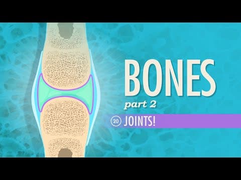
In a synovial joint the bones are separated by a joint cavity filled with synovial fluid and enclosed in a joint capsule. The articulating surfaces are covered with smooth articular cartilage and are not joined directly, so the joint is freely movable. Most joints of the limbs are synovial, and they are the joints used in almost every sporting movement.
Feature | Fibrous | Cartilaginous | Synovial |
|---|---|---|---|
What joins the bones | Dense fibrous tissue | Cartilage (usually fibrocartilage) | Joint capsule and ligaments; the bones are separated by a fluid-filled cavity |
Joint cavity | No | No | Yes |
Amount of movement | None or almost none (immovable) | Slight (slightly movable) | Free (freely movable) |
Stability | Very high | High | Lower; varies with the class of joint |
Main function | Protection; fixing bones together | Shock absorption with limited flexibility | Movement |
Examples | Skull sutures; distal tibiofibular joint (syndesmosis) | Intervertebral joints; pubic symphysis | Knee, hip, shoulder, elbow, wrist, ankle |
Structure of a synovial joint
Every synovial joint has the same basic structures, each with a specific function. Together they allow free movement with very low friction while keeping the bones in position.

Structure | Description | Function |
|---|---|---|
Articular cartilage | Smooth hyaline cartilage on the ends of the bones | Reduces friction between bones; absorbs and spreads load |
Joint capsule | A sleeve of dense fibrous tissue that encloses the joint and attaches to the bones around it | Holds the bones together; encloses the synovial fluid; adds stability |
Synovial membrane | A thin membrane lining the inside of the capsule (but not the cartilage) | Secretes synovial fluid into the joint cavity |
Synovial fluid | A thick, slippery fluid filling the joint cavity | Lubricates the joint, nourishes the articular cartilage and helps absorb shock |
Ligaments | Bands of collagen outside (and sometimes inside) the capsule | Join bone to bone; stabilise the joint and limit excessive movement |
Bursae | Small fluid-filled sacs lined with synovial membrane, between a tendon, ligament or skin and a bone | Reduce friction where structures rub over one another |
Menisci and pads | Wedges or discs of fibrocartilage between the articular surfaces (knee); pads of fat in some joints | Improve the fit of the bones, absorb shock and spread load, adding stability |
The synovial membrane is a thin layer of tissue that lines the inner surface of the joint capsule and secretes synovial fluid. The fluid is slippery because of large molecules dissolved in it, so it reduces friction between the cartilage surfaces to very low levels. It also carries oxygen and nutrients to the cartilage, which has no blood supply of its own.
A warm-up makes synovial joints work better. As the joint moves and warms, more synovial fluid is secreted and it becomes less viscous, so it lubricates the joint more effectively, and the movement helps fluid and nutrients pass into the articular cartilage. This is one reason why warm-ups include movements through the full range used in the sport.
The knee has two menisci, medial and lateral, between the femur and the tibia. A meniscus can be torn when the knee twists while bent and bearing weight, for example when a footballer turns sharply with the foot planted. The inner part of each meniscus has almost no blood supply, so tears there heal poorly, which is why a meniscus injury can end a season.
 YouTube
Classes of synovial joints
Synovial joints are divided into six classes according to the shape of their articulating surfaces. The shape decides the axes about which the bones can rotate and therefore which movements the joint allows. A joint that moves about one axis is uniaxial, about two axes biaxial and about three axes multiaxial; this is sometimes described as the joint's number of degrees of freedom of rotation.

A hinge joint has a convex surface on one bone fitting into a concave surface on the other, like the hinge of a door. It allows flexion and extension only, in the sagittal plane about a frontal axis. The elbow (between the humerus and the ulna) and the joints between the phalanges are hinge joints. The knee is usually classed as a hinge joint, though it also allows a small amount of rotation when it is flexed.
In a pivot joint a rounded or peg-like part of one bone rotates within a ring formed by another bone and a ligament. It allows rotation only, about a longitudinal axis. The atlanto-axial joint between the first two cervical vertebrae lets the head turn from side to side, and the radio-ulnar joints allow pronation and supination of the forearm.
A ball-and-socket joint has a ball-shaped head on one bone fitting into a cup-shaped socket on the other. It is multiaxial, allowing flexion and extension, abduction and adduction, medial and lateral rotation, horizontal flexion and extension, and circumduction. The hip and shoulder are the two ball-and-socket joints, and they are the most mobile joints in the body.
In a saddle joint each articulating surface is concave in one direction and convex in the other, like a rider sitting on a saddle. It is biaxial, allowing flexion and extension, abduction and adduction, circumduction and opposition. The carpometacarpal joint at the base of the thumb is the main example, and it gives the human thumb its ability to grip.
A condyloid joint (ellipsoid joint) has an oval convex surface fitting into an oval concave one. It is biaxial, allowing flexion and extension, abduction and adduction and circumduction, but not rotation. The wrist (between the radius and the carpals) and the knuckles (metacarpophalangeal joints) are condyloid joints.
In a gliding joint (plane joint) the articulating surfaces are nearly flat, so the bones slide over one another in small movements in several directions. The joints between the carpals, between the tarsals and between the articular processes of the vertebrae are gliding joints. Each allows only a little movement, but together they give flexibility to the wrist, foot and spine.
Class | Shape of surfaces | Axes | Movements allowed | Examples |
|---|---|---|---|---|
Hinge | Convex into concave, like a door hinge | 1 (frontal) | Flexion, extension | Elbow; knee; interphalangeal joints |
Pivot | Peg rotating within a ring | 1 (longitudinal) | Rotation | Atlanto-axial joint; radio-ulnar joints |
Condyloid (ellipsoid) | Oval convex into oval concave | 2 | Flexion, extension, abduction, adduction, circumduction | Wrist; metacarpophalangeal joints |
Saddle | Both surfaces concave one way and convex the other | 2 | Flexion, extension, abduction, adduction, circumduction, opposition | Carpometacarpal joint of the thumb |
Ball-and-socket | Ball into a cup | 3 | Flexion, extension, abduction, adduction, rotation, horizontal flexion and extension, circumduction | Hip; shoulder |
Gliding (plane) | Nearly flat | Small slides in several directions | Gliding | Intercarpal and intertarsal joints; between vertebral articular processes |
Stability and mobility
Joint stability is the ability of a joint to keep its articulating surfaces in their correct position and resist dislocation or injury. Range of motion (ROM) is the amount of movement possible at a joint, usually measured in degrees. The two are linked by a trade-off: the features that increase stability tend to reduce range of motion, and the reverse.
Range of motion is measured with a goniometer or from video, and it differs between joints, movements and people.
Shoulder and hip compared
The shoulder and the hip are both ball-and-socket joints, but they sit at opposite ends of the stability–mobility trade-off, which matches their roles. The hip carries body weight and must be stable; the shoulder positions the hand and must be mobile.

Factor | Hip | Shoulder (glenohumeral joint) |
|---|---|---|
Shape of the socket | Deep acetabulum that covers much of the head of the femur, deepened further by a rim of fibrocartilage (the labrum) | Shallow glenoid fossa covering only a small part of the head of the humerus, with a smaller labrum |
Ligaments and capsule | Thick, strong capsule reinforced by some of the strongest ligaments in the body | Loose, thin capsule that allows a large range; weaker ligaments |
Muscles | Large, powerful muscles around the joint (gluteals, hip flexors, adductors) | Stability depends heavily on the rotator cuff muscles holding the head in the socket |
Range of motion | Large, but less than the shoulder | The largest of any joint in the body |
Stability | Very stable; dislocates only with very large forces (for example a car crash) | Less stable; the most frequently dislocated major joint, for example in rugby tackles or falls on an outstretched arm |
Factors that affect stability and range of motion
Shape of the articulating surfaces. Deep sockets and closely fitting surfaces give stability (hip); shallow ones give range (shoulder). Menisci and labra deepen sockets.
Ligaments and the joint capsule. Short, tight ligaments and a thick capsule increase stability but limit range; ligaments that have been overstretched in a previous sprain leave the joint less stable.
Muscle tone and strength. Muscles crossing a joint pull the bones together. Strong rotator cuff muscles stabilise the shoulder, and strengthening the muscles around the ankle or knee is a key part of preventing repeat injury. Tight muscles limit range of motion.
Age. Range of motion is greatest in childhood and tends to decrease with age as connective tissues become stiffer and less hydrated, and as people become less active.
Sex. On average, females have a greater range of motion than males at many joints, partly because of differences in connective tissue and in hormones such as oestrogen and relaxin, which increase ligament laxity.
Training and activity. Regular flexibility training increases range of motion, and strength training around a joint increases stability. Inactivity reduces both. Movements practised through a large range (gymnastics, dance) increase it.
Temperature. Warm muscles and connective tissues stretch more easily than cold ones, which is one reason for warming up before stretching.
Body bulk. Large muscle mass or body fat can physically block the end of a movement, for example limiting elbow flexion in a very muscular arm.
A small proportion of people have hypermobility, a range of motion at several joints well beyond the normal range. It can help in gymnastics, dance and swimming, but it may come with less joint stability and a higher risk of sprains and dislocations, so hypermobile athletes are often given extra strength training around their joints.
Health lens | Performance lens | |
|---|---|---|
Stability | Strengthening muscles around the knee and ankle reduces falls in older adults and repeat sprains after injury. | A stable shoulder lets a thrower or swimmer apply large forces without dislocation, so prehabilitation programmes strengthen the rotator cuff. |
Range of motion | Maintaining range of motion keeps everyday tasks (dressing, climbing stairs) possible in old age and in arthritis. | Large hip and shoulder range lets a hurdler, gymnast or swimmer reach positions that improve technique. |
Connective tissue adaptation | Weight-bearing activity builds bone density, reducing the risk of osteoporosis. | Gradual increases in load let tendons and ligaments adapt and reduce overuse injury. |
The way training changes these tissues links this subtopic with the training principles of progressive overload and recovery. Bone, tendon and ligament adapt to load as muscle does, but more slowly, so a programme that increases muscle strength quickly may overload tissues that have not yet adapted.
Case study: the playing arm of tennis and squash players
Racket sports offer an unusually clean test of Wolff's law. A right-handed tennis or squash player loads the bones of the playing arm with thousands of impacts and strong muscle contractions in every session, while the other arm does relatively little. Because both arms belong to the same person, they share the same genes, diet, hormones and general activity, so a difference between them can be put down mainly to the extra loading of the playing arm.
A team at the UKK Institute in Tampere, Finland, used this design in a study of 105 female national-level tennis and squash players and 50 healthy female controls, published in 1995. Bone mineral content (BMC), the mass of mineral in a section of bone, was measured in both arms by dual-energy X-ray absorptiometry (DXA) at four sites: the proximal humerus, the humeral shaft, the radial shaft and the distal radius. The outcome was the percentage difference between the dominant (playing) and non-dominant arm.

The controls had dominant-arm BMC 3–5% higher than the other arm, the effect of everyday use. In the players the difference was 8.5–16.2%, significantly larger at every site (p < 0.001). The players were also grouped by when they had started playing relative to menarche, the first menstrual period, which marks a stage of puberty. The earlier they had started, the larger the difference.
Group (starting age relative to menarche) | Side-to-side BMC difference: lowest to highest mean reported across groups and sites (%) |
|---|---|
Started before or at menarche | 10.5 to 23.5 |
Started more than 15 years after menarche | 2.4 to 9.6 |
Controls (non-players) | 3.2 to 4.6 |
A related study by the same group measured the humerus of tennis players in more detail and found that long-term playing increased BMC, bone density and the thickness of the compact bone wall in the playing arm, with only a small change in the width of the bone. The relative differences were much larger in young players who had started in childhood than in older women who had taken up tennis as adults, whose differences were only slightly larger than those of their controls.
The group then followed 64 female players and 27 controls for five years, during which most players cut their training sharply (the young starters from about 4.7 to 1.4 sessions a week). The extra bone was largely kept. At follow-up the humeral shaft difference was still 22% in the young starters, 10% in those who had started later and 3.5% in the controls, and the change in training was only weakly related to the change in bone.
Sources: Kannus et al., Annals of Internal Medicine, 1995; Haapasalo et al., Journal of Bone and Mineral Research, 1996; Kontulainen et al., Journal of Bone and Mineral Research, 2001.
Analysis
The results fit Wolff's law. Repeated, high-magnitude loading of the playing arm (the impact of the ball on the racket and the pull of the forearm and shoulder muscles) stimulated bone formation, so the loaded bones became denser and their compact walls thicker, making them more resistant to bending. The much larger effect in players who started before or around puberty suggests that the growing skeleton is especially responsive to loading, which is why bone-building activity in childhood and adolescence matters for lifelong bone health.
Bone gained while young also seems to be largely kept when training is reduced, which may help protect against osteoporosis and fractures later in life.
Strengths of the design | Limitations |
|---|---|
Each player acts as her own control, so genes, diet, hormones and general activity are the same for both arms. | The main study was cross-sectional, so it cannot fully rule out other explanations for the starting-age effect. |
DXA is a precise, standard method of measuring bone mineral. | DXA measures mineral in a two-dimensional image, so it only estimates the bone's true density and strength. |
A large sample of national-level players was compared with a control group, and a five-year follow-up measured players again after their training fell. | All participants were Finnish women, so the findings may not apply to men or other populations; starting age relative to menarche was recalled by the players, which may introduce error. |
On balance, the side-to-side design gives strong evidence that loading caused the extra bone, while the evidence that starting before puberty matters is suggestive rather than conclusive.
Explain why comparing the playing arm with the non-playing arm of the same player is a stronger design than comparing players with non-players.
Suggest why the effect of playing was much greater in players who started before or at menarche.
Using Wolff's law, predict how the bones of a tennis player's playing arm would change during several months in a plaster cast, and suggest how this could affect a return to play.
Exam practice
Questions on connective tissues and joints often use a diagram of a synovial joint or a skeleton to label, a table to complete with joint types and movements, or a comparison of two joints. Extended questions ask how structure relates to function, especially the trade-off between stability and mobility.
Identification questions are short but precise.
The multiple-choice questions test structures and their functions.
Joint classes are often tested by the movement they allow.
A third question tests the connective tissues.
Data-based question
Paper 1B-style question (hypothetical data). A sports science student investigated whether a 10-minute warm-up increases range of hip flexion. Ten university footballers performed a straight-leg raise while lying on their back, and the angle of hip flexion was measured with a goniometer before and after a warm-up of jogging and dynamic leg swings.
Measurement | Before warm-up | After warm-up |
|---|---|---|
Mean hip flexion (°) | 72 | 79 |
Standard deviation (°) | 9 | 8 |
Structured questions
"Outline" questions on the synovial joint need one function per structure.
Comparison questions on two joints need both joints mentioned in every point.
Questions on Wolff's law ask for the principle and an application.
Extended response
Summary
Connective tissues are made of cells in a matrix of collagen fibres and ground substance, and the structure of each one matches its function.
Bone has a dense compact outer layer and a porous spongy interior; a long bone has a diaphysis, epiphyses, periosteum, marrow and articular cartilage.
Wolff's law: bone becomes denser and stronger when loaded more than usual and loses mass when unloaded.
Ligaments join bone to bone and stabilise joints; tendons join muscle to bone, transmit force and store elastic energy; both heal slowly.
Articular cartilage reduces friction and spreads load; fibrocartilage absorbs large forces; fascia surrounds muscles and transmits force.
Fibrous joints are immovable, cartilaginous joints slightly movable and synovial joints freely movable.
A synovial joint has articular cartilage, a joint capsule lined with synovial membrane, synovial fluid, ligaments and often bursae and menisci.
The six classes of synovial joint (hinge, pivot, condyloid, saddle, ball-and-socket, gliding) allow different movements because of their shape.
Stability and range of motion trade off: the hip is stable and the shoulder mobile; both depend on bony shape, ligaments, muscles, age, sex and training.
Self-check
- To do:
I can outline the structure and function of bone, ligaments, tendons, cartilage and fascia.
- To do:
I can describe the structure of a long bone and explain Wolff's law with an example.
- To do:
I can distinguish between fibrous, cartilaginous and synovial joints.
- To do:
I can label a synovial joint and give the function of each structure.
- To do:
I can describe the six classes of synovial joint with their movements and examples.
- To do:
I can compare the stability and mobility of the shoulder and hip.
- To do:
I can explain the factors that affect joint stability and range of motion.
- To do:
I can apply connective tissue and joint structure to sporting and health examples.