Explain how specific brain areas are linked to specific functions, describe the evidence from lesion, split-brain and imaging research, and evaluate the limitations of the argument that behaviour is localised.
Each is in the Psychology glossary, with every other term in the subject.
A small almond-shaped structure inside each temporal lobe that detects threat and triggers fear, and helps to form emotional memories.
A loss or impairment of the ability to produce or understand language, caused by brain damage, usually to the left hemisphere.
A region of the left inferior frontal lobe, named after Paul Broca, involved in producing speech and processing grammar; damage to it is linked to slow, effortful speech.
The thin, folded outer layer of grey matter covering the two cerebral hemispheres, responsible for perception, language, memory, thinking and voluntary movement.
One of the two halves of the cerebrum, left and right, joined by the corpus callosum; each mainly controls and receives sensation from the opposite side of the body.
The arrangement in which each cerebral hemisphere controls movement of, and receives sensation from, the opposite side of the body.
A thick band of nerve fibres that joins the left and right cerebral hemispheres and allows them to share information.
Lashley’s proposal that intact areas of the cortex can take over the functions of damaged areas, so that the amount of damage matters more than its location.
Describe what localisation of function means and name the main brain areas linked to language, memory, emotion, movement and planning.
Explain how lesion studies, split-brain research, electrical stimulation and brain imaging are used to study localisation.
Describe the evidence from Broca’s and Wernicke’s patients and explain the difference between the two forms of aphasia.
Explain lateralisation of function with reference to split-brain research and handedness.
Apply localisation of function to behaviour in health, development, relationships and learning.
Discuss the limitations of the argument that behaviour is localised, including distributed networks, plasticity, individual differences and culture.
Evaluate research on localisation with reference to measurement, causality, perspective and responsibility.
Localisation of function is the idea that particular areas of the brain carry out particular psychological functions, such as producing speech, recognising faces or forming new memories. It explains why a stroke in one part of the brain can take away a person’s ability to speak while leaving their memory untouched, and why damage elsewhere can do the opposite.
Localisation is one of the founding ideas of the biological approach, which explains behaviour through the brain, nervous system, genes and chemical messengers. Research on it raises questions of measurement, because brain function is usually inferred indirectly from damage or blood flow; of causality, because an area that is active during a task may not be the cause of it; and of perspective, because explaining a complex behaviour by one small region is a form of
With a free account, Augment remembers where you stopped and what you have practiced.
The lobe at the front of each cerebral hemisphere, involved in voluntary movement, speech production, planning, decision-making and self-control.
Brain tissue made up mainly of neuron cell bodies, dendrites and synapses, which forms the cerebral cortex; its volume is measured in structural brain scans.
A curved structure inside each medial temporal lobe that is essential for forming new declarative memories and for spatial memory.
A small structure at the base of the brain that regulates hunger, thirst, body temperature and sleep, and controls hormone release through the pituitary gland.
The specialisation of the two cerebral hemispheres for different functions, such as language being processed mainly in the left hemisphere in most people.
An area of damaged or destroyed brain tissue, caused by injury, disease or surgery; studying the effects of lesions is a main method of linking brain areas to functions.
The theory that specific areas of the brain are responsible for specific psychological functions, so that damage to one area produces a predictable loss of that function.
The lobe at the back of each cerebral hemisphere, which contains the visual cortex and processes information from the eyes.
The lobe at the top and back of each cerebral hemisphere, involved in processing touch, body position and spatial information.
A discredited 19th-century theory that personality traits are located in specific brain areas whose size can be read from bumps on the skull.
The front part of the frontal lobe, involved in executive functions such as planning, working memory, decision-making, impulse control and social behaviour; it is the last brain region to mature.
A person whose corpus callosum has been cut, usually to control severe epilepsy, so that the two hemispheres can no longer share information directly.
The lobe at the side of each cerebral hemisphere, involved in hearing and language comprehension and, on its inner surface, in memory and emotion through the hippocampus and amygdala.
A region of the left posterior temporal lobe, named after Carl Wernicke, involved in understanding language; damage to it is linked to fluent but meaningless speech and poor comprehension.
Brain tissue made up mainly of myelinated axons, which carries signals between regions of grey matter; its volume increases through childhood and adolescence.
The human brain contains about 86 billion nerve cells, but they are not arranged at random. Groups of cells in some regions respond to sounds, others to touch, others to the planning of movements. Localisation of function is the claim that this specialisation goes beyond simple sensation and movement: higher functions such as language, memory and emotion also depend on identifiable regions.
The strongest form of the theory says that each function sits in one place, like rooms in a house. A weaker and more accurate form says that each region makes a specialised contribution, but most behaviours need several regions working together. Most of the debate about localisation is about which of these two versions the evidence supports.
The opposite view is holism: the idea that psychological functions emerge from the activity of the whole brain, so that the location of damage matters less than its amount. Modern neuroscience combines the two views. Areas are specialised, but they work as parts of connected networks.
Most of the functions studied in localisation research depend on the cerebral cortex, the thin, folded outer layer of the brain. The cortex is made of grey matter, which contains the cell bodies of neurons and the synapses where they connect. Beneath it lies white matter, bundles of long nerve fibres covered in a fatty insulating layer, which carry signals between regions.
The two hemispheres look almost identical, but they are not equal partners. Each controls the opposite side of the body, a principle called contralateral control: the left motor cortex moves the right hand, and information from the right visual field reaches the left hemisphere first. The corpus callosum, a band of about 200 million nerve fibres, joins the hemispheres so that each normally knows what the other is doing.
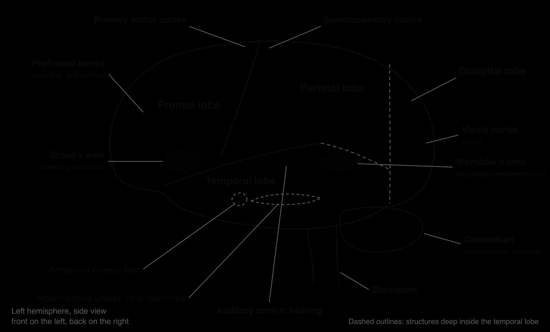
Each hemisphere is divided into four lobes, named after the skull bones that cover them. The frontal lobe at the front is involved in movement, speech production, planning and self-control. The parietal lobe behind it processes touch and spatial information. The occipital lobe at the back processes vision, and the temporal lobe at the side processes sound, language comprehension and, on its inner surface, memory and emotion.
The diagram is schematic: real brains vary in the exact size and position of each area, and the boundaries between lobes are not visible lines. It shows the areas that the evidence for localisation is built on. Below the cortex sit structures that are older in evolutionary terms, such as the hippocampus, the amygdala and the hypothalamus, which together are sometimes called the limbic system.
Area | Where it is | Main functions | Classic evidence |
|---|---|---|---|
Prefrontal cortex | Front of the frontal lobe | Planning, decision-making, working memory, impulse control, social behaviour | Phineas Gage; patients with frontal damage |
Primary motor cortex | Strip at the back of the frontal lobe | Voluntary movement of the opposite side of the body | Penfield’s electrical stimulation |
Broca’s area | Left inferior frontal lobe | Speech production and grammar | Broca (1861) |
Somatosensory cortex | Strip at the front of the parietal lobe | Touch, pressure, temperature and pain from the opposite side of the body | Penfield’s electrical stimulation |
Visual cortex | Occipital lobe | Processing what is seen | Partial blindness after occipital damage |
Auditory cortex and Wernicke’s area | Temporal lobe; Wernicke’s area in the left posterior temporal lobe | Hearing; understanding language | Wernicke (1874) |
Hippocampus | Inside the medial temporal lobe | Forming new declarative memories; spatial memory | H.M.: Scoville and Milner (1957) |
Amygdala | Inside the temporal lobe, in front of the hippocampus | Detecting threat; fear; emotional memory | Patient S.M.: Feinstein et al. (2011) |
Hypothalamus | Below the thalamus, at the base of the brain | Hunger, thirst, body temperature, hormone release through the pituitary gland | Animal lesion and stimulation studies |
Cerebellum | Below the occipital lobe | Coordination, balance, learning motor skills | Clumsy movement after cerebellar damage |
Researchers cannot damage or remove a brain area in a healthy person to see what is lost. Psychologists therefore study localisation in four main ways, each with its own strengths and its own measurement problems. The method used shapes what a study can conclude about cause and effect.
A lesion is an area of damaged tissue, caused by a stroke, injury, tumour, infection or surgery. In a lesion study, researchers compare what a patient can and cannot do with where the damage is. If damage to area X consistently removes function Y and leaves other functions intact, area X is probably needed for Y. In the 19th century the location of the damage could be found only after death, at post-mortem; today it is mapped with brain scans while the patient is alive.
Lesion evidence is strongest when it shows a double dissociation: damage to area A impairs function 1 but not function 2, while damage to area B impairs function 2 but not function 1. Broca’s and Wernicke’s patients form the classic double dissociation for language production and comprehension. A single dissociation is weaker, because the damaged area might simply handle the harder of two tasks.
In the 1930s to 1950s the neurosurgeon Wilder Penfield operated on patients with severe epilepsy while they were awake, because the brain itself feels no pain. Before removing tissue he applied a weak electrical current to points on the exposed cortex and asked the patient what they felt. Stimulating one strip of the frontal lobe made parts of the opposite side of the body twitch; stimulating a strip just behind it produced tingling sensations. These maps of the motor and somatosensory cortex showed an orderly, localised layout, with large areas for the hands, lips and tongue.
Some patients with severe epilepsy had their corpus callosum cut to stop seizures spreading between the hemispheres. Because the two halves could no longer share information, researchers could present information to one hemisphere at a time and test what each could do on its own. This research is the main evidence for lateralisation, the specialisation of each hemisphere for particular functions.
Brain imaging allows localisation to be studied in healthy people. Structural magnetic resonance imaging shows the size and shape of brain areas. Functional magnetic resonance imaging (fMRI) and positron emission tomography (PET) show which areas become more active during a task, by measuring changes in blood oxygen or in the use of glucose. Imaging studies have confirmed many lesion findings, but they have also shown that most tasks activate several areas at once.
Method | What it shows | Strength for localisation | Limitation |
|---|---|---|---|
Lesion (case) study | Which functions are lost when an area is damaged | Shows that an area is necessary for a function | Natural lesions are rarely neat; few cases; the brain may reorganise after damage |
Electrical stimulation | What happens when an area is briefly activated | Direct, precise mapping in the living brain | Only possible during surgery, usually in people with epilepsy |
Split-brain testing | What each hemisphere can do alone | Isolates the contribution of each hemisphere | Very few patients, all with a long history of epilepsy |
fMRI and PET | Which areas are more active during a task | Healthy participants; whole brain at once | Correlational: activity does not prove that an area is needed |
The idea that the mind has parts located in the brain is older than scientific psychology. Around 1800 the anatomist Franz Joseph Gall and his follower Johann Spurzheim proposed phrenology: the brain consisted of about 27 “organs”, each responsible for a trait such as cautiousness, friendliness or a talent for language, and a well-developed organ pushed out the skull above it. A trained phrenologist could therefore, they claimed, read character from the bumps on a person’s head.
Phrenology had no valid measurement. Its practitioners looked for cases that fitted their map and explained away those that did not, which is a clear example of confirmation bias. Skull shape does not follow the shape of the brain beneath it, and the “faculties” were everyday character words rather than functions defined by research. Even so, phrenology introduced the question that later research could test: does each mental function depend on a particular part of the brain?
When | Who | Contribution to the debate |
|---|---|---|
c. 1800 | Gall and Spurzheim | Phrenology: traits sit in brain “organs” that shape the skull. Unscientific, but it framed the question. |
1861 | Paul Broca | Loss of speech production linked to a lesion in the left frontal lobe: the first convincing clinical evidence of localisation. |
1874 | Carl Wernicke | Loss of language comprehension linked to the left posterior temporal lobe; language explained as connected centres. |
1930s–1950s | Wilder Penfield | Electrical stimulation maps of the motor and somatosensory cortex in awake patients. |
1950 | Karl Lashley | Rats’ maze memory depended on how much cortex was removed, not where: equipotentiality. |
1957 onward | Scoville and Milner; Sperry and Gazzaniga | H.M. and memory; split-brain patients and lateralisation. |
1990s onward | Brain imaging | Functions studied in healthy brains; most tasks rely on networks of areas. |
 YouTube
The table shows a pattern that runs through the whole topic. Each new method first seemed to confirm strict localisation, and then, as evidence built up, showed that functions are shared out more widely than the first studies suggested. The theory has therefore been refined over time: the claim that areas are specialised has survived, while the claim that each function sits in one place has been weakened.
Language is the classic case of localisation, because damage to two different areas of the left hemisphere produces two different kinds of language problem. A loss of language ability caused by brain damage is called aphasia. The study of aphasia gave psychology its first evidence that a complex mental ability depends on specific regions.
In 1861 the French surgeon Paul Broca examined a patient, Louis Victor Leborgne, who for about 21 years had been unable to say anything except the syllable “tan”, although he could understand what was said to him. When Leborgne died, Broca found a lesion on the surface of the left frontal lobe. A second patient, Lazare Lelong, could say only five words and had damage in a similar place. Over the following years Broca collected further cases and concluded that speech production depends on the left inferior frontal lobe, now called Broca’s area.
People with Broca’s aphasia speak slowly and with great effort. Their sentences contain the main content words but leave out small grammatical words such as “the”, “is” and “to”, so that speech sounds like a telegram: “Walk… dog… park.” Comprehension of simple sentences is largely intact, and most patients are painfully aware of their difficulty.
In 1874 the German neurologist Carl Wernicke described patients with the opposite pattern. They spoke fluently, with normal rhythm and grammar, but their speech made little sense and often contained wrong or invented words. They also had great difficulty understanding what others said. Their damage lay in the left posterior temporal lobe, next to the auditory cortex, in a region now called Wernicke’s area.
Feature | Broca’s (expressive) aphasia | Wernicke’s (receptive) aphasia |
|---|---|---|
Damaged area | Left inferior frontal lobe | Left posterior temporal lobe |
Speech | Slow, effortful, halting; few grammatical words | Fluent and well-paced, but often meaningless, with wrong or invented words |
Comprehension | Mostly intact for simple sentences | Poor |
Awareness of the problem | Usually aware and frustrated | Often unaware that speech makes little sense |
Example | “Walk… dog… park.” | “I went to the tib with the kalter and then it was gone.” |
Wernicke did more than add a second centre. He argued that language depends on connections between centres: sounds are recognised in the auditory cortex, given meaning in Wernicke’s area, and passed forward to Broca’s area, where speech is planned before the motor cortex moves the mouth. The two areas are joined by a bundle of white matter fibres, the arcuate fasciculus. In the 1960s the neurologist Norman Geschwind developed this into the Wernicke–Geschwind model.
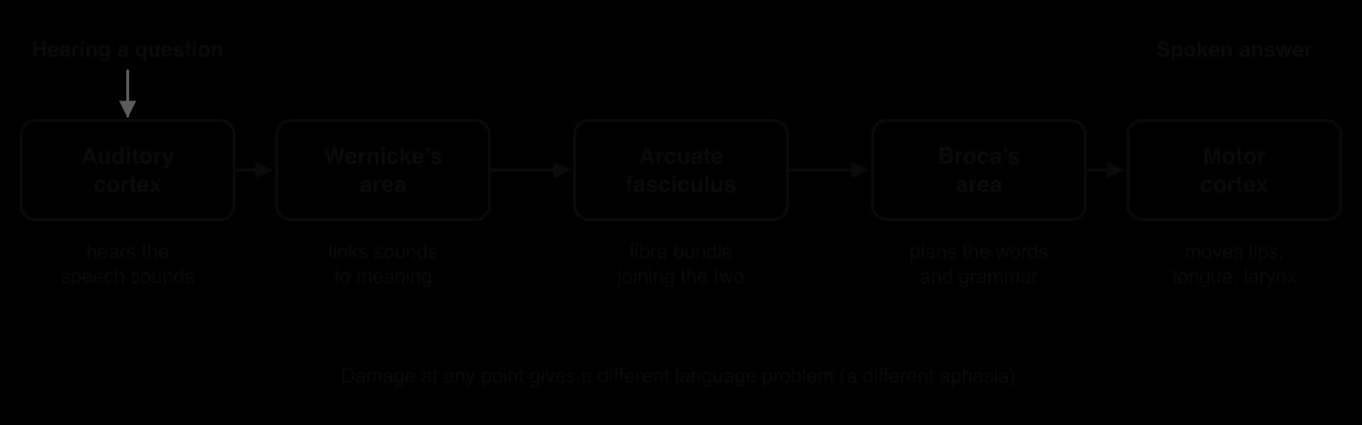
The model makes a testable prediction. If the arcuate fasciculus is damaged but both areas are intact, patients should understand speech and speak fluently but be unable to repeat what they hear. This pattern, called conduction aphasia, does occur, which supported the model. Later research has shown that the real language network is larger, with more pathways and contributions from both hemispheres, but the idea that language depends on connected regions rather than a single centre has lasted.
Broca noticed that his patients’ damage was almost always on the left side. This was the first evidence of lateralisation: the two hemispheres, although similar in structure, are specialised for different functions. In most people the left hemisphere is dominant for language, while the right hemisphere contributes more to some visuospatial tasks, recognising faces and processing the emotional tone of speech.
The clearest evidence for lateralisation comes from split-brain patients. From the 1960s, Roger Sperry and his student Michael Gazzaniga at the California Institute of Technology tested a small number of people whose corpus callosum had been cut to control severe epilepsy. In daily life these patients seemed almost normal, because both eyes and ears still sent information to both hemispheres. Carefully controlled laboratory tests, however, showed that each hemisphere was now working on its own.
Each eye sends information from the left visual field to the right hemisphere and from the right visual field to the left hemisphere. Sperry’s team asked a patient to stare at a central point while a word or picture was flashed for a fraction of a second to one side, too briefly for the eyes to move. The patient then had to say what they had seen, or find the object by touch with one hand behind a screen.
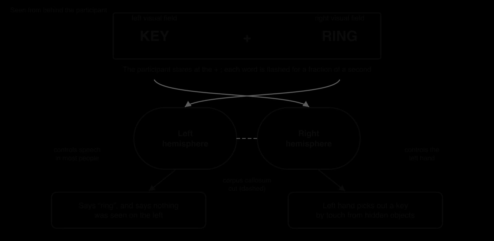
When an image was flashed to the right visual field, the left hemisphere received it and the patient could name it. When it was flashed to the left visual field, the patient usually said they had seen nothing, yet their left hand, controlled by the right hemisphere, could pick out the correct object. The right hemisphere had seen the image and understood it well enough to act on it, but could not put it into words.
Sperry (1968) concluded that each hemisphere can perceive, learn and remember independently, and that speech is normally lateralised to the left. He shared the Nobel Prize in Physiology or Medicine in 1981 for this work.
Lateralisation is a tendency, not a rule. Knecht et al. (2000) measured which hemisphere was dominant for language in 326 healthy adults in Germany, using functional transcranial Doppler ultrasound, which records changes in blood flow to each hemisphere while a person thinks of words. Most people were left-dominant, but the proportion with language in the right hemisphere rose steadily with left-handedness.
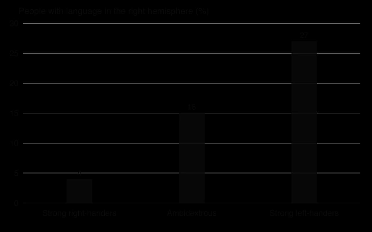
The study was done with healthy volunteers rather than patients, so the variation cannot be blamed on brain damage. The finding matters for localisation because it shows that the location of a function differs between individuals. A statement such as “language is in the left hemisphere” describes most people, not everyone.
Popular books and online quizzes often claim that people are either “left-brained” (logical, analytical) or “right-brained” (creative, emotional). Nielsen et al. (2013) tested this directly with resting-state brain scans of 1,011 people aged 7 to 29. They found lateralised hubs in each hemisphere, such as language areas on the left and attention areas on the right, but no evidence that some individuals have a generally stronger left or right hemisphere. Lateralisation therefore applies to specific networks, and it cannot be used to sort people into “left-brained” and “right-brained” types.
 YouTube
Localisation research extends well beyond language. Three further functions show both the strengths of the theory and its limits: memory, which turns out to depend on different structures for different kinds of memory; fear, which depends heavily on one small structure; and self-control, which depends on the frontal lobes.
The hippocampus is a curved structure on the inner surface of each temporal lobe. The best-known evidence for its role comes from Henry Molaison, known in research for decades as H.M. In 1953, at the age of 27, he had large parts of both medial temporal lobes removed, including the front part of the hippocampus and most of the amygdala, to treat severe epilepsy. Scoville and Milner (1957) reported that afterwards he could no longer form new long-term memories of facts and events, a condition called anterograde amnesia, although his short-term memory and his intelligence were normal.
H.M. is also evidence of the limits of localisation. He could still learn new motor skills: when Brenda Milner taught him to trace a star while looking only at its mirror image, his performance improved over three days, although each day he did not remember having practised.
Procedural memory therefore depends on other structures, such as the cerebellum and parts of the basal ganglia. Memory consists of several systems that depend on different brain areas. Later MRI by Corkin et al. (1997) also showed that the surgery had spared part of the hippocampus and damaged surrounding cortex, so H.M.’s amnesia cannot be pinned on the hippocampus alone.
The amygdala is an almond-shaped structure in front of the hippocampus. Feinstein et al. (2011) studied S.M., a woman whose amygdalae had been destroyed on both sides by a rare genetic disease. The researchers took her to a pet shop with snakes and spiders, through a “haunted house” attraction and showed her frightening film clips. She showed no fear at all, approached the snakes with curiosity and reported almost none of the fear that other visitors felt, while her other emotions were normal.
S.M. seemed to show that fear is localised in the amygdala. Two years later, Feinstein et al. (2013) asked S.M. and two other patients with the same damage to breathe air containing 35% carbon dioxide, which produces a feeling of suffocation. All three felt fear and had panic attacks.
The amygdala appears to be needed for fear of external threats, such as a snake, but not for fear triggered from inside the body. The same emotion can therefore depend on different brain systems in different situations.
The prefrontal cortex (PFC) is involved in executive functions: planning, holding information in mind, switching between tasks, controlling impulses and making social judgements. Much of the early evidence came from people with frontal damage. In 1848 an iron rod passed through the left frontal lobe of a railway foreman, Phineas Gage. His doctor, John Harlow, reported in 1868 that the formerly reliable Gage became impulsive, rude and unable to keep to plans.
Gage is a useful example of how evidence can be distorted. Macmillan and Lena (2010) re-examined the historical records and found that the dramatic accounts grew over time. Gage later worked for several years as a stagecoach driver in Chile, a job that needed planning and self-control, so he probably adapted far better than most textbooks claim. The case supports a role for the frontal lobes in personality, but it is weak evidence for strict localisation, and his recovery points to neuroplasticity.
Localisation of function is used in all four contexts of the course, often together with factors from other approaches. The table gives one example from each, with the point it makes.
Context | Behaviour | How localisation is used | Limit of the explanation |
|---|---|---|---|
Health and well-being | Recovery from stroke | The symptoms (for example aphasia or weakness on one side) show which area was damaged and guide rehabilitation. | Recovery depends on plasticity and therapy, not only on where the damage is. |
Human development | Risk-taking in adolescence | The prefrontal cortex matures later than reward-related areas, which may explain impulsive choices. | Peers, culture and opportunity also shape risk-taking. |
Human relationships | Responding to threat or social rejection | The amygdala responds to threatening faces; frontal areas help control emotional reactions. | Social and cultural norms shape what counts as a threat. |
Learning and cognition | Remembering new information | The hippocampus is needed to form new declarative memories. | Different kinds of memory depend on different structures, and recall involves widespread networks. |
In each case localisation explains part of the behaviour, but a full explanation also needs cognitive and sociocultural factors. An adolescent’s late-maturing prefrontal cortex does not on its own explain why one teenager drives dangerously and another does not; the presence of friends, the norms of the group and the chance to take the risk all matter. This is why localisation is best treated as one level of explanation within an interactionist account.
The evidence that brain areas are specialised is strong. The argument that a behaviour is located in one area, so that the area “contains” the behaviour, is much weaker. Six limitations are well supported by research.
Complex behaviours draw on many connected areas. Language involves Broca’s and Wernicke’s areas, the white matter pathways between them, the motor and auditory cortex and contributions from the right hemisphere. Brain imaging shows that even a simple task, such as naming a picture, activates regions in several lobes at once. Clinical studies since the 1970s have also found that damage limited to Broca’s area usually causes only a temporary speech problem, while lasting Broca’s aphasia follows larger lesions that include deeper tissue. Modern scans of Broca’s own patients point the same way.
A useful comparison is an orchestra. Each section plays a different part, and losing the violins changes the music in a predictable way, but the music is not “in” the violin section. In the same way, an area can make a specialised contribution without being the place where a behaviour is stored.
If a function can move to a new area after damage, it cannot be fixed to one location. Danelli et al. (2013) studied E.B., a boy in Italy whose left hemisphere was surgically removed (a left hemispherectomy) when he was two and a half years old. After an initial loss of language he recovered within two years, and by the age of 14 his language was close to normal, with fMRI showing a language network in his right hemisphere that resembled the usual left-hemisphere pattern.
Recovery is most complete when damage happens early, and adults recover less fully, but this process of functional reorganisation shows that the location of a function depends partly on experience and history.
In a long series of studies, Karl Lashley trained rats to run mazes and then removed different areas of their cortex. Lashley (1950) reported that the loss of maze memory depended on how much cortex was removed rather than where it was removed from. He proposed two principles: mass action (the cortex works as a whole for complex learning) and equipotentiality (intact areas can take over the work of damaged ones).
Lashley’s findings challenge strict localisation for complex learning, but they have their own weaknesses. Rats can solve a maze using vision, smell, touch and movement, so each lesion may have removed a different route to the same solution. The findings are also hard to extrapolate from rats to human language or memory, which is a general limitation of animal models.
The same function is not always in the same place. Knecht et al. (2000) found right-hemisphere language in 4% of strong right-handers but 27% of strong left-handers. The size and exact position of Broca’s and Wernicke’s areas also vary considerably between people, which is one reason why surgeons map language areas in each patient before operating rather than relying on a standard map.
Most localisation research has studied speakers of alphabetic languages such as English and German. Siok et al. (2004) used fMRI with Chinese children, some with reading difficulties and some without. Chinese dyslexia was linked to reduced activity in the left middle frontal gyrus, an area that coordinates the visual form, sound and meaning of characters, rather than the left temporoparietal areas usually linked with dyslexia in English readers. Which brain areas support reading depends partly on the writing system a person learns, so localisation is shaped by culture as well as biology.
Much of the evidence for localisation comes from case studies of people with damaged brains, whose brains may have reorganised after injury and who may have had other conditions, such as long-term epilepsy. Natural lesions rarely respect the boundaries of the areas researchers want to study. Imaging evidence in healthy people is correlational, and the history of phrenology is a reminder that a neat map can be produced by selective attention to cases that fit.
Limitation | What it means | Evidence |
|---|---|---|
Distributed networks | Behaviours need many connected areas, not one centre | Language network; Dronkers et al. (2007); imaging of naming tasks |
Plasticity | Functions can move after damage | E.B.: Danelli et al. (2013) |
Equipotentiality | The amount of damage can matter more than its location | Lashley (1950) |
Individual differences | Locations vary between people | Handedness and language: Knecht et al. (2000) |
Culture | Experience shapes which areas a skill uses | Chinese reading: Siok et al. (2004) |
Method | Case studies and imaging have limits on what they can show | Phineas Gage; reverse inference in fMRI |
The six concepts give a structure for evaluating both the theory and the research behind it. Four of them are especially useful for evaluating localisation.
Concept | Key question | Application to localisation |
|---|---|---|
Measurement | How is the function of an area measured? | Indirectly: from what is lost after damage, from blood flow on scans or from responses to stimulation. |
Causality | Does the area cause the behaviour? | Lesions show necessity in one person; imaging shows only correlation; networks make single causes unlikely. |
Perspective | Is one brain area a sufficient explanation? | Localisation is reductionist; behaviour also depends on cognition, culture and experience. |
Responsibility | Is research on brain-damaged patients ethical? | Patients may be unable to give informed consent, and decades of testing raise questions of dignity and privacy. |
No method observes a function directly. Lesion studies infer what an area does from what is lost, but a lost ability might reflect damage to fibres passing through the area or the effects of swelling and disease elsewhere. fMRI measures the BOLD signal, a change in blood oxygen that follows neural activity by several seconds, so it is an indirect and slow measure of what neurons are doing. Using several methods together, a form of triangulation, strengthens a conclusion: Broca’s area is linked to speech production by lesion, stimulation and imaging evidence, which makes the link more credible than any one method could.
Lesion studies come closest to showing cause, because damage comes before the loss of function. Even so, they show only that an area is needed in that brain, not that it produces the behaviour alone. Imaging studies are correlational: an area that is active during a task may be monitoring or suppressing rather than producing it. Brain stimulation in healthy people, such as transcranial magnetic stimulation, can briefly disrupt an area and test whether performance falls, which gives stronger evidence of a causal role.
Explaining a behaviour such as aggression or depression by “an overactive amygdala” or “a weak prefrontal cortex” reduces a complex behaviour to one part of the brain. This kind of biological reductionism can be useful, because it produces precise and testable claims, but it leaves out the cognitive processes and social contexts that shape when and how the brain responds. Siok et al. (2004) show that even the brain basis of a skill can depend on culture, which supports an interactionist perspective in which brain, mind and environment influence one another.
The founding evidence for localisation came from a small number of European and North American patients, chosen because their symptoms were striking. Unusual cases are more likely to be reported and remembered than ordinary ones, which can make localisation look more precise than it is. Modern imaging research still relies mostly on WEIRD samples, and research such as Siok et al. (2004) shows that findings from English speakers do not always generalise to readers of other writing systems.
Many key cases involved people who could not easily give or remember informed consent. Leborgne could not speak; H.M. could not remember agreeing to studies and was tested for more than 50 years; in later years consent was given on his behalf, and how it was obtained has since been questioned. Split-brain surgery was carried out for medical reasons, but the research that followed made use of patients in a vulnerable situation.
Researchers protected anonymity with initials such as H.M. and S.M., but the brains of Broca’s patients are still kept in a university collection in Paris, and photographs of them are widely published, which raises questions about dignity after death.
Two pieces of research on the same two patients, separated by almost 150 years, belong together. Broca’s original observations founded the theory of localisation; the MRI study by Dronkers et al. (2007) tested how accurate those observations were with modern technology.
Broca aimed to identify the part of the brain responsible for articulate speech by studying patients who had lost the ability to speak but could still understand language. Dronkers, Plaisant, Iba-Zizen and Cabanis (2007) aimed to find the exact location and extent of the lesions in the preserved brains of Broca’s two patients, and to check whether other areas had been damaged too.
Broca used the clinical case method. Louis Victor Leborgne had been a patient at the Bicêtre Hospital in Paris for many years. He had lost the ability to speak about 21 years earlier, could say little except “tan”, but understood speech and communicated with gestures; over time he also developed paralysis of the right side of his body.
Broca examined him shortly before his death in April 1861 and carried out the post-mortem. Months later he examined Lazare Lelong, an 84-year-old man who could say only five words, including “yes”, “no” and a version of his own name. Broca kept both brains whole rather than slicing them, so he could see only their surfaces, and gave them to a museum in Paris, the Musée Dupuytren.
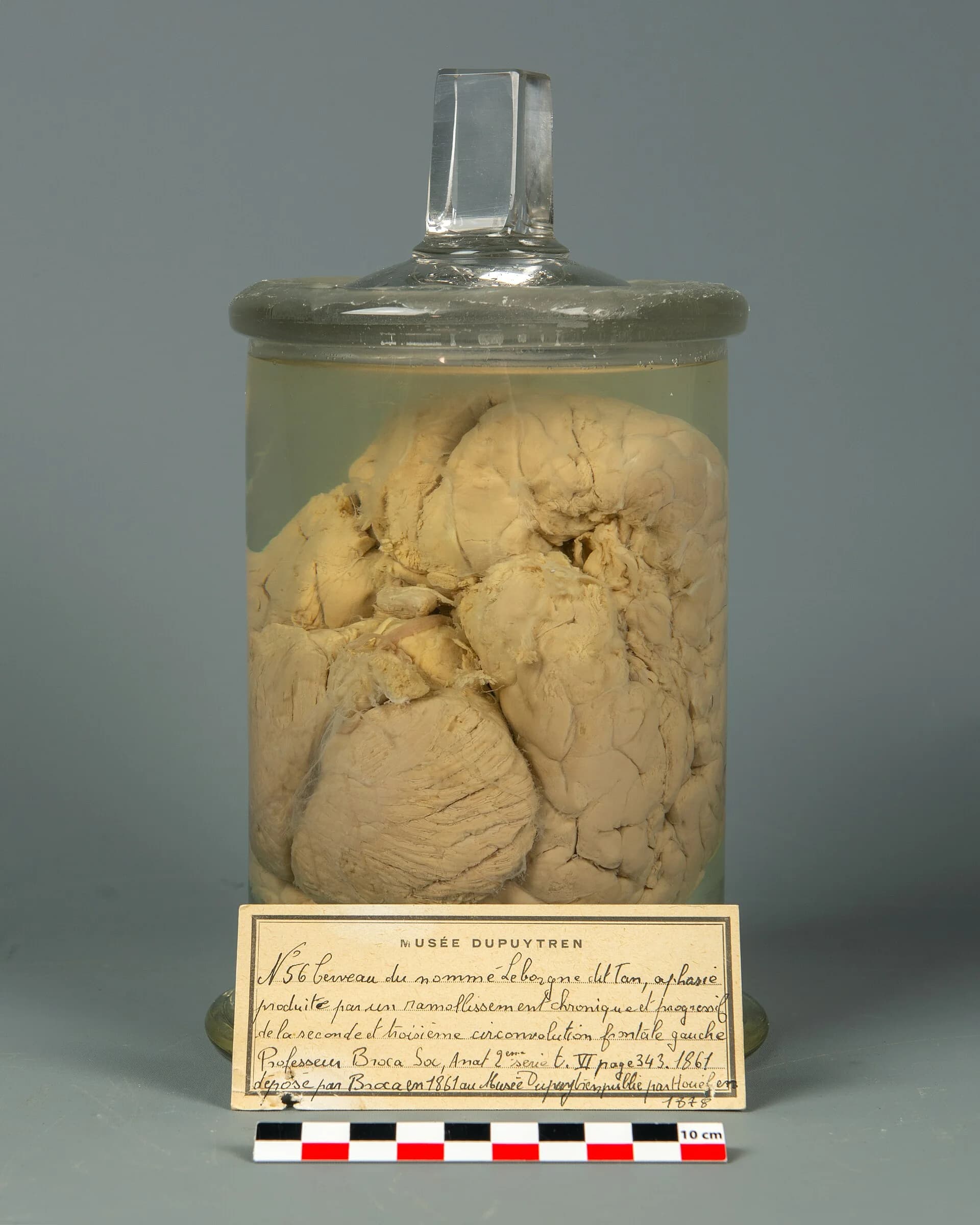
Dronkers et al. scanned both preserved brains with high-resolution volumetric MRI. The method produced thin images through the whole of each brain, so the researchers could trace the lesions below the surface and compare them with the area that modern research calls Broca’s area.
Leborgne (“Tan”) | Lelong | |
|---|---|---|
Speech | Almost only the syllable “tan” | Five words |
Comprehension | Largely intact | Largely intact |
What Broca saw (1861) | Lesion on the surface of the left inferior frontal lobe | Lesion on the surface of the left inferior frontal lobe |
What MRI showed (2007) | Damage extended far into medial regions beneath the surface | Damage extended into medial regions beneath the surface |
Broca found damage on the surface of the left inferior frontal lobe in both patients, in the region later named after him. Dronkers et al. found that both lesions extended significantly into deeper, medial parts of the brain, well beyond the surface damage Broca had described. They also found inconsistencies between the area Broca originally identified and the region that modern studies label “Broca’s area”.
Broca concluded that articulate speech depends on a specific area of the left frontal lobe, the first convincing evidence that a higher mental function is localised and, as more cases accumulated, that it is lateralised to the left. Dronkers et al. concluded that his patients’ loss of speech was probably caused by damage to a wider set of regions, including deeper tissue that is now known to be important for speech. Their findings support localisation in the weaker sense (a region of the left frontal lobe and its connections is needed for speech production) rather than the strong sense (speech is located in one surface area).
Broca’s post-mortem method could see only the brain’s surface, so it measured the lesion incompletely. The 2007 MRI study shows how a better measurement tool can change the interpretation of a classic finding. Both studies rely on a sample of two, and the original symptoms were described by a single clinician, so the measurement of what each patient could and could not do was limited by 19th-century standards.
Leborgne’s condition progressed over two decades, and by the time of his death the damage was extensive. The speech loss came first, which suggests the frontal damage began early, but it is impossible to know which parts of the lesion caused which symptoms. Natural lesions do not respect anatomical boundaries, so lesion evidence alone cannot show that one small area produces speech.
Neither patient could consent to his brain being studied or displayed, and Leborgne could hardly communicate at all. Historians have since identified Leborgne by his full name and traced his life, which restores his identity but also removes the anonymity usually given to patients. The research shows how ethical standards for case studies have changed: today patients or their families give informed consent, and identities are protected.
Sources: Broca, P. (1861). Remarques sur le siège de la faculté du langage articulé. _Bulletin de la Société Anatomique de Paris, 6, 330–357; Dronkers, N. F., Plaisant, O., Iba-Zizen, M. T. and Cabanis, E. A. (2007). Paul Broca’s historic cases: high resolution MR imaging of the brains of Leborgne and Lelong. Brain, 130(5), 1432–1441._
Questions on localisation of function appear mainly in Paper 1. Section A can ask you to explain localisation, or one limitation of the argument that behaviour is localised, with an example. Section B gives a short scenario from one of the four contexts and asks you to apply localisation to it. Localisation can also support a Section C essay, for example on biological factors in cognitive processes, and research on it can appear as an unseen study in Paper 2.
A Section A question may focus on the second half of the topic instead: “Explain one limitation of the argument that behaviour is localised.” The same structure applies: explain the limitation, give an example and show how the example demonstrates it.
Localisation of function is the theory that specific brain areas carry out specific psychological functions; modern research supports a weaker version in which areas make specialised contributions within networks.
Localisation is studied through lesion studies, electrical stimulation, split-brain testing and brain imaging; lesions show that an area is necessary, while imaging shows only correlation.
Broca’s area (left frontal lobe) is linked to speech production and Wernicke’s area (left temporal lobe) to comprehension; damage produces two different aphasias.
Split-brain research shows that speech is lateralised to the left hemisphere in most people, but lateralisation varies with handedness and there is no evidence for “left-brained” or “right-brained” people.
The hippocampus is needed for new declarative memories, the amygdala for fear of external threats and the prefrontal cortex for executive functions, but each conclusion has limits (H.M.’s spared procedural memory, S.M.’s panic, Gage’s recovery).
Limitations of the argument that behaviour is localised: distributed networks, plasticity and recovery, equipotentiality, individual differences, culture and methodological problems.
Dronkers et al. (2007) found that the lesions in Broca’s patients extended deep beyond the surface area Broca described, so speech depends on a wider network.
Localisation research raises issues of measurement (indirect methods), causality (necessary is not sufficient), perspective (reductionism) and responsibility (consent of brain-damaged patients).
I can describe localisation of function and name the main areas linked to language, memory, emotion, movement and planning.
I can explain how lesion studies, stimulation, split-brain testing and brain imaging are used to study localisation, and what each can show.
I can describe Broca’s and Wernicke’s findings and explain the difference between the two aphasias.
I can explain lateralisation using split-brain research and the link between handedness and language.
I can apply localisation of function to a behaviour in each of the four contexts.
I can discuss at least four limitations of the argument that behaviour is localised, each with evidence.
I can evaluate localisation research with reference to measurement, causality, perspective and responsibility.