Notes on A2.3 Viruses
Viruses are tiny infectious particles made of genetic material in a protein coat, which can reproduce only inside living cells. They cause diseases such as influenza, COVID-19 and AIDS, and the bacteriophages that infect bacteria are being developed as treatments for infections that antibiotics cannot cure.
Viruses carry very few genes because they use the metabolism of their host cells for almost everything. Their structure is simple, but the ways they store their genes, the cycles they follow inside cells and the speed at which they evolve vary widely.
Structural features common to all viruses
What a virus is
A virus particle, or virion, is a package of genetic material inside a protein coat. It has no cytoplasm, no ribosomes and no metabolism of its own, so it cannot grow, respire or make proteins outside a cell. A virus can multiply only by entering a living host cell and using that cell's ribosomes, enzymes, ATP and raw materials. Viruses are therefore described as obligate intracellular parasites.
Every virus shares a small set of structural features:
Small, fixed size. Most virions are 20–300 nm across, far smaller than bacteria and below the resolution of the light microscope. Unlike cells, virions do not grow: each particle is assembled at its final size.
Nucleic acid as genetic material. The genome is either DNA or RNA, never both. It carries a small number of genes, from as few as four in some tiny RNA viruses to a few hundred.
A protein capsid. The capsid is a coat built from many copies of one or a few protein subunits. It protects the genome and, in many viruses, carries the proteins that bind to the host cell.
No cytoplasm and few or no enzymes. Some viruses carry one or two enzymes needed at the very start of infection, such as reverse transcriptase in HIV, but none has the enzymes for metabolism or a ribosome for protein synthesis.
Because viruses lack cells and cannot carry out the functions of life on their own, most biologists do not classify them as living organisms. They do, though, have genes, they evolve by natural selection, and they use the same genetic code as cells, so they are part of the biological world in every other sense. They are outside the cell theory (A2.2): viruses are not made of cells and do not arise by cell division but by assembly inside a host.
Diversity of structure in viruses
Viruses are much more varied than cells in the way their genetic material is stored. Cells always use double-stranded DNA as their genome. Viruses use every possible form of nucleic acid:
Feature | Variation among viruses | Examples |
|---|---|---|
Type of nucleic acid | DNA or RNA | Lambda phage (DNA); coronaviruses, influenza, HIV (RNA) |
Number of strands | Single-stranded or double-stranded | Lambda (double-stranded DNA); SARS-CoV-2 (single-stranded RNA) |
Shape of the genome | Linear or circular; one molecule or several segments | Influenza has eight separate RNA segments |
Sense of RNA | Positive-sense RNA can be translated directly; negative-sense RNA must first be copied into its complement | SARS-CoV-2 (positive); influenza (negative) |
Reverse transcription | Retroviruses copy their RNA into DNA | HIV |
Capsid shape | Icosahedral (20-sided), helical, or complex (head and tail) | Lambda (head and tail); nucleocapsid of coronaviruses (helical) |
Envelope | Present or absent | Coronaviruses and HIV (enveloped); lambda (not enveloped) |
An enveloped virus is surrounded by a viral envelope, a phospholipid bilayer outside the capsid. The envelope is not made by the virus: it is a piece of the host cell's plasma membrane (or another host membrane), taken by the new virion as it buds out of the cell. Viral glycoproteins inserted into this membrane stick out as spikes and bind to receptors on the next host cell. Detergents and alcohol-based hand gels break up the lipid envelope, which is why they inactivate enveloped viruses such as coronaviruses more easily than non-enveloped ones.
Three viruses show this diversity clearly: bacteriophage lambda, the coronaviruses and HIV.
Bacteriophage lambda
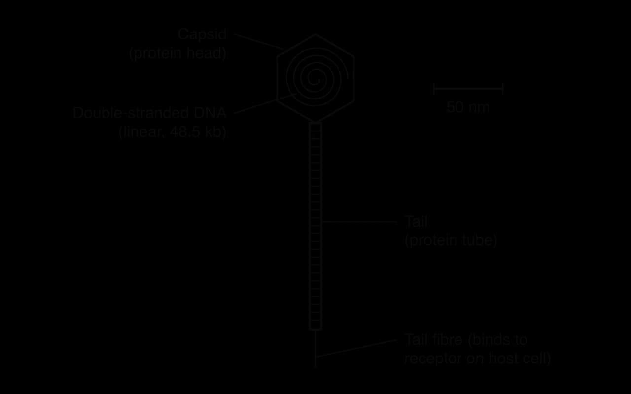
A bacteriophage (phage) is a virus that infects bacteria. Bacteriophage lambda (λ) infects Escherichia coli. Its head is an icosahedral protein capsid about 55 nm across, holding one linear molecule of double-stranded DNA of about 48,500 base pairs (48.5 kb). Attached to the head is a long, flexible protein tail about 150 nm long, ending in a tail fibre that binds to a specific protein (LamB) in the outer membrane of E. coli. Lambda has no envelope.
Electron micrographs show the same structure in real lambda particles, prepared by negative staining, in which a heavy-metal stain surrounds the particle and shows its outline.
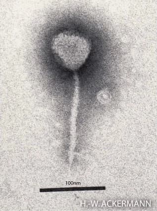
The head-and-tail shape works like a syringe. The tail fibre holds the phage on the surface of the bacterium while the DNA passes down the hollow tail, so the genome enters the cell and the protein coat does not.
 YouTube
Coronaviruses
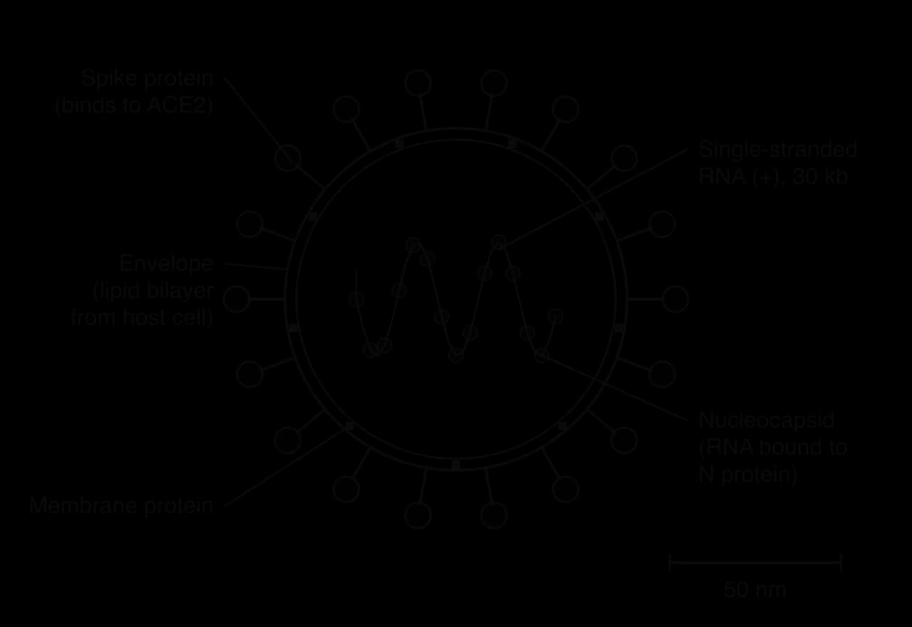
Coronaviruses are a family of enveloped RNA viruses that infect birds and mammals. They include SARS-CoV-2, the cause of COVID-19, and several viruses that cause common colds. The genome is one long molecule of single-stranded, positive-sense RNA of about 30,000 nucleotides (30 kb), among the largest of all RNA virus genomes. Positive-sense means the RNA can act directly as mRNA and be translated by the host's ribosomes as soon as it enters the cell. The RNA is wound into a helical nucleocapsid with a nucleocapsid (N) protein, and this lies inside an envelope studded with spike (S) proteins. In SARS-CoV-2 the spike protein binds to a receptor called ACE2 on cells lining the airways.
HIV, a retrovirus
The human immunodeficiency virus (HIV) is an enveloped retrovirus. Its capsid is cone-shaped and holds two copies of a single-stranded RNA genome of about 9,700 nucleotides, together with the enzyme reverse transcriptase. Glycoproteins in the envelope (gp120) bind to the CD4 receptor on helper T-cells of the immune system.

Inside the host cell, reverse transcriptase uses the viral RNA as a template to make a DNA copy, the reverse of normal transcription. A second viral enzyme, integrase, then inserts this DNA into a chromosome of the host cell, where it stays for the life of the cell. Each time the cell transcribes the viral genes, it makes new viral RNA and proteins. C3.2 describes how HIV destroys helper T-cells and causes AIDS.
Feature | Bacteriophage lambda | Coronavirus (SARS-CoV-2) | HIV |
|---|---|---|---|
Host | Escherichia coli (bacterium) | Mammals, including humans | Humans (helper T-cells) |
Genetic material | Double-stranded DNA, linear, 48.5 kb | Single-stranded positive-sense RNA, about 30 kb | Two copies of single-stranded RNA, about 9.7 kb |
Enzymes carried | None needed | None needed at entry | Reverse transcriptase, integrase, protease |
Capsid | Icosahedral head with a tail | Helical nucleocapsid | Cone-shaped |
Envelope | Absent | Present, with spike proteins | Present, with gp120 glycoproteins |
Receptor on host | LamB protein of the outer membrane | ACE2 | CD4 |
The lytic cycle
Viruses depend on their host cells for every life function: energy from the host's ATP, nucleotides and amino acids from the host's cytoplasm, and the host's ribosomes for protein synthesis. The lytic cycle is the sequence of steps by which a virus enters a cell, takes over its metabolism to make new virions and then destroys the cell, releasing them. In bacteriophage lambda it has five stages.
Attachment. The tail fibre of a free lambda virion collides with an E. coli cell and binds to the LamB protein in its outer membrane. Binding is specific: lambda can only infect bacteria with this receptor.
Injection (penetration). The tail tip penetrates the cell wall and the phage DNA is injected through the hollow tail into the cytoplasm. The capsid and tail stay outside the cell as an empty "ghost".
Synthesis. The linear DNA forms a circle. The host's RNA polymerase transcribes the phage genes and the host's ribosomes translate them, using host ATP, nucleotides and amino acids. Phage proteins take control: some copy the phage DNA many times, and others are the protein subunits of new heads and tails.
Assembly. Head proteins come together into empty capsids, and a copy of the phage DNA is packed into each. Tails are added to complete new virions.
Lysis and release. Late phage genes code for enzymes that break down the plasma membrane and the peptidoglycan cell wall. The weakened cell bursts (lyses), releasing about 100 new phages, each able to infect another cell.
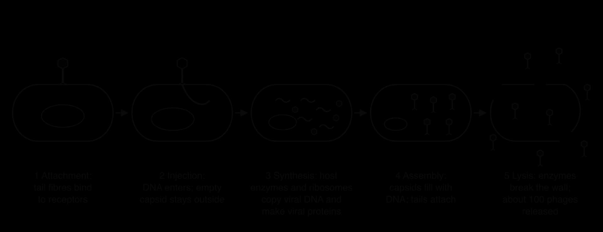
The timing of the lytic cycle is shown by a one-step growth experiment, in which a culture of bacteria is infected with phages all at once and the number of free phages in the medium is measured at intervals. For a while there are few free phages, because the new virions are still being made inside the cells. Then all the infected cells lyse at about the same time and the number of free phages rises steeply to a new plateau, about 100 times higher.
The lysogenic cycle
Some phages, including lambda, have a second option after injecting their DNA. Instead of making new virions at once, the phage can insert its DNA into the host chromosome and stay there, inactive, for many generations. This is the lysogenic cycle.
Attachment and injection happen exactly as in the lytic cycle, and the lambda DNA forms a circle.
Integration. A phage enzyme, integrase, inserts the phage DNA into the bacterial chromosome at a specific site. The integrated phage DNA is called a prophage.
Repression. One phage gene, coding for a repressor protein (cI), stays switched on. The repressor binds to the phage DNA and prevents transcription of the genes for the lytic cycle, so no phage proteins for replication or new capsids are made and the cell is not harmed.
Replication with the host. Every time the bacterium replicates its chromosome and divides by binary fission, the prophage is copied as part of it. After many divisions a whole population of bacteria carries the prophage.
Induction. If the host cell is damaged, for example by ultraviolet light that damages its DNA, the repressor is destroyed. The prophage is cut out (excised) of the chromosome and the phage enters the lytic cycle, making new virions and lysing the cell.
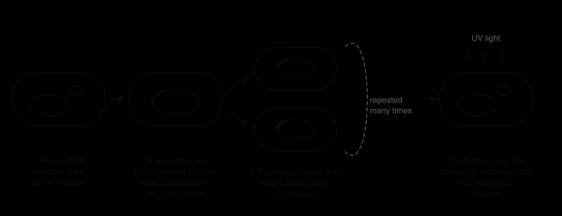
A phage that can follow either cycle is a temperate phage; lambda is the best-studied example. A virulent phage, such as T4, can only follow the lytic cycle. The "decision" between the cycles depends on conditions in the cell: lysogeny is favoured when many phages infect each cell or when the bacteria are starved, situations in which new phages would have few healthy hosts to infect. Whichever way the switch goes, it depends on the expression of phage genes inside the host, which is why the lambda switch became a classic model of gene regulation.
Feature | Lytic cycle | Lysogenic cycle |
|---|---|---|
Phage DNA | Stays separate from the host chromosome | Integrated into the host chromosome as a prophage |
Viral genes for replication | Expressed at once | Repressed |
New virions | Made within about an hour | Not made until induction |
Effect on host cell | Killed by lysis | Survives and divides normally |
Spread of phage DNA | By release of many new virions | By host cell division (vertical transmission) |
Phage types | Virulent and temperate phages | Temperate phages only (e.g. lambda) |
Both cycles begin in the same way, with attachment and injection of the phage DNA, and both depend entirely on the enzymes, ribosomes and raw materials of the host cell. They differ in what happens to the phage DNA after it enters, and so in whether the host cell survives.
 YouTube
Case study: engineered phages against a drug-resistant infection
Phages kill bacteria, so they can in principle treat bacterial infections that antibiotics no longer control. The first use of engineered phages to treat a human mycobacterial infection, reported in 2019, shows why the difference between the lytic and lysogenic cycles matters.
The patient was a 15-year-old with cystic fibrosis who had received a double lung transplant. After the transplant the patient developed a widespread infection with Mycobacterium abscessus, a bacterium resistant to most antibiotics, with infected skin nodules, an infected surgical wound and infection in the liver. Drugs to suppress the immune system, needed so that the new lungs were not rejected, made the infection harder to fight.
Researchers screened phages from a collection of more than 10,000, isolated by school and university students, and found three that could kill the patient's strain of bacteria. Only one, phage Muddy, was naturally lytic. The other two, ZoeJ and BPs, are temperate: on entering a cell they can integrate as prophages instead of killing it, and a lysogen is protected against further infection by the same phage. Such phages are useless for treatment. The team therefore deleted the repressor gene from each, so that the phages could no longer establish lysogeny and were forced into the lytic cycle.
Phage | Natural life cycle | Change made |
|---|---|---|
Muddy | Lytic | None |
ZoeJ | Temperate | Repressor gene deleted, so it can only follow the lytic cycle |
BPs | Temperate | Repressor gene deleted; a host-range mutant chosen to infect the patient's strain well |
Source: Dedrick et al., Nature Medicine 25: 730–733, 2019.
The three-phage cocktail was given by intravenous injection twice a day, at about 10⁹ phage particles of each phage per dose, for at least 32 weeks, and was also applied directly to the wound and skin lesions. The treatment caused no side effects. The surgical wound closed, liver function improved, and the skin nodules gradually healed. Bacteria taken from the skin lesions during treatment stayed sensitive to all three phages. Using a cocktail of several phages, like using several drugs at once, makes it less likely that bacteria resistant to all of them will appear.
The case shows the total dependence of phages on their hosts (they could act only where live M. abscessus cells were present) and the importance of the lytic cycle: only phages that lyse their host cells kill the bacteria. It also shows a limit of the method, since each patient's bacterial strain needs its own matched phages.
Evidence for several origins of viruses
Why viruses probably evolved after cells
Viruses leave no fossils, so their origins have to be worked out from the structure and genes of viruses alive today. Several lines of evidence are used:
Dependence on cells. Every virus is an obligate intracellular parasite that needs a host cell's ribosomes and metabolism. A virus could not have multiplied before there were cells, so viruses as they exist now are likely to have evolved after the first cells.
The universal genetic code. Viral genes use the same genetic code as cells and are translated by host ribosomes. This shows that viruses share a common origin with cellular life, or at least evolved within it.
Extreme diversity. Viruses use double- and single-stranded DNA and RNA, positive and negative sense, linear, circular and segmented genomes, and reverse transcription. No single ancestor could easily explain all of these. Most biologists conclude that viruses arose several times, independently, from different cellular sources: viruses are polyphyletic.
Genes shared with hosts. Many viral genes are similar to genes of their hosts, which suggests they were picked up from host cells at different times.
Shared features such as protein capsids do not prove a single origin. A capsid built from many copies of one protein is a simple solution to the problem of protecting a short genome with very few genes, so similar capsids may have evolved separately in unrelated viruses. This is convergent evolution, the independent evolution of similar structures in unrelated groups (A4.1), at the level of molecules.
Hypotheses for the origin of viruses
Three main hypotheses are proposed. They are not mutually exclusive: different groups of viruses may have arisen in different ways.
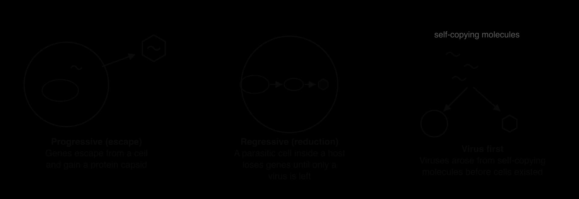
The progressive hypothesis (escape hypothesis). Pieces of genetic material, such as plasmids or transposable elements that can already move within or between cells, escaped from cells and gained the genes for a capsid, becoming able to move between cells as particles. Retroviruses, whose genes resemble those of retrotransposons in host genomes, may have arisen this way.
The regressive hypothesis (reduction hypothesis). Viruses descend from small cells that lived as parasites inside other cells. Because the host supplied everything, the parasites lost one gene after another until only the genes for replication and a capsid remained. Mitochondria show that cells living inside cells can lose most of their genes.
The virus-first hypothesis. Viruses descend from self-replicating molecules that existed before the first cells. The main objection is that all known viruses need cells.
Giant viruses blur the line between viruses and cells and have revived the regressive hypothesis. Mimivirus, described in 2003, infects amoebae, is large enough to be seen with a light microscope and has a genome of about 1.2 million base pairs, larger than that of some bacteria. It carries genes for some steps of protein synthesis that are normally found only in cells, as might be expected if it descends from a cell that lost most, but not all, of its cellular genes.
Genes also move in the other direction. About 8% of the human genome consists of endogenous retroviruses, the remains of retroviral DNA inserted into the chromosomes of egg or sperm cells of ancestors and inherited ever since. The syncytin genes, needed to form the human placenta, came from the envelope genes of such retroviruses. The boundary between viral and cellular genes is therefore not fixed.
Rapid evolution in viruses
Why viruses evolve so fast
Viruses evolve faster than almost any cellular organism. Evolution by natural selection needs variation, inheritance and differences in survival, and viruses have all three in extreme amounts:
High mutation rates. RNA viruses copy their genomes with RNA polymerases (or, in retroviruses, reverse transcriptase) that have no proofreading. Errors are not corrected, so many new virions carry at least one mutation. DNA polymerases in cells proofread their work and make far fewer errors.
Very short generation times. A lytic cycle can be completed in hours, so a virus can pass through many generations within one infection.
Very large numbers of offspring. Each infected cell releases hundreds or thousands of virions, and an infected person can carry billions. Even rare mutations therefore appear many times every day.
Reassortment. Viruses with segmented genomes, such as influenza, can swap whole segments when two different strains infect the same cell.
Strong natural selection acts on this variation. The host's immune system and antiviral drugs kill most virions, and any variant that escapes them leaves more offspring. New variants that evade immunity or resist a drug can therefore spread through a population in months. Such changes can be followed as they happen by sequencing samples of a virus taken a few months apart.
Influenza: a new vaccine every year
Influenza viruses are enveloped RNA viruses whose genome is split into eight segments of negative-sense RNA. The envelope carries two glycoproteins, haemagglutinin (H), which binds to host cells, and neuraminidase (N), which releases new virions from the cell. These are the main targets of antibodies, and strains are named after them, such as H1N1 or H3N2.
Antigenic drift is the gradual change in H and N caused by point mutations in their genes. Antibodies made against last year's virus bind less well to this year's, so people can be infected again. Drift is why influenza vaccines must be updated regularly: the World Health Organization reviews the circulating strains twice a year, once for each hemisphere, and recommends which strains the next vaccine should contain.
Antigenic shift is a sudden, major change that occurs when two different influenza strains infect the same cell and their segments are mixed (reassortment). The new virus may carry an H or N that human immune systems have never met, which can cause a pandemic.
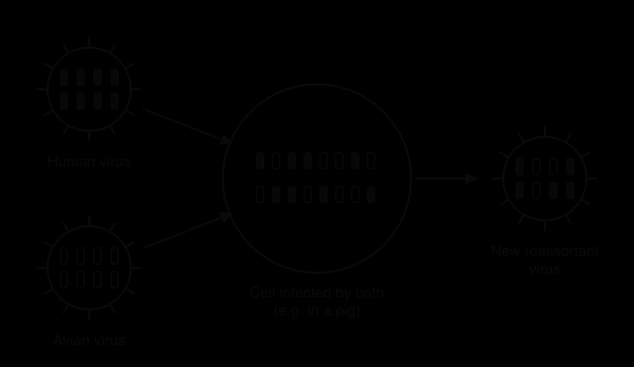
The 2009 H1N1 pandemic virus is an example of reassortment. Its eight segments came from influenza viruses of pigs, birds and humans that had mixed in pigs over several years before the virus spread to people.
Feature | Antigenic drift | Antigenic shift |
|---|---|---|
Cause | Point mutations in H and N genes | Reassortment of segments from two different strains |
Speed | Gradual, every season | Sudden, rare |
Size of change in antigens | Small | Large: new H and/or N |
Consequence | Seasonal epidemics; vaccine updated each year | Possible pandemic |
HIV: drug resistance and combination therapy
HIV shows all the features of fast viral evolution. An infected person who is not being treated produces roughly 10 billion (10¹⁰) new virions every day. Reverse transcriptase makes many errors and does not proofread, so the virus in one person becomes a swarm of slightly different variants within months.
When the first anti-HIV drug, zidovudine (AZT), was used alone in the late 1980s, resistant variants appeared within months in many patients: a mutation giving resistance was already present in some virions, and the drug killed the others. The solution, introduced from 1996, is antiretroviral therapy using a combination of usually three drugs that act on different viral enzymes, such as reverse transcriptase, integrase and protease. A variant would need resistance mutations against all three drugs at once, which is extremely unlikely, so the virus is held at very low levels for decades. Combination therapy must be taken every day for life, because it does not remove the viral DNA integrated into the chromosomes of long-lived host cells.
Rapid evolution has also prevented an effective HIV vaccine so far. The envelope glycoproteins vary greatly between strains and change within each patient, and the parts of the virus that do not vary are hidden from antibodies.
SARS-CoV-2 showed the same process at a global scale. The Omicron variant, designated a variant of concern by the World Health Organization in November 2021, carried more than 30 changes in its spike protein compared with the original virus, which allowed it to infect many people who were already immune.
Exam practice
Viruses are an HL-only subtopic, assessed in Paper 1A multiple-choice questions on virus structure and the order of the lytic cycle, in Paper 1B or Paper 2 Section A data questions (one-step growth curves, influenza strain data, drug resistance graphs), and in Paper 2 Section B extended responses on the lytic and lysogenic cycles or on the evolution of viruses and its consequences for medicine. Common command terms are outline, compare and contrast, explain and evaluate.
Diagrams can earn credit in extended responses if they are clearly labelled, but each step must still be described in words.
The surface proteins that vary so quickly in influenza, HIV and SARS-CoV-2 are the same molecules that antibodies and vaccines target, which links viral evolution directly to immunity.
Self-check
- To do:
I can list the structural features common to all viruses and explain why viruses are obligate intracellular parasites.
- To do:
I can describe the diversity of viral genomes, capsids and envelopes, using lambda, coronaviruses and HIV as examples.
- To do:
I can explain how a retrovirus uses reverse transcriptase.
- To do:
I can outline the five stages of the lytic cycle of bacteriophage lambda and interpret a one-step growth curve.
- To do:
I can outline the lysogenic cycle, including integration, the prophage and induction.
- To do:
I can compare and contrast the lytic and lysogenic cycles.
- To do:
I can evaluate the evidence that viruses have several origins and outline the progressive and regressive hypotheses.
- To do:
I can explain why viruses evolve rapidly and the consequences for treating influenza and HIV.
- To do:
I can distinguish antigenic drift from antigenic shift.