
Use microscopes to study cells, calculate magnification, compare prokaryote, animal, fungal and plant cells, and (HL) explain the endosymbiotic origin of eukaryotes.
Each is in the Biology glossary, with every other term in the subject.
How close a measurement or the mean of several measurements is to the true value of the quantity being measured.
The division of a prokaryote cell into two genetically identical daughter cells after its circular DNA has been copied; mitochondria and chloroplasts divide in a similar way.
The theory that all living organisms are composed of one or more cells, that the cell is the smallest unit of life, and that cells arise only from pre-existing cells.
A strong, freely permeable layer outside the plasma membrane that gives shape and support and stops the cell bursting; made of peptidoglycan in bacteria, cellulose in plants and chitin in fungi.
A short cylinder of microtubules found in pairs near the nucleus of animal cells, which organises microtubules and forms the base of cilia and flagella.
A tough nitrogen-containing polysaccharide that forms the cell walls of fungi and the exoskeletons of arthropods.
A double-membrane organelle of plants and algae in which photosynthesis takes place; it contains thylakoids stacked into grana in a fluid stroma, with 70S ribosomes and circular DNA.
A DNA molecule carrying genes; in eukaryotes chromosomes are linear and bound to histone proteins, while the main prokaryote chromosome is a naked circular loop.
A short hair-like projection from the surface of a eukaryote cell, with a core of microtubules, that beats to move the cell or to move fluid over the cell surface; cilia are usually many and short.
A phloem cell with a nucleus and many mitochondria that lies beside a sieve tube element, is connected to it by plasmodesmata and keeps it alive.
A vacuole in freshwater protists such as Paramecium that collects excess water entering by osmosis and expels it from the cell.
One of the folds of the inner mitochondrial membrane that project into the matrix and increase the surface area for the reactions that produce ATP.
Electron microscopy of samples frozen so rapidly that water forms glass-like ice, with many images combined by computer to give three-dimensional structures of proteins at near-atomic resolution.
A photosynthetic bacterium that releases oxygen; chloroplasts are thought to have evolved from a cyanobacterium taken in by endosymbiosis.
Everything inside the plasma membrane except the nucleus: a watery fluid (cytosol) containing dissolved substances, enzymes, ribosomes and, in eukaryotes, organelles.
A network of protein filaments (microtubules and microfilaments) in the cytoplasm of eukaryote cells that maintains shape, holds organelles in place and moves structures.
Reasoning from a general statement or theory to a specific prediction, such as predicting that a newly discovered organism will be made of cells.
A microscope that uses a beam of electrons focused by electromagnets to form an image; its very short wavelength gives a resolution below 1 nm, but specimens must be dead and in a vacuum.
The process by which a prokaryote cell taken into a larger cell survived and became a permanent part of it; mitochondria and chloroplasts are thought to have originated in this way.
An organism whose cells have a nucleus enclosed in a double membrane and membrane-bound organelles; animals, plants, fungi and protists are eukaryotes.
The removal from an organism of waste products of its metabolism, such as carbon dioxide, urea or excess water; the removal of undigested food (egestion) is not excretion.
A transparent scale with arbitrary divisions placed in the eyepiece of a microscope; once calibrated with a stage micrometer it is used to measure specimens.
The circular area of a specimen that is visible through the eyepiece at one time; its diameter decreases as the magnification increases.
A long whip-like projection from a cell that moves it through liquid; eukaryote flagella have a microtubule core and are usually one or two per cell, while prokaryote flagella are simpler protein fibres.
A dye that absorbs light of one wavelength and emits light of a longer wavelength, so that the structures it binds to glow against a dark background in a fluorescence microscope.
An electron microscopy technique in which a specimen is frozen rapidly and split so that the fracture runs through the centre of membranes, showing the proteins embedded in them.
The processes carried out by all living organisms: metabolism, nutrition, growth, response to stimuli, movement, excretion, homeostasis and reproduction.
A stack of flattened, curved membrane sacs (cisternae) that receives proteins from the rough ER in vesicles, modifies and sorts them, and sends them out in new vesicles.
A bacterium with a thick cell wall of peptidoglycan and no outer membrane, which stains purple with the Gram stain; Bacillus and Staphylococcus are examples.
A stack of thylakoids in a chloroplast; grana are joined to each other by unstacked thylakoid membranes called intergranal lamellae.
A basic protein around which eukaryote DNA is wound to package it into chromosomes; prokaryote DNA is not associated with histones.
One of the thin, branching threads that make up the body of most fungi; aseptate hyphae have no cross walls, so many nuclei share one continuous cytoplasm.
A tentative, testable explanation of an observation, from which predictions can be made and checked by experiment or further observation.
A light microscopy technique in which antibodies carrying a fluorescent marker bind to one specific protein, showing where that protein is located in a cell or tissue.
Reasoning from many specific observations to a general statement or theory, such as concluding that all organisms are made of cells after observing cells in many different organisms.
A microscope that uses visible light focused by glass lenses to form a magnified image; its resolution is limited to about 0.2 µm, but it can be used to view living cells in colour.
A small membrane-bound vesicle in animal cells containing hydrolytic (digestive) enzymes, which break down food, worn-out organelles and engulfed bacteria.
The number of times larger an image is than the real object; magnification = image size ÷ actual size, with both sizes in the same units.
A thin solid filament made of the protein actin, part of the cytoskeleton; microfilaments change cell shape and are involved in cell movement and division of the cytoplasm.
A photograph or digital image taken through a microscope; an electron micrograph is taken with an electron microscope and a light micrograph with a light microscope.
A hollow cylinder made of the protein tubulin, part of the cytoskeleton; microtubules form the spindle in cell division and the core of cilia and flagella.
A tiny finger-like fold of the plasma membrane on the surface of some animal cells, such as those lining the small intestine, that increases the surface area for absorption.
The fluid inside the inner membrane of a mitochondrion, containing enzymes of the Krebs cycle and link reaction, 70S ribosomes and naked circular DNA.
A double-membrane organelle in which aerobic cell respiration produces ATP; the inner membrane is folded into cristae around a fluid matrix, which holds 70S ribosomes and circular DNA.
The condition of an organism made of many cells that stay together, communicate and are specialised for different functions; it has evolved independently many times.
The double membrane that surrounds the nucleus of a eukaryote cell, perforated by nuclear pores.
A protein-lined opening through both membranes of the nuclear envelope that controls movement of molecules such as mRNA and proteins between the nucleus and cytoplasm.
The region of a prokaryote cell that contains its main DNA molecule, a naked, circular loop not enclosed by a membrane.
A dense region inside the nucleus of a eukaryote cell where ribosomal RNA is made and ribosome subunits are assembled.
The organelle of eukaryote cells that contains the chromosomes, enclosed by a double membrane with pores; it controls the cell through the genes that are transcribed.
The polymer of sugars cross-linked by short chains of amino acids that forms the cell walls of bacteria; it is thick in Gram-positive bacteria.
The partially permeable membrane of phospholipids and proteins that surrounds the cytoplasm of every cell and controls what enters and leaves; also called the cell membrane.
A small, circular DNA molecule in many prokaryotes, separate from the main loop of DNA; plasmids carry a few genes, such as antibiotic resistance, and are not essential.
How close repeated measurements of the same quantity are to each other; precise measurements show little spread, though they may still be inaccurate.
An organism whose cells have no nucleus and no membrane-bound organelles; its DNA is a naked loop in the cytoplasm. Bacteria and archaea are prokaryotes.
Observations described in words rather than measured, for example the colour, shape or texture of cells seen with a microscope.
Observations recorded as numbers with units, obtained by measuring or counting, for example the length of a cell in micrometres.
A biconcave blood cell packed with haemoglobin that carries oxygen; in mammals the mature cell has no nucleus and no mitochondria.
The smallest distance between two points at which they can still be seen as separate points; it determines how much detail a microscope can show.
A small structure of RNA and protein, not bounded by a membrane, that synthesises polypeptides; prokaryotes, mitochondria and chloroplasts have 70S ribosomes, while the eukaryote cytoplasm has larger 80S ribosomes.
A network of flattened membrane sacs (cisternae) with 80S ribosomes attached to the outer surface, which synthesise proteins that are then transported in vesicles, often for secretion.
A line drawn on a micrograph or drawing and labelled with the actual length it represents, so that sizes can be worked out whatever the image is enlarged to.
A living phloem cell with no nucleus and very little cytoplasm, joined end to end with others through perforated sieve plates to form a sieve tube that transports sap.
A network of membrane tubules without ribosomes that synthesises lipids such as phospholipids and steroids and, in liver cells, detoxifies drugs.
A coloured chemical that binds to particular cell structures and increases contrast, such as iodine for starch in plant cells or methylene blue for nuclei in animal cells.
A skeletal muscle cell: a very long, cylindrical, multinucleate fibre formed by the fusion of many cells, with a striped (striated) appearance due to its regularly arranged proteins.
The fluid inside the inner membrane of a chloroplast, surrounding the thylakoids, where the enzymes of the Calvin cycle are found.
A specimen placed in a drop of liquid (water or stain) on a glass slide and covered with a cover slip, for viewing with a light microscope in a single session.
In science, a well-tested explanation of a wide range of observations that is supported by a large body of evidence and is used to make predictions; it is more than a guess.
A flattened membrane sac inside a chloroplast that holds chlorophyll and is the site of the light-dependent reactions of photosynthesis.
A membrane-bound sac in the cytoplasm; plant cells usually have one large permanent sap vacuole, bounded by the tonoplast, which stores solutes and keeps the cell turgid.
A small sac of membrane that transports or stores substances inside a cell; secretory vesicles carry proteins to the plasma membrane for release by exocytosis.
Cells are the units from which every living organism is built. Knowing what cells contain, and how their structures are seen and measured with microscopes, is the basis for almost every other part of biology, from enzymes and membranes to disease and inheritance.
All cells share a few basic structures, which shows the unity of life, but they also differ widely, from bacteria a few micrometres long to plant cells with walls and chloroplasts. The two broad types, prokaryote and eukaryote, are distinguished by whether the DNA is enclosed in a nucleus.
39 exam-style questions on this subtopic, written in the style of the IB papers. The first has its markscheme open. Counted on
Evolution has been occurring over an immense length of time.
Discuss the evidence that the last universal common ancestor (LUCA) evolved in the vicinity of hydrothermal vents.
7 marks
a. hydrothermal vents release hydrogen and other reduced compounds, a source of chemical energy;
b. they are rich in iron and sulfur minerals that can catalyse reactions forming carbon compounds;
c. deep vents were shielded from intense UV radiation on the early Earth;
d. fossilized remains of life / microfossils have been found in ancient seafloor vent precipitates;
e. among the oldest evidence of life on Earth;
f. genomic analysis: conserved genes shared by bacteria and archaea, probably present in LUCA;
g. include genes for using H2 / fixing CO2 and for proteins with iron–sulfur clusters;
h. no genes for photosynthesis or using oxygen, consistent with a dark, anaerobic habitat;
i. reconstructed ancient proteins are stable at high temperatures, suggesting a hot environment;
j. (against) fossil filaments could be formed by non-biological processes / rocks are altered by heat and pressure;
k. (against) high temperatures make RNA and other polymers unstable;
l. (against) other sites, e.g. shallow pools on land, have also been proposed;
m. (against) exact early-Earth conditions cannot be replicated and the first cells did not fossilize, so the hypothesis is hard to test;
[7 max]
For full marks, answers must include at least one point against the hypothesis.
The official deck for this part of the syllabus.
What is a cell?
Is a single ribosome or a single DNA molecule alive? Explain your answer.
With a free account, Augment remembers where you stopped and what you have practiced.
Every living organism, from a single bacterium to a blue whale, is made of cells. A cell is a small compartment of living material surrounded by a membrane, and it is the smallest unit that can carry out all the processes of life on its own. Structures smaller than a cell, such as a single ribosome or a molecule of DNA, are not alive by themselves.
The cell theory brings these ideas together in three statements:
All living organisms are composed of one or more cells.
Cells are the smallest units of life.
Cells arise only from pre-existing cells, by cell division.
The third statement means that new cells cannot appear from non-living material today. Every cell in a human body descends, by repeated division, from a single fertilised egg, and every fertilised egg descends from cells of the parents. Cells therefore form an unbroken line of descent going back billions of years, which is a central idea of continuity and change in biology.
In everyday speech a "theory" is often a guess or a hunch ("my theory is that the bus is late because of the rain"). In science a theory is something much stronger: a broad explanation that accounts for a large number of observations, has been tested many times and has not been contradicted by the evidence. Theories allow scientists to make predictions. A hypothesis is narrower and more tentative: a proposed explanation for one observation that can be tested.
The cell theory was reached by inductive reasoning, which means generalising from many specific observations. Robert Hooke saw and named "cells" in thin slices of cork in 1665. In the nineteenth century Matthias Schleiden (1838) concluded that all plant tissues are made of cells, Theodor Schwann (1839) extended the conclusion to animals, and Rudolf Virchow (1855) argued that every cell comes from another cell. No single observation proved the theory; it became accepted because thousands of observations of different organisms all fitted it.
Once a theory is accepted it can be used for deductive reasoning, which runs in the opposite direction: from a general statement to a specific prediction. If all organisms are made of cells, then a newly discovered organism from a deep-sea vent should also be made of cells. Each time a prediction like this is confirmed, confidence in the theory grows. A well-supported counter-example would force the theory to be revised, which is how scientific knowledge changes.
Some structures test the edges of the cell theory. Skeletal muscle fibres and some fungal hyphae are not divided into separate cells, and viruses are not cells at all. These cases (atypical eukaryote cells, and viruses in A2.3) show that a theory can remain useful even when some structures are hard to fit into it.
Most cells are between about 1 µm and 100 µm across, far too small to see with the naked eye, which cannot separate details closer than about 0.1 mm. Cell biology therefore depends on microscopes. Two units are used for the sizes of cells and their parts: the micrometre (1 µm = 0.001 mm) and the nanometre (1 nm = 0.001 µm).
Unit | Symbol | In metres | Typical use |
|---|---|---|---|
millimetre | mm | 10⁻³ m | Large cells such as a human egg (0.1 mm), small organisms |
micrometre | µm | 10⁻⁶ m | Cells (bacteria about 2 µm, red blood cells about 8 µm) and large organelles |
nanometre | nm | 10⁻⁹ m | Membranes (about 7–10 nm thick), ribosomes, viruses, molecules |
Moving one row down the table multiplies the number by 1,000: 1 mm = 1,000 µm and 1 µm = 1,000 nm. Mixing up these conversions is the most frequent source of error in microscope calculations.
A light microscope (also called a compound microscope) uses two sets of glass lenses. The objective lens, close to the specimen, forms a magnified image, and the eyepiece lens magnifies that image again. Light from a lamp passes up through the condenser, which focuses it onto the specimen, and the iris diaphragm controls how much light reaches it.
The total magnification of a light microscope is the magnification of the eyepiece multiplied by the magnification of the objective lens in use:
A typical school microscope has a ×10 eyepiece and objectives of ×4, ×10 and ×40, giving total magnifications of ×40, ×100 and ×400.
A temporary mount is a thin specimen in a drop of liquid on a glass slide, covered with a cover slip. It is used for viewing cells with a light microscope during one session. The specimen must be thin, ideally one cell layer thick, so that light can pass through it.
Place a drop of water or stain in the centre of a clean slide.
Add a thin specimen: for example, a piece of the epidermis peeled from the inner surface of an onion bulb leaf with forceps, or cells gently scraped from the inside of the cheek with a clean cotton bud.
Lower a cover slip onto the drop at an angle, using a mounted needle, so that air bubbles are pushed out instead of trapped.
Remove excess liquid with a paper towel at the edge of the cover slip.
Most cell structures are colourless and transparent, so a stain is added to increase contrast. Each stain binds to particular molecules: iodine solution stains starch blue-black and makes plant cell walls and nuclei easier to see, and methylene blue stains the nuclei of animal cells, such as cheek cells, dark blue. A stain can be drawn under a cover slip that is already in place by adding a drop at one edge and drawing it through with paper towel held at the opposite edge.
Focusing always starts with the lowest-power objective, because it gives the largest field of view and the most space between the lens and the slide.
With the ×4 objective in place, look from the side and use the coarse focus knob to bring the objective close to the slide without touching it.
Looking through the eyepiece, turn the coarse focus knob to move the lens and slide apart until the specimen comes into focus.
Use the fine focus knob to make the image sharp, then move the slide so that the part of interest is in the centre of the field of view.
Turn the nosepiece to a higher-power objective and use only the fine focus knob. With high-power objectives the lens is very close to the slide, and the coarse focus knob could push it through the cover slip.
Magnification is the number of times larger an image is than the actual object. The magnification, the size of the image and the actual size of the object are linked by one equation:
Rearranging gives the other two forms: actual size = image size ÷ magnification, and image size = actual size × magnification. Image size and actual size must be in the same units before dividing. Image sizes are usually measured with a ruler in millimetres, so they are converted to micrometres (× 1,000) when the actual size is in micrometres.
A scale bar is a line on a micrograph or drawing labelled with the actual length it represents. Unlike a stated magnification, a scale bar stays correct when the image is enlarged or reduced, because the bar changes size with the image. To find the actual size of a structure, measure both the structure and the scale bar on the image in the same units, then use the ratio:
An eyepiece graticule is a small transparent scale, usually numbered 0–100, fixed inside the eyepiece. It is seen on top of the specimen, so it can be used to measure cells. Its divisions are arbitrary: the length of specimen covered by one division depends on which objective lens is in use. The graticule must therefore be calibrated for each objective using a stage micrometer, a slide engraved with a scale of known length (often 1 mm divided into 100 divisions of 0.01 mm, or 10 µm).

The field of view is the circular area of the specimen visible through the eyepiece. Increasing the magnification shows a smaller area in more detail: if the magnification increases by a factor of four, the diameter of the field of view decreases by a factor of four. A quick way to estimate cell size is to measure the diameter of the field of view at low power (for example by placing a transparent ruler on the stage) and count how many cells fit across it.
A photograph taken through a microscope, a micrograph, is a permanent record of what was seen. Many microscopes now have a built-in digital camera, and a phone camera held over the eyepiece also works. A micrograph is only useful for measurement if it carries a scale bar or a stated magnification.
Biological drawings follow strict conventions so that they are clear and accurate:
Use a sharp pencil and single, continuous lines; no shading, colouring or sketchy lines.
Draw what is actually seen, in correct proportion, not a textbook diagram.
Draw label lines with a ruler; they must not cross and must end exactly on the structure. Keep labels outside the drawing, ideally in a column.
Give a title, and state the magnification or add a scale bar.
Observations made with a microscope are of two kinds. Quantitative data are numbers with units, obtained by measuring or counting, such as a cell length of 70 µm or 12 dividing cells in a field of view. Qualitative data are descriptions in words, such as "the nucleus stains dark blue" or "the cells are roughly rectangular". Both are useful: qualitative observations often suggest a question, and quantitative data allow it to be tested.
Measurements can be judged by their accuracy, how close they are to the true value, and their precision, how close repeated measurements are to each other. An uncalibrated graticule can give very precise readings that are all inaccurate, because each reading is wrong by the same factor.
Making an image larger does not always show more detail. Above a certain magnification a light microscope image just becomes blurred, like a digital photograph enlarged until it shows its pixels. The limit is set by resolution, the smallest distance at which two separate points can still be seen as two.
Resolution depends on the wavelength of the radiation used to form the image: the shorter the wavelength, the better (smaller) the resolution. Visible light has wavelengths of roughly 400–700 nm, which limits a light microscope to a resolution of about 0.2 µm (200 nm). Mitochondria (about 1 µm long) are just visible, but ribosomes (about 25 nm) and the structure of membranes are not.

An electron microscope uses a beam of electrons instead of light, focused by electromagnets instead of glass lenses. Electrons have a far shorter wavelength than light, so electron microscopes resolve details below 1 nm and can produce useful magnifications of several hundred thousand times. The invention of the electron microscope in the 1930s allowed biologists to see the internal membranes of cells, ribosomes and viruses for the first time, a clear case of new technology leading to new discoveries.
Transmission electron microscopes (TEM) pass electrons through a very thin section of a specimen stained with heavy metals. They give two-dimensional images of the inside of cells and organelles.
Scanning electron microscopes (SEM) scan electrons across the surface of a specimen coated with a thin layer of metal. They give three-dimensional images of surfaces, such as the shape of pollen grains or the surface of a cell.
The electron beam must travel in a vacuum, so specimens must be dead and dehydrated. Electron micrographs are black and white; colours in published images have been added afterwards (false colour). Electron microscopes are also large and expensive, and preparing specimens can create artefacts, structures that were not present in the living cell.
Feature | Light microscope | Electron microscope |
|---|---|---|
Radiation used | Visible light | Beam of electrons |
Lenses | Glass | Electromagnets |
Resolution | About 0.2 µm | Below 1 nm |
Useful magnification | Up to about ×1,500 | Several hundred thousand times |
Specimens | Living or dead | Dead only (in a vacuum) |
Image | Natural colour or stains | Black and white (false colour may be added) |
Preparation and cost | Quick and cheap | Complex, slow and expensive |
Freeze-fracture is a way of preparing specimens for electron microscopy. A sample is frozen very rapidly and then struck with a blade, and the fracture tends to split membranes down the middle, between the two layers of phospholipids. A thin replica of the exposed surface is made with metal and carbon and viewed in the electron microscope. Freeze-fracture images show the inside of membranes covered with particles, which are proteins that span the membrane. This was important evidence for the fluid mosaic model of membranes (B2.1).
In cryogenic electron microscopy (cryo-EM), a solution of protein molecules is frozen so quickly that the water turns into a glass-like solid instead of ice crystals, so the molecules keep their natural shape. Thousands of images of individual molecules lying at different angles are taken with an electron microscope, and computers combine them into a three-dimensional model, often at near-atomic resolution. Cryo-EM can show proteins that are hard to crystallise, such as large membrane proteins, and even different shapes of the same protein as it works. Its developers, Jacques Dubochet, Joachim Frank and Richard Henderson, received the 2017 Nobel Prize in Chemistry.
Fluorescence has made light microscopy much more powerful. A fluorescent stain absorbs light of one colour (often ultraviolet or blue) and emits light of a longer wavelength, so the structures it binds to glow against a dark background. Some fluorescent stains bind to DNA, for example, and make every nucleus in a tissue stand out.
Immunofluorescence adds specificity. Antibodies that bind to one particular protein, such as tubulin, are linked to a fluorescent marker. The antibodies attach only where that protein is, so the position of the protein in the cell lights up; different proteins can be shown at the same time in different colours. A related method uses green fluorescent protein (GFP), first found in the jellyfish Aequorea victoria. The gene for GFP can be joined to the gene for a protein of interest, so that living cells make the protein with a glowing tag attached. Osamu Shimomura, Martin Chalfie and Roger Tsien shared the 2008 Nobel Prize in Chemistry for this work.
Cells vary enormously in size, shape and function, but every cell, prokaryote or eukaryote, has the same four basic components:
DNA as the genetic material. All cells store their genetic information in DNA, using the same genetic code.
Cytoplasm. The cytoplasm is everything inside the plasma membrane apart from the nucleus. It is mainly water, in which ions, sugars, amino acids and many other substances are dissolved, so that the chemical reactions of the cell can take place in solution.
A plasma membrane. The plasma membrane (also called the cell membrane) is a thin barrier made mainly of lipids, with proteins embedded in it. It separates the cytoplasm from the surroundings and controls what enters and leaves.
Ribosomes and enzymes. Ribosomes make proteins, and enzymes catalyse the reactions of metabolism.
These shared features are evidence for unity in living things. They are most simply explained by common ancestry: all cells alive today are thought to descend from one ancestral population of cells that already had DNA, ribosomes and a lipid membrane (A2.1).
A prokaryote is an organism whose cell has no nucleus. The name means "before the nucleus". Bacteria and archaea are prokaryotes. Their cells are small, usually 1–5 µm long, and their internal structure is simple: there are no membrane-bound organelles, so the cytoplasm is a single compartment.
The standard example of prokaryote structure is a Gram-positive eubacterium such as Bacillus subtilis, a rod-shaped soil bacterium, or Staphylococcus aureus, a spherical bacterium that grows in clusters on human skin. A Gram-positive bacterium has a thick cell wall of peptidoglycan and no outer membrane; it stains purple with the Gram stain, a test used to identify bacteria in hospital laboratories.

The structures of a Gram-positive bacterium are:
Cell wall. A thick cell wall of peptidoglycan (a polymer of sugars cross-linked by short chains of amino acids) lies outside the plasma membrane. It is freely permeable but very strong, so it maintains the shape of the cell and stops it bursting when water enters by osmosis.
Plasma membrane. A phospholipid bilayer with proteins, controlling entry and exit of substances.
Cytoplasm. A single compartment containing enzymes, ribosomes and the DNA.
Naked DNA in a loop. The main chromosome is one circular DNA molecule, not associated with histone proteins ("naked"), found in an area called the nucleoid. Many bacteria also contain plasmids, small extra loops of DNA carrying a few genes, such as genes for antibiotic resistance.
70S ribosomes. Smaller than eukaryote cytoplasmic ribosomes (80S). The "S" is a Svedberg unit, a measure of how fast a particle sinks in a centrifuge, which depends on its size.
Prokaryote structure varies. Some bacteria have flagella for swimming, pili for attaching to surfaces, or a slimy capsule outside the wall, and some groups, such as the mycoplasmas and the phytoplasmas that infect plants, have no cell wall at all.
 YouTube
A eukaryote ("true nucleus") is an organism whose cells have a nucleus surrounded by a double membrane. Animals, plants, fungi and protists such as Paramecium are eukaryotes. Eukaryote cells are usually much larger than prokaryote cells, typically 10–100 µm across.
The key feature of eukaryote cells is compartmentalisation: the cytoplasm is divided by membranes into separate compartments called organelles, each with its own conditions and enzymes. The features shared by all eukaryote cells are:
A plasma membrane enclosing cytoplasm that is divided into compartments, with 80S ribosomes.
A nucleus enclosed by a nuclear envelope, a double membrane with nuclear pores. Inside are chromosomes, linear DNA molecules wound around proteins called histones.
Membrane-bound organelles: mitochondria, endoplasmic reticulum, the Golgi apparatus and a variety of vesicles and vacuoles, including lysosomes.
A cytoskeleton of microtubules and microfilaments, protein fibres that maintain the shape of the cell and move organelles and vesicles.

The structures in the diagram of an animal cell are described in the table. B2.2 Organelles and compartmentalisation explains how each structure is adapted to its function and how the organelles work together.
Structure | Appearance in electron micrographs | Function |
|---|---|---|
Nucleus | Large, round, with a double membrane (nuclear envelope) crossed by pores; darker patches of chromatin and a dense nucleolus | Holds the chromosomes; site of DNA replication and transcription; the nucleolus makes ribosome subunits |
Mitochondrion | Oval, 1–3 µm long, double membrane; inner membrane folded into cristae around the matrix | Aerobic cell respiration, producing ATP |
Rough endoplasmic reticulum (rER) | Flattened membrane sacs (cisternae) studded with ribosomes | Synthesis of proteins, mainly for secretion or for membranes |
Smooth endoplasmic reticulum (sER) | Network of membrane tubules without ribosomes | Synthesis of lipids such as phospholipids and steroid hormones |
Golgi apparatus | Stack of curved, flattened sacs with vesicles at the edges | Modifies, sorts and packages proteins from the rER into vesicles |
Vesicles | Small membrane-bound spheres | Transport materials; secretory vesicles release proteins by exocytosis |
Lysosomes | Small vesicles with dense contents | Contain digestive enzymes that break down food, worn-out organelles and bacteria |
Free ribosomes (80S) | Small dark granules in the cytoplasm, about 25 nm | Synthesise proteins that are used inside the cell |
Centrioles | Pair of short cylinders at right angles near the nucleus | Organise microtubules; form the bases of cilia and flagella |
Microvilli | Finger-like folds of the plasma membrane | Increase surface area for absorption (e.g. in the small intestine) |
The diagram of a plant cell shows the same nucleus, mitochondria, ER and Golgi apparatus as an animal cell, together with a cellulose cell wall, chloroplasts for photosynthesis and a large permanent vacuole filled with cell sap.

Apart from the four structures common to all cells, prokaryote and eukaryote cells differ in almost every feature. The comparison table sets the differences out row by row.
Feature | Prokaryote cell | Eukaryote cell |
|---|---|---|
Typical size | 1–5 µm | 10–100 µm |
Nucleus | Absent; DNA in the nucleoid | Present, with a double membrane and pores |
DNA | One circular molecule, naked (no histones); plasmids often present | Several linear chromosomes bound to histones; no plasmids in most cells |
Ribosomes | 70S | 80S in the cytoplasm (70S in mitochondria and chloroplasts) |
Membrane-bound organelles | Absent | Present (mitochondria, ER, Golgi, lysosomes, chloroplasts in plants) |
Cytoskeleton | Simple protein filaments only | Microtubules and microfilaments |
Cell wall | Usually present, made of peptidoglycan | Cellulose in plants, chitin in fungi, absent in animals |
Cell division | Binary fission | Mitosis (and meiosis for sexual reproduction) |
The generalisation that prokaryote cells are tiny and have no internal membranes was built from observing thousands of species. In 2009 the microbiologist Olivier Gros found long white filaments growing on sunken mangrove leaves in shallow water in Guadeloupe, in the Caribbean. They turned out to be single bacterial cells, described in 2022 as Thiomargarita magnifica. Each cell is a single tube that averages more than 9 mm long and can reach about 2 cm, the size of a human eyelash.

A typical bacterium such as E. coli is about 2 µm long, so an average T. magnifica cell is about 9,000 µm ÷ 2 µm = 4,500 times longer. Most of the volume of the cell is a single large central vacuole, which pushes the cytoplasm into a thin layer around the edge. Its DNA is also unusual. Instead of one naked loop in a nucleoid, the cell holds many copies of its genome (more than half a million) packaged with ribosomes inside small membrane-bound compartments that the researchers called "pepins".
Feature | Typical bacterium (E. coli) | Thiomargarita magnifica |
|---|---|---|
Cell length | About 2 µm | Average more than 9 mm; up to about 2 cm |
Visible to the naked eye | No | Yes |
DNA | One naked loop in the nucleoid | More than half a million genome copies inside membrane-bound "pepins" |
Membranes inside the cell | None | Pepin membranes and a large central vacuole |
Source: Volland et al., Science 376: 1453–1458, 2022.
The discovery shows how a single well-studied example can test a generalisation reached by inductive reasoning. T. magnifica is still a prokaryote: it is a bacterium, and its DNA is not in a nucleus with a double membrane and pores. It does show, though, that statements such as "prokaryotes have no internal membranes" describe most prokaryotes rather than all of them. It also raises a question about size. Most cells are microscopic because substances must diffuse across the plasma membrane and through the cytoplasm, and this becomes too slow in a large cell (B2.3 explains the role of the surface area to volume ratio). The huge vacuole of T. magnifica keeps its living cytoplasm thin, so no part of it is far from the surface.
A unicellular organism is a single cell that must carry out every function of life by itself. Biologists usually list eight functions of life:
Function | Meaning |
|---|---|
Metabolism | All the chemical reactions in the organism, catalysed by enzymes |
Nutrition | Obtaining food: energy and the materials needed for growth |
Growth | An increase in size or dry mass |
Response to stimuli | Detecting changes in the environment and reacting to them |
Movement | Changing position or moving parts of the body |
Excretion | Removing waste products of metabolism |
Homeostasis | Keeping internal conditions within limits |
Reproduction | Producing offspring, sexually or asexually |
Two freshwater protists show how one cell performs all eight functions in different ways: Paramecium, a heterotroph, and Chlamydomonas, an autotroph.
Paramecium (for example Paramecium caudatum) is a slipper-shaped protist, about 0.2–0.3 mm long, found in ponds. Its surface is covered with thousands of short cilia that beat in coordinated waves.

Nutrition: cilia sweep bacteria and other small organisms down the oral groove to the cytostome, where they are enclosed in food vacuoles by endocytosis.
Metabolism: lysosomes fuse with the food vacuoles and their enzymes digest the food; the products are absorbed into the cytoplasm and used in cell respiration and to build new molecules.
Growth: the cell uses the absorbed materials to increase in size until it is large enough to divide.
Response: if the cell bumps into an obstacle or meets a harmful chemical, the cilia reverse their beat, the cell backs away, turns and swims off in a new direction.
Movement: beating of the cilia drives the cell through the water.
Excretion: carbon dioxide and ammonia from metabolism diffuse out across the plasma membrane.
Homeostasis: pond water is more dilute than the cytoplasm, so water constantly enters by osmosis. Two contractile vacuoles collect this water and expel it, keeping the water content of the cell constant (D2.3).
Reproduction: usually asexual, by dividing in two (mitosis of the micronucleus followed by division of the cell); two individuals can also exchange genetic material by conjugation.
Chlamydomonas (for example Chlamydomonas reinhardtii) is a unicellular green alga, about 10 µm long, that lives in fresh water and damp soil. It has a cell wall, one large cup-shaped chloroplast and two flagella.

Nutrition: autotrophic. The chloroplast carries out photosynthesis, making sugars from carbon dioxide and water using light energy; starch is stored around the pyrenoid. In the dark some species can also absorb organic molecules such as acetate.
Metabolism: enzymes catalyse photosynthesis, cell respiration and the synthesis of all the molecules the cell needs.
Growth: the cell grows using the sugars it makes and mineral ions absorbed from the water.
Response: the eyespot detects light, and the cell swims towards moderate light and away from very intense light (phototaxis).
Movement: the two flagella beat with a breaststroke-like action, pulling the cell forwards.
Excretion: oxygen from photosynthesis and carbon dioxide from respiration diffuse out of the cell.
Homeostasis: contractile vacuoles near the base of the flagella expel excess water.
Reproduction: asexual, by mitosis producing several daughter cells inside the parent cell wall; under stress such as a shortage of nitrogen, sexual reproduction occurs by fusion of gametes.
Animals, fungi and plants are all eukaryotes, but their cells differ in ways that match how each group lives. Fungi were once grouped with plants because many are not mobile and have cell walls, but their cells differ from plant cells in several important ways.
Feature | Animal cells | Fungal cells | Plant cells |
|---|---|---|---|
Cell wall | Absent | Present, made of chitin | Present, made of cellulose |
Vacuoles | Small and usually temporary (e.g. food vacuoles, vesicles) | Often large and permanent | Usually one large permanent sap vacuole, bounded by the tonoplast |
Chloroplasts and other plastids | Absent | Absent | Chloroplasts in green tissues; other plastids such as amyloplasts (starch storage) and chromoplasts (pigments) |
Centrioles | Present | Absent in most fungi | Absent in flowering plants and conifers |
Cilia and flagella | Present in some cells (e.g. sperm, airway epithelium) | Absent in most fungi; flagellate spores in chytrids | Absent in flowering plants; swimming sperm of mosses and ferns have flagella |
Carbohydrate store | Glycogen | Glycogen | Starch |
Nutrition | Heterotrophic (ingestion) | Heterotrophic (secrete enzymes and absorb) | Autotrophic (photosynthesis) |
The large sap vacuole of plant cells stores water, ions, sugars and pigments, and the pressure of the cell sap pushing against the cell wall keeps the cell turgid, which supports non-woody plants. Animal cells, lacking a wall, rely instead on the cytoskeleton for their shape and can change shape, as phagocytes do when they engulf bacteria.
The typical eukaryote cell has one nucleus. Several important cell types are exceptions, and each exception is linked to its function.
Aseptate fungal hyphae. The body of many fungi is a network of thin threads called hyphae. In some fungi, such as the bread mould Rhizopus, the hyphae have no cross walls (septa), so a single continuous mass of cytoplasm with hundreds of nuclei fills each tube. Substances can move freely along the hypha as it grows.
Skeletal muscle fibres. A striated muscle fibre forms when many embryonic muscle cells fuse end to end. The result is a very long cylinder, often several centimetres, with many nuclei just under its plasma membrane (the sarcolemma). The fibre can contract along its whole length as one unit.

Mammalian red blood cells. A red blood cell loses its nucleus (and its mitochondria) as it matures in the bone marrow. This leaves more room for haemoglobin, so each cell can carry more oxygen, and allows the cell to be a flexible biconcave disc that squeezes through capillaries. Without a nucleus the cell cannot make new proteins or repair itself, so it lives only about 120 days.
Phloem sieve tube elements. A sieve tube element is a living plant cell that loses its nucleus and most of its organelles as it develops, leaving a clear channel for the flow of sap. The end walls become sieve plates with large pores. Each sieve tube element depends on a companion cell beside it, which has a nucleus and many mitochondria and is connected through plasmodesmata. The companion cell carries out the metabolism needed to keep the sieve tube element alive.

Light and electron micrographs can be used to decide whether a cell is a prokaryote, a plant cell or an animal cell, by looking for a few key structures.
Cell type | Features that identify it |
|---|---|
Prokaryote | Small (1–5 µm); no nucleus, only a lighter nucleoid region; no membrane-bound organelles; cell wall present; cytoplasm full of small ribosomes |
Plant cell | Cell wall outside the plasma membrane; large central sap vacuole; chloroplasts (in green tissue); nucleus often pushed to the side; often regular, box-like shape |
Animal cell | No cell wall; no large central vacuole; no chloroplasts; nucleus usually central; irregular or rounded outline; may show microvilli |
In electron micrographs the structures to recognise are the nucleoid region and cell wall of prokaryotes, and in eukaryotes the nucleus, mitochondria, chloroplasts, sap vacuole, Golgi apparatus, rough and smooth ER, chromosomes, ribosomes, cell wall, plasma membrane and microvilli. Chromosomes are visible as dense, separate threads only in cells that are dividing; in non-dividing cells the DNA is spread out as darker and lighter patches of chromatin in the nucleus.
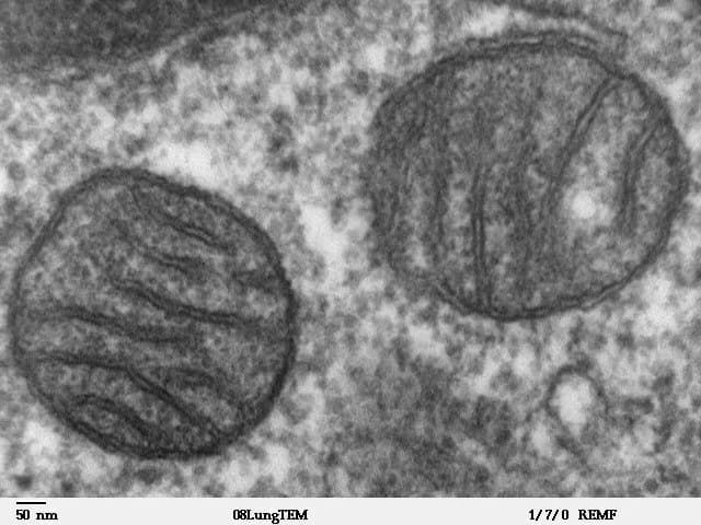
Drawings of organelles from electron micrographs follow the same conventions as all biological drawings, with one addition: annotations. A label names a structure; an annotation is a short note beside the label that states its function or a feature. The drawings of a mitochondrion and a chloroplast show the structures to include.

The chloroplast is drawn in the same way. Its internal membranes are the thylakoids, flattened sacs that are stacked into grana and lie in a fluid called the stroma.

Annotations for the structures most often drawn:
Structure | Annotation (function) |
|---|---|
Nucleus | Double membrane with pores allows mRNA out; contains chromosomes that carry the genes |
Mitochondrion | Cristae give a large area for the membrane enzymes of aerobic respiration that produce ATP |
Chloroplast | Thylakoids in grana absorb light; stroma holds the enzymes that fix carbon dioxide |
Sap vacuole | Stores water, ions and sugars; pressure of sap against the wall keeps the cell turgid |
Golgi apparatus | Modifies proteins from the rER and packages them into vesicles |
Rough ER | Ribosomes on its surface synthesise proteins that enter the cisternae for transport |
Smooth ER | Synthesises lipids, including phospholipids and steroids |
Chromosomes | Condensed DNA and histones, visible during division, carrying genes to daughter cells |
Cell wall | Resists the pressure of water entering by osmosis, preventing bursting |
Plasma membrane | Partially permeable; controls entry and exit of substances |
Secretory vesicles | Carry proteins to the plasma membrane and release them by exocytosis |
Cilia | Many short beating projections with a microtubule core; move fluid or the cell |
Flagella | One or a few long whip-like projections; propel the cell |
All eukaryotes share a long list of complex features: a nucleus with a double membrane and pores, linear chromosomes with histones, 80S ribosomes, a cytoskeleton of microtubules, mitosis and meiosis, and sexual reproduction. Such complex features are very unlikely to have evolved more than once, so all eukaryotes are thought to descend from a single unicellular common ancestor that already had a nucleus and reproduced sexually.
The host cell in which eukaryote features arose was probably related to archaea. Genome sequencing of deep-sea sediments has found a group called the Asgard archaea, whose genes include several previously thought to be found only in eukaryotes, such as genes for cytoskeleton proteins.
Endosymbiosis is the process in which one cell lives permanently inside another and becomes part of it. The endosymbiotic theory states that mitochondria and chloroplasts evolved from free-living prokaryotes that were taken in by a larger cell and not digested:
An anaerobic host cell took in an aerobic bacterium, a proteobacterium, by endocytosis.
The bacterium was not digested but survived and divided inside the host. It used oxygen to release much more energy from food than the host could, and supplied the host with ATP, while the host supplied food and protection, so the relationship benefited both partners (mutualism).
Over many generations the bacterium lost genes it no longer needed, many of its genes moved to the host nucleus, and it became a mitochondrion. All eukaryotes descend from this ancestor, so almost all eukaryotes have mitochondria.
Later, in one lineage, a cell that already had mitochondria took in a photosynthetic cyanobacterium, which became the chloroplast. The descendants of this cell include the red and green algae and the land plants.

Mitochondria and chloroplasts still have many features of bacteria, which are hard to explain unless they descend from bacteria:
Evidence | Explanation |
|---|---|
Double membrane | The inner membrane is the original bacterial plasma membrane; the outer membrane comes from the host's membrane when the bacterium was taken in by endocytosis |
Own DNA: naked and circular | Like a bacterial chromosome, and unlike the linear chromosomes with histones in the nucleus |
70S ribosomes | The same size as bacterial ribosomes, not the 80S ribosomes of the eukaryote cytoplasm |
Division by binary fission | New mitochondria and chloroplasts form only by division of existing ones, in a process like bacterial binary fission; a cell cannot make them from scratch |
Similar size | Mitochondria (1–3 µm) and chloroplasts (2–10 µm) are the size of bacteria |
Antibiotic sensitivity | Antibiotics that block bacterial ribosomes, such as chloramphenicol, also block protein synthesis in mitochondria and chloroplasts, but have little effect on 80S ribosomes |
Gene sequences | Mitochondrial genes are most similar to those of proteobacteria; chloroplast genes are most similar to those of cyanobacteria |
Endosymbiosis giving rise to a new organelle has also been observed in a modern lineage. In 2024 researchers showed that a nitrogen-fixing cyanobacterium living inside the marine alga Braarudosphaera bigelowii divides in step with the host cell and imports proteins made by the host. It now counts as an organelle, named the nitroplast, and gives the alga the ability to fix nitrogen, which no eukaryote was previously known to do.
 YouTube
A multicellular organism such as a human starts as a single cell, the zygote, and becomes a body of about 200 different cell types, including neurons, muscle fibres and red blood cells. The process by which unspecialised cells become specialised is differentiation. Almost every cell in the body contains the same genome, all the genes of the organism. Cells differ because they use, or express, different sets of those genes.
Gene expression means a gene being transcribed and translated to make its protein. In a pancreatic β-cell the gene for insulin is expressed, while in a skin cell it is present but switched off. The proteins a cell makes determine its structure and function, so the pattern of gene expression determines the cell type.
Changes in gene expression are often triggered by the cell's environment. Cells in an embryo receive chemical signals from neighbouring cells, and the concentration of a signal depends on how far the cell is from its source. A cell's position in the embryo therefore helps decide which genes it switches on and so which cell type it becomes (B2.3 describes stem cells and these signals in more detail).
Differentiated cells are organised into tissues (groups of similar cells with the same function), tissues into organs, and organs into organ systems. This is a clear example of the levels of organisation in biology, from molecules to cells to whole organisms.
Multicellularity means being made of many cells that stay together, communicate and divide the work between them. Multicellular organisms have evolved independently from unicellular ancestors many times, including in animals, land plants (from green algae), fungi, red algae and brown algae such as kelps.
Multicellularity brings several advantages:
Larger body size. A larger organism can avoid being eaten by small predators, can eat larger prey and can reach light or resources beyond the reach of single cells.
Cell specialisation. Different cells can become specialised for different functions (division of labour), each doing one job efficiently, such as nerve cells for signalling and muscle cells for contraction.
Longer life. Damaged or worn-out cells can be replaced while the organism survives.
Multicellularity can evolve quickly when it is favoured by natural selection. In a laboratory experiment published in 2012, researchers grew the unicellular yeast Saccharomyces cerevisiae in tubes and each day kept only the cells that settled to the bottom fastest. Within about 60 days, multicellular "snowflake" clusters had evolved in which daughter cells stay attached to the parent after division. The clusters settled faster because they were heavier, and some cells in each cluster died in a way that let the cluster split into smaller clusters, a simple form of reproduction by a multicellular body.
Cell structure is assessed in every paper. Paper 1A multiple-choice questions ask candidates to identify structures in micrographs, compare prokaryote and eukaryote cells and calculate magnification. Paper 1B and Paper 2 Section A use micrographs and data to ask for calculations of actual size, magnification or scale bar length, and drawings of organelles. Paper 2 Section B extended responses may ask candidates to compare cell types, explain the functions of life in a unicellular organism or, at HL, evaluate the evidence for endosymbiosis. Command terms include calculate, identify, draw, label, annotate, distinguish, compare and contrast, outline and explain.
In drawing questions, credit depends on accuracy and clarity: lines must be continuous, labels ruled, and only structures that are visible in the micrograph should be drawn. When asked to annotate, add a function to each label.
I can state the three parts of the cell theory and explain how it was developed by inductive reasoning.
I can explain the difference between a theory and a hypothesis, and between qualitative and quantitative data.
I can prepare a temporary mount, stain it and focus a light microscope correctly.
I can calculate magnification, actual size, scale bar length and field of view, and calibrate an eyepiece graticule.
I can distinguish magnification from resolution, compare light and electron microscopes, and outline freeze-fracture, cryo-EM and immunofluorescence.
I can draw and label a prokaryote cell and list the structures common to all cells.
I can describe the features of eukaryote cells and compare animal, fungal and plant cells.
I can explain how Paramecium and Chlamydomonas carry out the functions of life.
I can describe atypical cells: aseptate hyphae, skeletal muscle fibres, red blood cells and sieve tube elements.
I can identify cells and organelles in micrographs and draw and annotate organelles.
I can explain the endosymbiotic theory and evaluate the evidence for it. (HL)
I can explain differentiation in terms of gene expression and outline why multicellularity has evolved repeatedly. (HL)
Outline approaches used to estimate the dates of the first living cells and of LUCA.
4 marks
a. fossils of early cells / stromatolites found in ancient rocks;
b. rocks dated by radiometric dating / decay of long-lived radioisotopes;
c. ratios of carbon-12 to carbon-13 in ancient carbon indicate carbon fixed by organisms;
d. molecular clock: number of differences in base / amino acid sequences of shared genes;
e. assuming a constant rate of change;
f. calibrated using fossil / geological dates;
g. estimates suggest life appeared more than 3.5 billion years ago / within about 1 billion years of Earth forming;
[4 max]
Describe the evidence that mitochondria originated by endosymbiosis.
4 marks
a. mitochondria have 70S ribosomes like prokaryotes (not 80S);
b. mitochondria have their own naked / circular DNA;
c. mitochondria divide by binary fission / replicate independently of the cell;
d. mitochondria have a double membrane, consistent with engulfment;
e. mitochondria are similar in size to bacteria;
f. new mitochondria arise only from pre-existing mitochondria;
g. mitochondrial DNA / ribosomal RNA sequences resemble those of certain bacteria;
[4 max]
One additional mark is available for the construction of the answer.
The boundary between living and non-living things is not always clear.
Outline the processes of life carried out by unicellular organisms.
4 marks
Outline the roles of mRNA, ribosomes and tRNA when a viral gene is translated inside a host cell.
4 marks
Discuss whether viruses should be considered living organisms.
7 marks
Carbohydrates perform a wide range of functions in living organisms.
Outline how glucose is produced in photosynthesis.
4 marks
Describe the differences between the cells of plants, animals and fungi.
4 marks
Explain how the structures of different carbohydrates are related to their functions.
7 marks