The part of the immune system that responds specifically to particular pathogens, using lymphocytes and antibodies, and builds up a memory so that later responses are faster and stronger.
The clumping together of cells or pathogens when antibodies bind to antigens on their surfaces, for example when mismatched red blood cells are transfused.
Acquired immunodeficiency syndrome: the condition that develops when HIV has destroyed so many helper T-lymphocytes that the body can no longer fight opportunistic infections.
The ability of a strain of bacteria to survive and reproduce in the presence of an antibiotic that would normally kill it; it evolves by natural selection.
A molecule, usually a protein or glycoprotein on the surface of a pathogen or cell, that is recognised by the immune system and stimulates the production of specific antibodies.
A lymphocyte that, when activated by its specific antigen and a helper T-lymphocyte, divides to form plasma cells that secrete one type of antibody, and memory cells.
Notes on C3.2 Defence against disease
43 min readLast reviewed
The human body is constantly exposed to microorganisms, and a few of them cause disease. The body defends itself in layers: barriers that keep most pathogens out, white blood cells that destroy any that get in, and a system that recognises each pathogen specifically and responds faster when the same pathogen returns.
Understanding these defences explains how vaccines work, why antibiotics cure bacterial infections but not colds, and how diseases spread through populations. Defence against disease is an example of interaction at every level of organisation: between molecules (antigens and antibodies), between cells (lymphocytes signalling to each other), between organisms (a host and its pathogen) and between populations (the spread of an epidemic).
Questions on C3.2
44 exam-style questions on this subtopic, written in the style of the IB papers. The first has its markscheme open. Counted on
Question 1 · BIO-002458 marks
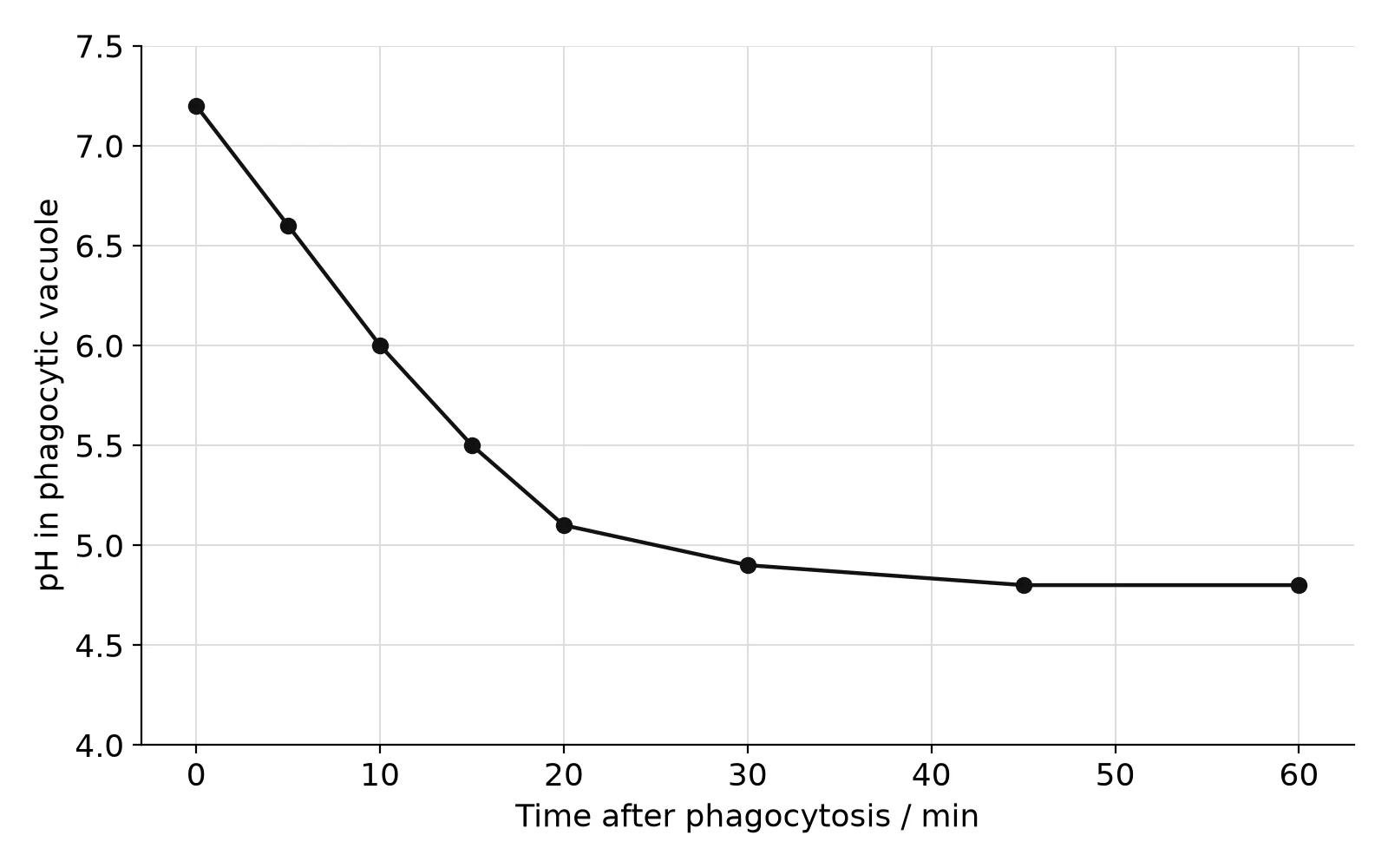
Mouse macrophages were allowed to engulf bacteria by phagocytosis. The pH inside the phagocytic vacuoles was measured using a pH-sensitive fluorescent dye bound to the bacteria.
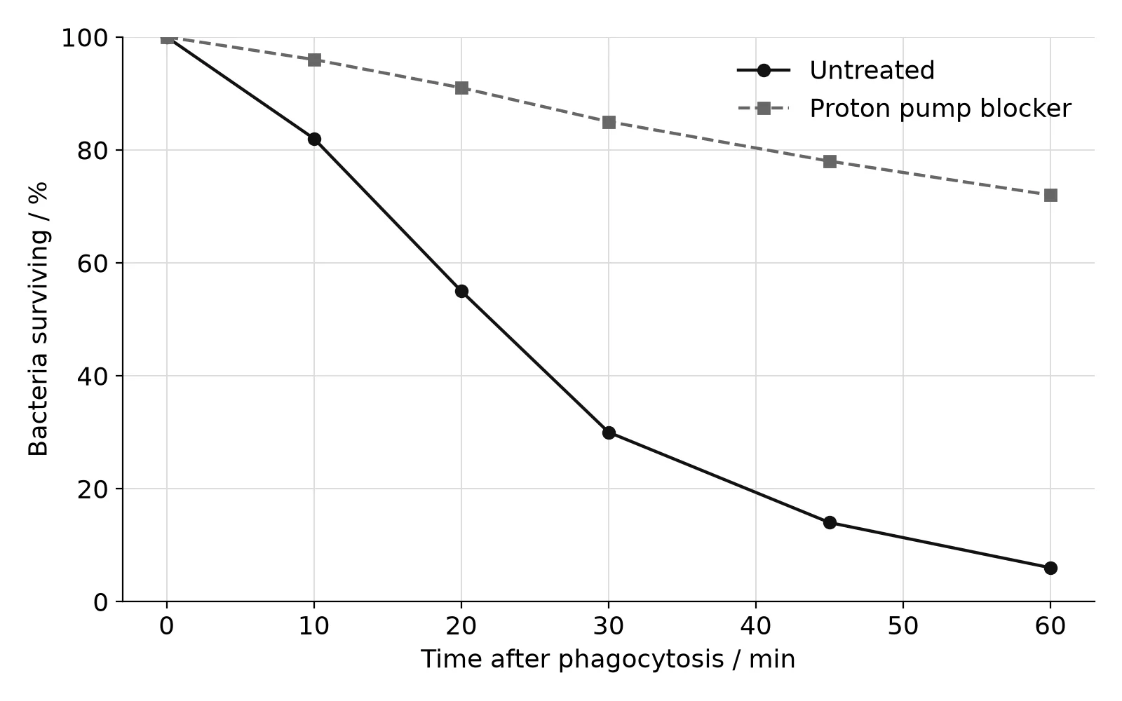
In a second experiment, the percentage of engulfed bacteria still alive was measured in untreated macrophages and in macrophages treated with a drug that blocks the proton pumps in lysosome and vacuole membranes. In treated macrophages the pH in the vacuoles stayed above 6.5.
(a)
State the pH in the phagocytic vacuoles 15 minutes after phagocytosis.
[1]1 mark
Markscheme (a)
5.5;
(b)
Calculate the difference between the percentage of bacteria surviving in untreated and treated macrophages at 60 minutes.
[1]1 mark
Markscheme (b)
Flashcards for C3.2
The official deck for this part of the syllabus.
C3.2 Defence against disease · front
What is an infectious disease?
C3.2 Defence against disease · front
Give four examples of infectious diseases.
C3.2 Defence against disease254 cards
Carry on with C3.2 in Augment
With a free account, Augment remembers where you stopped and what you have practiced.
The average number of people infected by one infected person in a population where everyone is susceptible (R₀); the herd immunity threshold is 1 − 1/R₀.
The activation of only those B-lymphocytes whose antibodies match a particular antigen, followed by their division by mitosis to form a large clone of identical cells.
The number of deaths from all causes during a period above the number expected from previous years; it estimates the total effect of an event such as a pandemic.
A lymphocyte that is activated by antigen presented by a macrophage and then activates B-lymphocytes that recognise the same antigen; it is the cell infected by HIV.
The protection of a whole population, including non-immune individuals, from an infectious disease when a large enough proportion of the population is immune that transmission is greatly reduced.
The part of the immune system that responds in the same, non-specific way to broad categories of pathogen and does not change during an organism's life.
A type of white blood cell with a large nucleus that is part of the adaptive immune system; B-lymphocytes produce antibodies and T-lymphocytes help control the response.
Short-term protection from antibodies made by another organism, such as antibodies passed from mother to baby or given by injection; no memory cells are formed.
The first response of the adaptive immune system to an antigen; it is slow to start and produces a relatively small amount of antibody, but forms memory cells.
A preparation containing antigens of a pathogen, or nucleic acid coding for them, that stimulates immunity to that pathogen without causing the disease.
An infectious disease that can be transmitted from other animal species to humans.
Pathogens as the cause of infectious diseases
An infectious disease is a disease caused by a pathogen that can be passed from one host to another. Measles, tuberculosis, malaria and COVID-19 are infectious diseases. Diseases such as cancer, type 2 diabetes and coronary heart disease are not infectious, because they are not caused by a pathogen and cannot be caught from another person.
A wide range of organisms can infect humans. Most pathogens belong to four groups:
Type of pathogen
Features
Examples of human diseases
Viruses
Not cells; a protein capsid around DNA or RNA; can only reproduce inside a host cell
Influenza, measles, HIV/AIDS, COVID-19, rabies
Bacteria
Prokaryotic cells with a cell wall and 70S ribosomes
Some animals, such as parasitic worms, also cause disease. Archaea are absent from the table, because no species of archaea is known to cause disease in humans, even though archaea live in the human gut. Many pathogens are also species-specific: they infect one host species, or a few, because they can only enter cells that have particular receptor molecules on their surface.
A virus is not a living cell, but it is included as a pathogen because it causes disease. A virus has no metabolism of its own. It enters a host cell and uses the cell's ribosomes, enzymes and ATP to make copies of itself, often destroying the cell as new virus particles are released.
Careful observation and the germ theory of disease
Until the late nineteenth century, many doctors believed that diseases were caused by "miasma", bad air from rotting matter, or by an imbalance in the body. The germ theory, which states that many diseases are caused by microorganisms that pass from person to person, was established by the work of Louis Pasteur and Robert Koch in the 1860s–1880s. Before that, careful observations of epidemics had already shown that disease spread in ways that could be controlled.
In the 1840s Ignaz Semmelweis worked in the maternity hospital in Vienna, which had two clinics. In the First Clinic, babies were delivered by doctors and medical students, who often came straight from dissecting dead bodies. In the Second Clinic, babies were delivered by midwives, who did not do dissections. Many more mothers in the First Clinic died of childbed fever (puerperal fever), an infection after childbirth. Semmelweis concluded that "cadaverous particles" were carried on the doctors' hands, and from May 1847 he required everyone to wash their hands in a solution of chlorinated lime before examining patients.
Year
First Clinic (doctors): births
First Clinic: deaths
First Clinic: mortality / %
Second Clinic (midwives): mortality / %
1844
3,157
260
8.2
2.3
1845
3,492
241
6.9
2.0
1846
4,010
459
11.4
2.8
1847 (handwashing from May)
3,490
176
5.0
1.0
1848
3,556
45
1.3
1.3
Source: Semmelweis, The Etiology, Concept and Prophylaxis of Childbed Fever, 1861.
The data on maternal deaths in the two clinics show that before handwashing the mortality in the First Clinic was several times higher than in the Second Clinic, and that by 1848 the two clinics had the same low mortality. Semmelweis had no microscope evidence of a pathogen, and many doctors rejected his conclusion, partly because it implied that they were causing their patients' deaths. His work was accepted only after the germ theory was established.
A second example comes from London in 1854. During a severe outbreak of cholera in the Soho district, the physician John Snow plotted the homes of the people who died on a street map. The deaths were clustered around a public water pump in Broad Street. Snow persuaded the local council to remove the pump handle, and the outbreak, which was already declining, came to an end. Snow's map is one of the first uses of epidemiology, the study of the patterns of disease in populations, and it showed that cholera was spread by contaminated water, not by bad air.
Skin and mucous membranes as a primary defence
The first line of defence against pathogens is to stop them entering the body. This primary defence is provided by the skin and by the mucous membranes that line the openings of the body.
The skin as a physical and chemical barrier
The outer layer of the skin is made of several layers of dead cells packed with keratin, a tough fibrous protein. New cells are produced continuously below and pushed outwards, so the surface is constantly shed and replaced. This tough, dry layer forms a physical barrier that most pathogens cannot cross unless the skin is cut, burnt or bitten.
The skin is also a chemical barrier. Sebaceous glands, attached to hair follicles, secrete an oily substance called sebum, which keeps the skin and hair supple and contains fatty acids that inhibit the growth of bacteria and fungi. Sebum and sweat make the surface of the skin slightly acidic, at about pH 5, which slows the growth of many pathogens. The skin is also covered with harmless bacteria and fungi, which compete with pathogens for space and nutrients.
Mucous membranes
The airways, the gut, the urinary and reproductive tracts and the inside of the eyelids are lined by mucous membranes. These are thin layers of living cells, so they are much easier for pathogens to cross than the skin. Their defences are chemical and mechanical:
Mucus is a sticky solution of glycoproteins secreted by goblet cells. It traps bacteria, viruses and dust particles.
Cilia on the cells lining the trachea and bronchi beat in waves and sweep mucus with its trapped particles up to the throat, where it is swallowed. Stomach acid (about pH 2) then kills most of the pathogens.
Lysozyme, an enzyme in tears, saliva and mucus, digests the cell walls of many bacteria.
Sealing cuts in skin by blood clotting
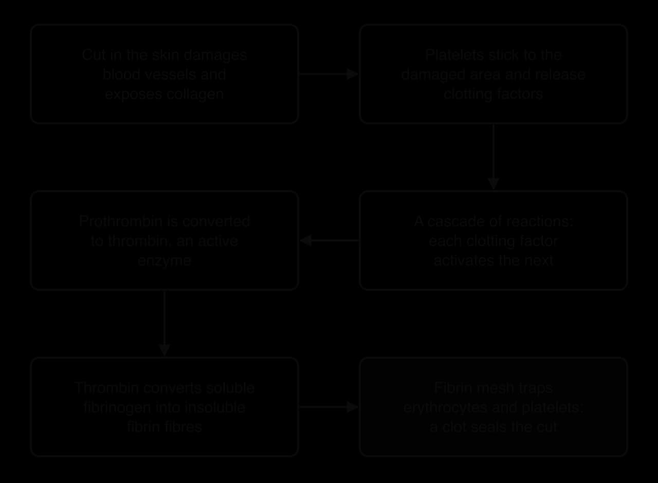
When the skin is cut, blood vessels are broken, blood is lost and pathogens can enter. Blood clotting seals the wound quickly. A clot stops further bleeding and blocks the entry of pathogens until new skin has grown over the wound.
Clotting depends on platelets. Platelets are small fragments of cells, without a nucleus, that circulate in the blood. When a vessel is damaged, the collagen fibres in its wall are exposed. Platelets stick to the collagen and to each other, forming a temporary plug, and they release chemicals called clotting factors. The damaged tissue releases clotting factors too.
The clotting factors set off a cascade: a series of reactions in which each activated factor activates the next one. Because each enzyme molecule can activate many molecules of the next factor, the signal is amplified at every step, and a large response follows a small trigger within seconds. Calcium ions and vitamin K are needed for several of the steps.
The cascade amplifies the signal from damaged tissue and platelets, so that thrombin is produced rapidly and converts fibrinogen to fibrin.
The last steps of the cascade are the most important:
The cascade converts an inactive plasma protein, prothrombin, into its active form, thrombin. Thrombin is an enzyme.
Thrombin converts fibrinogen, a soluble protein dissolved in the plasma, into fibrin, an insoluble protein that forms long fibres.
The fibrin fibres form a mesh across the wound. Erythrocytes (red blood cells) and platelets are trapped in the mesh, and the mass forms a clot.
The clot dries and hardens into a scab, which protects the wound while the skin repairs itself underneath.
Clotting factors circulate in inactive forms and are activated only at a wound, because a clot inside an unbroken vessel can block blood flow. A clot that forms inside a blood vessel is a thrombosis; in a coronary artery it can cause a heart attack (B3.2). People with haemophilia lack one of the clotting factors, so their blood clots very slowly.
The innate and adaptive immune systems
Pathogens that get past the primary defences are attacked by the immune system. Its cells are the white blood cells (leucocytes), which are made in the bone marrow. The immune system has two parts that work together: the innate immune system and the adaptive immune system.
Feature
Innate immune system
Adaptive immune system
Specificity
Non-specific: responds to broad categories of pathogen (for example, any bacterium)
Specific: responds to one particular pathogen, recognised by its antigens
Change during life
Does not change; responds in the same way every time
Builds up a memory of each pathogen, so the response becomes faster and stronger
Speed
Acts within minutes to hours
Primary response takes days to develop
Main cells
Phagocytes (such as neutrophils and macrophages)
Lymphocytes (B-lymphocytes and T-lymphocytes)
Inherited or acquired
Inherited and present from birth
Develops after exposure to each pathogen
The innate immune system is present in all animals and is the first to act. Its main cells are phagocytes, which engulf and digest pathogens. The adaptive immune system is found only in vertebrates. It recognises each pathogen by the molecules on its surface and produces antibodies that match them exactly, and it remembers pathogens it has met before.
Infection control by phagocytes
A phagocyte is a white blood cell that ingests pathogens by phagocytosis. The two main kinds are neutrophils, the most common white blood cells, which have a lobed nucleus and arrive first at an infection, and macrophages, larger cells that live in the tissues and also present antigens to lymphocytes.
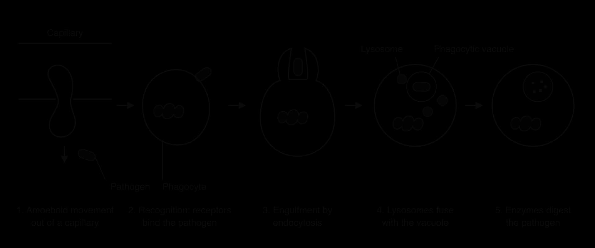
Phagocytes control infection in a sequence of steps:
Movement to the site of infection. Damaged cells and pathogens release chemicals that attract phagocytes. Phagocytes squeeze out of the blood through the walls of capillaries by amoeboid movement, changing their shape and extending parts of the cytoplasm (pseudopodia), and move through the tissue towards the infection.
Recognition. Receptors on the plasma membrane of the phagocyte bind to molecules found on the surface of pathogens but not on body cells, such as components of bacterial cell walls.
Engulfment. The phagocyte surrounds the pathogen with pseudopodia, and the plasma membrane fuses around it. The pathogen is taken into the cell by endocytosis, enclosed in a vesicle called a phagocytic vacuole.
Digestion. Lysosomes fuse with the phagocytic vacuole and release hydrolytic enzymes into it. The enzymes digest the pathogen, and the useful products are absorbed into the cytoplasm.
Phagocytosis is endocytosis of a whole pathogen. The phagocytic vacuole and lysosomes keep the digestive enzymes away from the rest of the cell.
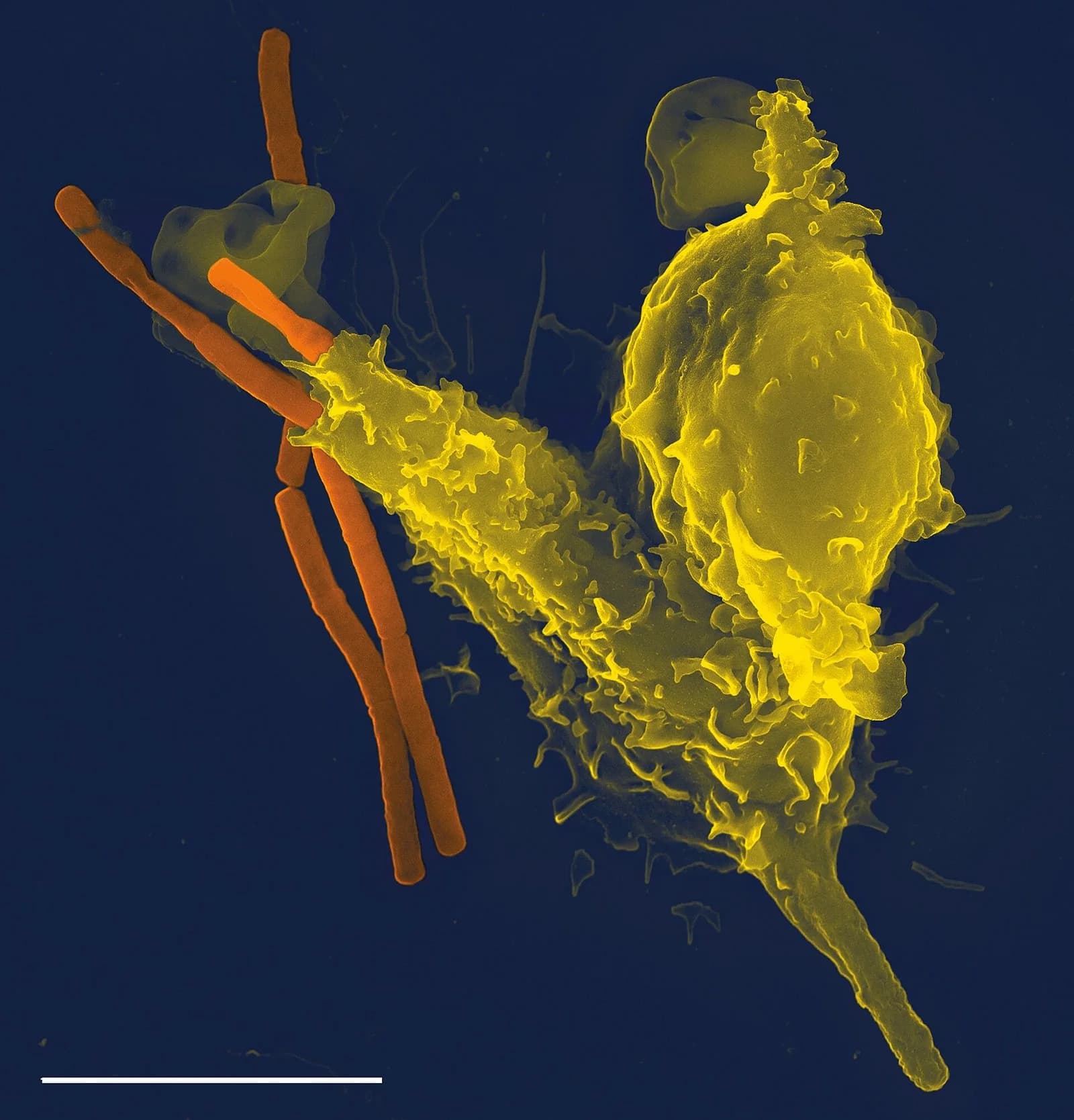
The scanning electron micrograph of a neutrophil shows a phagocyte in the act of engulfing bacteria. Its extended surface wraps around the rod-shaped cells before they are taken in.
A neutrophil engulfing rod-shaped anthrax bacteria (Bacillus anthracis). Neutrophils are about 12 µm across. Photo: Volker Brinkmann, CC BY 2.5, via Wikimedia Commons.
At a site of infection, phagocytes also release chemicals that cause inflammation: nearby capillaries widen and become leakier, so more blood, fluid and phagocytes reach the area. The pus that forms in an infected wound is mainly dead neutrophils and the remains of digested bacteria.
Lymphocytes and the production of antibodies
The adaptive immune response is carried out by lymphocytes, white blood cells with a large round nucleus and little cytoplasm. Lymphocytes circulate in the blood and lymph, and large numbers of them are held in the lymph nodes, small organs along the lymphatic vessels that filter the lymph. Lymph nodes in the neck and armpits swell during an infection because lymphocytes there are dividing rapidly.
There are two main types of lymphocyte. B-lymphocytes (B-cells) produce antibodies. T-lymphocytes (T-cells) include helper T-lymphocytes, which activate B-cells and coordinate the response. The two types cooperate: in most infections, B-cells only produce antibodies after they receive a signal from helper T-cells.
Antibodies
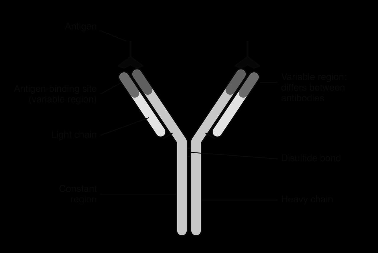
An antibody is a protein (an immunoglobulin) that binds specifically to one antigen. Each antibody molecule is Y-shaped and made of four polypeptide chains: two long heavy chains and two shorter light chains, held together by disulfide bonds. The tips of the two arms are the variable regions, which form two identical antigen-binding sites. The amino acid sequence of the variable region is different in each type of antibody, which gives each binding site a unique shape and chemical properties that match one antigen. The rest of the molecule, the constant region, is the same in all antibodies of one class.
Each antibody has two identical antigen-binding sites at the tips of its arms. Their shape is set by the variable regions, which differ between antibodies.
Antibodies help to destroy pathogens in several ways. Because each antibody has two binding sites, it can bind two pathogens at once, so antibodies clump pathogens together (agglutination), which makes them easier for phagocytes to engulf. Antibodies also mark pathogens for phagocytosis, block the sites that viruses use to enter cells, neutralise toxins and activate proteins in the plasma that make holes in bacterial membranes.
One B-cell, one antibody
Each B-lymphocyte produces just one type of antibody. During its development in the bone marrow, the genes for the variable region are cut and rejoined in a random way, so each B-cell ends up with a different combination. A copy of its antibody is placed in its plasma membrane, where it acts as a receptor. An individual has an immense number of B-cells, each making a different antibody, and together they can bind almost any antigen. For any particular antigen, though, only a very small number of B-cells have a matching antibody before an infection begins.
Antigens as recognition molecules
An antigen is a molecule that is recognised by the immune system and triggers the production of antibodies. Most antigens are proteins or glycoproteins, and many are located on the outer surface of pathogens: in the capsid or envelope of a virus, the cell wall or flagella of a bacterium, or the plasma membrane of a protist. Toxins released by bacteria can also act as antigens.
The body's own cells also carry antigens, but the immune system normally does not respond to them. During development, lymphocytes that would bind to the body's own molecules are destroyed, so the immune system distinguishes "self" from "non-self".
Antigens on red blood cells and blood transfusion
Antigens on the surface of red blood cells determine a person's blood group. In the ABO blood group system, red blood cells may carry antigen A, antigen B, both or neither, and the plasma contains antibodies against whichever of A and B the person does not have.
Blood group
Antigens on red blood cells
Antibodies in plasma
Can safely receive red cells from
A
A
Anti-B
A, O
B
B
Anti-A
B, O
AB
A and B
Neither
A, B, AB, O
O
Neither
Anti-A and anti-B
O only
If a person is given a transfusion of red cells with an antigen they do not have, the antibodies in their plasma bind to the transfused cells. The antibodies cause agglutination, so the red cells clump together and block small blood vessels, and the cells are then destroyed (haemolysis). This transfusion reaction can be fatal, which is why blood is always matched before a transfusion. Type O blood has no A or B antigens, so its red cells can be given to people of any ABO group in an emergency.
Activation of B-lymphocytes by helper T-lymphocytes
A B-cell does not produce large amounts of antibody until it is activated. Activation happens in three steps, and the B-cell itself needs two signals, its specific antigen and an activated helper T-cell, so that antibodies are only made when a genuine pathogen is present:
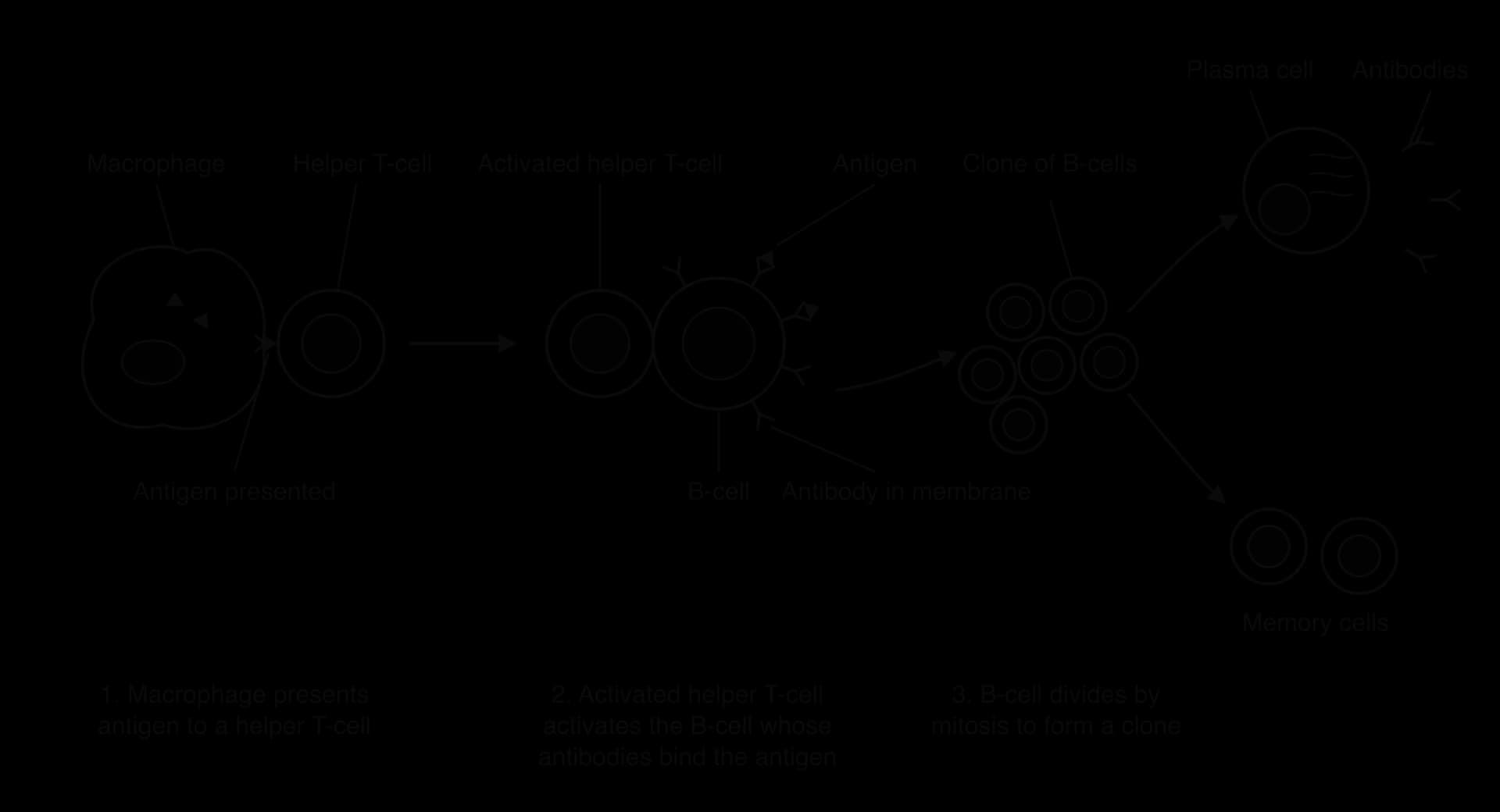
Antigen presentation to helper T-cells. A macrophage engulfs a pathogen and digests it. Fragments of the pathogen's antigens are then displayed on the macrophage's plasma membrane. A helper T-cell with a receptor that matches the antigen binds to it and is activated.
Direct contact with the antigen. A B-cell whose membrane-bound antibody matches the same antigen binds to it, takes it in and displays fragments on its surface.
Contact with an activated helper T-cell. The activated helper T-cell binds to the B-cell displaying the same antigen and releases signalling molecules (cytokines) that activate the B-cell.
Only activated B-lymphocytes divide to produce plasma cells, which secrete antibodies, or memory cells. The requirement for help from a T-cell activated by the same antigen is a safety check that makes it unlikely that antibodies are produced against the body's own molecules.
The adaptive response: helper T-cells activated by a macrophage activate the matching B-cell, which divides to form plasma cells and memory cells.
Clones of antibody-secreting plasma cells
Before an infection there are relatively few B-cells able to make any one antibody, far too few to fight a pathogen that is multiplying rapidly. Once a B-cell has been activated, it divides repeatedly by mitosis. All the cells produced are genetically identical, so they form a clone, and all of them make the same antibody. The process in which a pathogen's antigen selects the matching B-cells, which are then multiplied, is called clonal selection.
Most cells of the clone develop into plasma cells. A plasma cell is a B-cell specialised for producing antibody: it has an extensive rough endoplasmic reticulum and a large Golgi apparatus, and it can secrete thousands of antibody molecules per second. Plasma cells live for only a few days, but together they produce the large amounts of antibody that control the infection. The rest of the clone become memory cells.
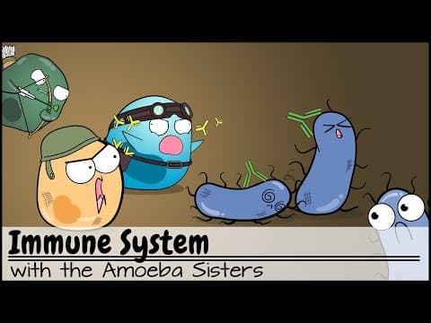
How the innate and adaptive immune systems work together, from phagocytes to B-cells, T-cells, antibodies and memory cells, is summarised in the video.
YouTube
Overview of the immune system: innate and adaptive defences, phagocytes, B-cells and T-cells, antibodies and memory cells.
Immunity and memory cells
Immunity is the ability of the body to eliminate an infectious disease, usually before any symptoms develop, because it has met the pathogen before. Immunity depends on memory cells: long-lived B-lymphocytes (and T-lymphocytes) produced during the first response, which stay in the body for years or decades and can make the specific antibody.
Primary and secondary immune responses
The first time a pathogen enters the body, the primary immune response is slow. It takes several days to find and activate the few matching B-cells and to build up a clone of plasma cells, so the concentration of antibody rises slowly and reaches a fairly low peak. During this time the pathogen multiplies and the person usually becomes ill.
If the same pathogen enters the body again, the secondary immune response is much faster and larger. There are now many memory cells that can make the right antibody, so a clone of plasma cells forms within a day or two. Antibody concentration rises sooner, reaches a much higher peak and stays high for longer. The pathogen is destroyed before it can cause disease, and the person is immune.
Primary response
Secondary response
General shape of the primary and secondary responses: after a second exposure, antibody is produced sooner, in much larger amounts and for longer.
The graph of antibody concentration shows the three differences between the two responses: the secondary response starts after a shorter delay, reaches a higher concentration of antibody, and lasts longer. Vaccination makes use of all three differences.
Transmission of HIV in body fluids
The human immunodeficiency virus (HIV) is a virus with an RNA genome that infects cells of the immune system. About 39.9 million people were living with HIV worldwide in 2023. HIV does not survive long outside the body and is not spread by touch, coughing, sharing food or insect bites. It is transmitted only when certain body fluids from an infected person (blood, semen, vaginal fluids and breast milk) enter the blood or tissues of another person.
The main routes of transmission are:
sexual intercourse without a condom, when the virus in semen or vaginal fluids crosses the mucous membranes of the vagina, rectum or penis, especially where they are damaged;
sharing hypodermic needles, for example among people who inject drugs, when traces of infected blood are left in the needle or syringe;
blood transfusion or blood products from an infected donor, now rare where donated blood is tested;
from mother to baby: across the placenta during pregnancy, through contact with blood during birth, or in breast milk after birth.
Each route can be blocked. Condoms prevent contact with infected fluids during sex; needle-exchange schemes provide sterile needles; donated blood is screened; and antiretroviral drugs taken by an HIV-positive mother during pregnancy and breastfeeding reduce the risk of transmission to the baby to a very low level. People with HIV whose antiretroviral treatment keeps the virus undetectable in their blood do not transmit it sexually.
Infection of lymphocytes by HIV and AIDS
HIV infects and destroys helper T-lymphocytes. The virus binds to a receptor protein called CD4 on the surface of these cells, enters them and uses reverse transcriptase to make a DNA copy of its RNA genome, which is inserted into the host cell's DNA (A2.3 Viruses). The infected cells produce new viruses and are killed in the process, and other infected cells are destroyed by the immune system itself.
Soon after infection there is a short illness like influenza, and then a long period, often of ten years or more without treatment, with few symptoms. During this time the number of helper T-cells falls slowly. Because helper T-cells are needed to activate B-cells, fewer and fewer plasma cells are produced, and the production of antibodies falls.
Helper T-cells
General shape of the fall in helper T-cells in a person with HIV who receives no treatment. AIDS is diagnosed when the count falls below about 200 cells per mm³ or opportunistic infections appear.
Eventually the immune system can no longer fight infections that a healthy immune system controls easily. This condition is AIDS (acquired immunodeficiency syndrome). A person with AIDS suffers from opportunistic infections, such as tuberculosis, pneumonia caused by the fungus Pneumocystis jirovecii, and rare cancers such as Kaposi's sarcoma. Opportunistic infections and cancers, rather than the direct effects of HIV, are the usual cause of death.
Antiretroviral therapy (ART) does not cure HIV, but it stops the virus from multiplying, so the number of helper T-cells recovers and AIDS does not develop. With treatment, a person diagnosed with HIV today can expect a nearly normal life span.
Antibiotics
An antibiotic is a chemical that kills bacteria or stops their growth, without harming the cells of the person or animal being treated. The first widely used antibiotic, penicillin, is made by the mould Penicillium. Alexander Fleming noticed in 1928 that bacteria did not grow near a Penicillium colony that had contaminated one of his culture plates. In nature, many antibiotics are made by saprotrophic fungi and soil bacteria, which secrete them to kill competing bacteria.
How antibiotics can kill bacteria without harming human cells
Antibiotics work by blocking processes that occur in bacteria but not in eukaryotic cells. Because the target is absent from human cells, or very different in them, the antibiotic can be given at a dose that kills the bacteria while leaving the patient's cells unharmed.
Process in bacteria
Difference from human cells
Example of antibiotic
Synthesis of the peptidoglycan cell wall
Human cells have no cell wall
Penicillin and related antibiotics
Protein synthesis on 70S ribosomes
Human cytoplasmic ribosomes are 80S, with a different structure
Streptomycin, tetracycline, erythromycin
DNA replication by bacterial enzymes (DNA gyrase)
Human cells use different enzymes
Ciprofloxacin
Synthesis of folate (needed to make nucleotides)
Humans obtain folate from the diet
Sulfonamides, trimethoprim
Penicillin, for example, inhibits the enzymes that build cross-links in the bacterial cell wall. A bacterium that grows without these cross-links has a weak wall, and it bursts by osmosis.
Why antibiotics do not work against viruses
Antibiotics have no effect on viruses. Viruses have no cell wall, no ribosomes and no metabolism of their own. They reproduce inside host cells using the host's own ribosomes, enzymes and ATP. There is therefore nothing for an antibiotic to block that would not also damage the host cell. Viral infections such as colds and influenza are not treated with antibiotics. A few antiviral drugs, such as the antiretrovirals used against HIV, work in a different way by blocking enzymes that only the virus uses, such as reverse transcriptase.
Evolution of antibiotic resistance
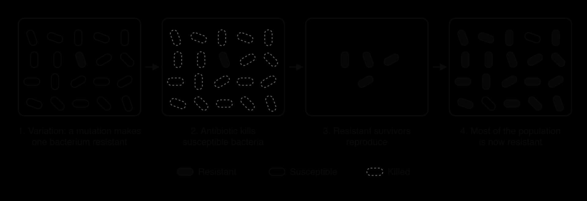
Antibiotic resistance is the ability of a strain of bacteria to survive and reproduce in the presence of an antibiotic that would normally kill it. Resistance evolves by natural selection:
Variation. In a large population of bacteria, a random mutation may give one bacterium a gene for resistance, for example a gene for an enzyme that breaks down the antibiotic, or a change in the protein that the antibiotic targets. Resistance genes can also be passed between bacteria on plasmids by horizontal gene transfer.
Selection. When the antibiotic is used, susceptible bacteria are killed, but the resistant bacteria survive.
Reproduction. The resistant bacteria reproduce by binary fission, passing the resistance gene to their offspring.
Change in the population. With repeated use of the antibiotic, the proportion of resistant bacteria increases until most of the population is resistant.
The antibiotic does not cause the mutation. It selects bacteria that were already resistant, which then reproduce.
Bacteria evolve resistance quickly because they reproduce rapidly, in some species every 20 minutes or so, and their populations are enormous, so even rare mutations appear often. Some strains are resistant to several antibiotics at once. Methicillin-resistant Staphylococcus aureus (MRSA), for example, is resistant to methicillin and most other penicillin-type antibiotics and causes serious infections in hospitals. Antimicrobial resistance was directly responsible for an estimated 1.27 million deaths worldwide in 2019.
Slowing the spread of resistance
The more an antibiotic is used, the stronger the selection for resistance. Careful use of antibiotics slows the spread of multi-resistant bacteria:
prescribing antibiotics only for bacterial infections, never for viral infections;
choosing a narrow-spectrum antibiotic that targets the specific bacterium where possible;
completing the prescribed course, so that partly resistant bacteria are not left to multiply;
reducing the use of antibiotics in farming, where they have been given to healthy animals to promote growth or prevent disease;
good hygiene in hospitals, such as handwashing and isolating infected patients, so resistant strains do not spread.
Discovering new antibiotics
Few new classes of antibiotic have been discovered since the 1980s, so new methods are being used to find them. Researchers search chemical libraries, collections of millions of different compounds, for molecules that kill bacteria. In one example, a computer model was trained on the structures of molecules tested against Escherichia coli and then used to screen chemical libraries for new candidates. It identified a compound, named halicin, that is structurally different from existing antibiotics and killed many drug-resistant bacteria in laboratory tests. Other approaches include culturing soil bacteria that could not be grown before, and testing compounds made by organisms in unusual habitats.
Why antibiotics act only on bacteria, how resistance is selected, and how vaccines prepare the immune system are summarised in the video.
YouTube
Antibiotics, antivirals and vaccines: why antibiotics work on bacteria but not viruses, how antibiotic resistance evolves, and how vaccines produce immunity.
Zoonoses
Most pathogens are species-specific, but some can pass from other animals to humans. A zoonosis is an infectious disease that can be transmitted from other species to humans. About 60% of known human infectious diseases are zoonotic, and so are most new diseases that have appeared in recent decades.
Zoonoses have various modes of transmission:
Zoonosis
Pathogen
Animal source
Mode of transmission to humans
Tuberculosis (bovine)
Bacterium, Mycobacterium bovis
Cattle
Drinking unpasteurised milk from infected cows; breathing droplets from infected animals
Rabies
Rabies virus
Dogs, bats and other mammals
Bites or scratches, when saliva containing the virus enters the wound
Japanese encephalitis
Japanese encephalitis virus
Pigs and wading birds
Bites from infected Culex mosquitoes, which carry the virus from animals to humans
COVID-19
Coronavirus, SARS-CoV-2
Most likely bats, possibly through an intermediate host
After the first spillover, spread between humans in respiratory droplets and aerosols
Rabies is almost always fatal once symptoms appear and kills tens of thousands of people each year, mostly in Asia and Africa, nearly all after bites from dogs. Japanese encephalitis is a vector-borne disease: the mosquito acts as a vector, an organism that carries the pathogen between hosts. Bovine tuberculosis is now rare in humans in countries where milk is pasteurised and cattle are tested.
Zoonoses are thought to be increasing because humans are coming into closer contact with wild and domestic animals. Destruction of forests brings people and livestock into the habitats of wild animals; the wildlife trade brings live animals of many species together in markets; and intensive farming keeps large numbers of genetically similar animals close together, which helps pathogens to spread and evolve. Global travel then carries new infections around the world in days.
Vaccines and immunisation
A vaccine is a preparation that stimulates the development of immunity to a specific pathogen without causing the disease. Vaccination (immunisation) is the process of giving a vaccine, usually by injection.
Vaccines contain antigens, or nucleic acids (DNA or mRNA) with base sequences that code for antigens:
Type of vaccine
What it contains
Example
Live attenuated
A weakened form of the pathogen that multiplies very little
Measles, mumps and rubella (MMR)
Inactivated
Whole pathogens killed by heat or chemicals
Injected polio vaccine, rabies vaccine
Subunit
Purified antigen molecules only
Hepatitis B, human papillomavirus (HPV)
mRNA
mRNA coding for an antigen, in lipid nanoparticles
Pfizer–BioNTech and Moderna COVID-19 vaccines
Viral vector
A harmless virus carrying DNA that codes for an antigen
Oxford–AstraZeneca COVID-19 vaccine
In an mRNA vaccine, the mRNA enters some of the person's cells, which use it to make the antigen protein (for COVID-19 vaccines, the spike protein of SARS-CoV-2) and display it. The mRNA is broken down within days and does not enter the nucleus or change the person's DNA.
How vaccines produce immunity
Whatever the type, a vaccine causes a primary immune response. Macrophages present the vaccine antigens, helper T-cells are activated, and the matching B-cells are activated and form clones of plasma cells and memory cells. Because the vaccine does not cause the disease, the person is not ill during the slow primary response. If the real pathogen later enters the body, memory cells cause a fast, large secondary response that destroys the pathogen before symptoms develop. Some vaccines need booster doses to increase the number of memory cells.
Herd immunity and the prevention of epidemics
An epidemic is a rapid increase in the number of cases of an infectious disease in a population, above the level normally expected. A pandemic is an epidemic that spreads across many countries or continents, as COVID-19 did from 2020.
An infectious disease can only spread if each infected person passes the pathogen to susceptible people. When a large enough proportion of a population is immune, most contacts of an infected person are immune too, so the chain of transmission is broken and the number of cases falls. This is herd immunity. It protects people who cannot be vaccinated or who did not develop immunity, such as newborn babies, people with weakened immune systems and people having cancer treatment.
When most people are immune, an infected person is unlikely to meet a susceptible person, so chains of transmission are broken.
The basic reproduction number and the herd immunity threshold
The basic reproduction number (R₀) is the average number of people that one infected person infects in a population where everyone is susceptible. If R₀ is above 1, the number of cases grows; if each infected person infects fewer than one other, the disease dies out. The proportion of the population that must be immune for herd immunity, the herd immunity threshold, depends on R₀:
herd immunity threshold=1−R01
Herd immunity threshold
The herd immunity threshold, 1 − 1/R₀, rises steeply at first and then levels off: the more infectious a disease, the closer to 100% the immune proportion must be.
Communicating about vaccines
Vaccination programmes only work if enough people choose to be vaccinated, so scientists and health agencies publish their research and explain it to the public. Vaccines are tested in large clinical trials before they are approved, and their safety is monitored afterwards. Serious side effects are very rare, but the risk is not zero. A decision to vaccinate weighs this small risk against the much greater risk of the disease itself, both to the individual and to others who rely on herd immunity. False claims spread quickly, as in the case of a 1998 study that wrongly linked the MMR vaccine to autism; the study was retracted in 2010, but vaccination rates fell in several countries and measles outbreaks followed.
Case study: the eradication of smallpox
Smallpox, caused by the variola virus, killed a high proportion of the people it infected and left many survivors scarred or blind. It is the only human disease that has been eradicated, meaning it no longer occurs anywhere in the world. When the World Health Organization began its Intensified Smallpox Eradication Programme on 1 January 1967, smallpox was causing an estimated 10–15 million cases and about 2 million deaths a year in 31 countries.
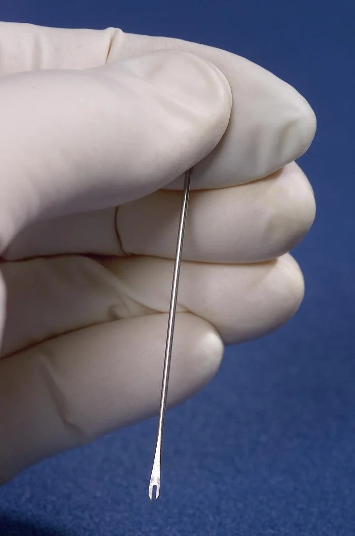
The programme combined mass vaccination with surveillance and containment. Health workers searched for every case. When one was found, the patient was isolated and everyone around them (family, neighbours and other contacts) was vaccinated. This "ring vaccination" surrounded each case with immune people, so the virus ran out of susceptible hosts, in the way the diagram of herd immunity shows. The vaccine, made from the related vaccinia virus, was given with a simple bifurcated needle, which needed very little training and very little vaccine per person.
The bifurcated needle holds a drop of vaccine between its two prongs and is pressed into the skin several times. It was used throughout the eradication programme. Photo: James Gathany, CDC, public domain, via Wikimedia Commons.
Date
Event
1 January 1967
WHO Intensified Smallpox Eradication Programme begins; about 10–15 million cases and 2 million deaths a year in 31 countries
26 October 1977
Rash of the last naturally occurring case, Ali Maow Maalin, a hospital cook in Merca, Somalia
9 December 1979
A global commission certifies that smallpox has been eradicated
8 May 1980
The World Health Assembly declares smallpox eradicated
Sources: World Health Organization; World Health Assembly resolution WHA33.3, 1980.
Several features of smallpox made eradication possible. Smallpox has no animal reservoir: the virus infects only humans, so once human transmission stopped, there was nowhere for it to survive. A zoonosis such as rabies, which circulates in dogs and bats, cannot be eradicated in the same way by vaccinating humans alone. In addition, infected people had a visible rash, so cases were easy to find; there were no symptom-free carriers; and recovery or vaccination gave long-lasting immunity, through memory cells, against the single type of virus.
Since 1980 routine smallpox vaccination has stopped. The virus survives only in two high-security laboratories, in the United States and Russia. The campaign is evidence that vaccination, careful surveillance and international cooperation can remove a disease from the human population entirely.
Evaluating data on the COVID-19 pandemic
COVID-19 was first identified in December 2019 and was declared a pandemic by WHO in March 2020. Data on cases, deaths, vaccination and excess mortality were published daily by many countries, and these data are often used to compare countries or to measure change over time. Two calculations are used most often: percentage change and percentage difference.
Percentage change
Percentage change compares the same quantity at two times, such as the number of cases in one country in two different weeks. It is always calculated relative to the original (earlier) value:
A positive answer is an increase and a negative answer a decrease.
Percentage difference
Percentage difference compares two values of the same kind measured at the same time, such as the number of cases in two countries, or in two groups of a trial. Neither value is the "original", so the difference is divided by the mean of the two values:
%difference=(a+b)/2∣a−b∣×100
Only comparable values should be compared. Cases and deaths are different quantities, and numbers of cases from countries of very different size should first be converted to rates (for example, cases per 100,000 people).
Evaluating a vaccine trial
The first large trial of the Pfizer–BioNTech mRNA vaccine (BNT162b2) gave two doses of vaccine or a placebo (a salt-water injection) to two groups of adults, allocated at random, so that neither participants nor investigators knew who had received which. Cases of COVID-19 were then counted from 7 days after the second dose.
Group
Participants analysed
Cases of COVID-19
Vaccine
18,198
8
Placebo
18,325
162
Source: Polack et al., New England Journal of Medicine, 2020.
Excess mortality
Reported COVID-19 deaths depend on how much testing a country did and how deaths were recorded, so they are not always comparable between countries. Excess mortality avoids this problem. It is the number of deaths from all causes during a period minus the number that would have been expected from the death rates of previous years. WHO estimated that there were about 14.9 million excess deaths worldwide in 2020 and 2021, compared with about 5.4 million deaths officially reported as caused by COVID-19.
Exam practice
Defence against disease is assessed in all parts of the papers. Paper 1A multiple-choice questions test the roles of the different cells (which cell secretes antibodies, which cells HIV infects), blood groups and the action of antibiotics. Paper 1B and Paper 2 data questions often use antibody-concentration graphs, vaccination and case data, or the results of clinical trials, and ask for percentage change or percentage difference. Paper 2 extended responses ask candidates to describe the process of antibody production, explain how vaccines give immunity or explain the evolution of antibiotic resistance. The command terms used most are state, outline, describe, explain, calculate and evaluate.
The practice question uses data for a hypothetical city that began a vaccination campaign against measles in week 2 of an outbreak.
Week
New cases of measles
Population vaccinated / %
1
1,240
78
5
310
93
Extended responses on antibody production, vaccination or resistance score best when the steps are in order and each cell or process is named precisely. A sequence of short, linked sentences is clearer than a long paragraph, and avoids repeating points.
A second useful exercise is to collect one set of real pandemic data, such as weekly cases in two countries, and practise both calculations on it.
Self-check
To do:
I can name the four main groups of pathogens and explain how Semmelweis and Snow helped establish disease control.
To do:
I can describe the skin and mucous membranes as primary defences.
To do:
I can outline the clotting cascade, including thrombin and the conversion of fibrinogen to fibrin.
To do:
I can distinguish between innate and adaptive immunity and describe phagocytosis.
To do:
I can explain how helper T-cells activate B-cells, and how clones of plasma cells and memory cells form.
To do:
I can explain antigens, antibodies and the results of mismatched blood transfusions.
To do:
I can sketch and explain primary and secondary immune responses.
To do:
I can outline the transmission of HIV and explain how it leads to AIDS.
To do:
I can explain why antibiotics do not work against viruses and how resistance evolves.
To do:
I can give examples of zoonoses and explain how vaccines and herd immunity prevent epidemics.
To do:
I can calculate percentage change and percentage difference from COVID-19 data.
66 % / percentage points;
(c)
Describe the changes in pH in the phagocytic vacuoles.
[2]2 marks
Markscheme (c)
a. pH falls / vacuole becomes more acidic over time; b. fastest fall in the first 15 / 20 minutes; c. levels off at about pH 4.8 after 30 / 45 minutes; d. overall fall of 2.4 pH units;
[2 max]
(d)
Using both graphs, deduce the relationship between the pH of the vacuole and the killing of bacteria.
[2]2 marks
Markscheme (d)
a. most bacteria are killed as the pH falls / killing is fastest after the pH falls below about 6; b. when the pH stays above 6.5 (blocker), few bacteria are killed; c. (so) acidification is needed for (most of the) killing of bacteria; d. some bacteria are still killed with the blocker, so other mechanisms also act (OWTTE);
[2 max]
(e)
Suggest why lysosomes fuse with the phagocytic vacuole rather than releasing their enzymes into the cytoplasm.
[2]2 marks
Markscheme (e)
a. hydrolytic enzymes are concentrated with the bacteria in a small volume; b. the cell's own molecules / organelles are protected from digestion; c. the low / acidic pH needed by lysosomal enzymes can be maintained in the vacuole but not in the cytoplasm;
[2 max]
Question 2 · BIO-0025015 marks
Eukaryotic cells are divided into compartments by membranes.
(a)
Describe the structure of the plasma membrane according to the fluid mosaic model.
[4]4 marks
(b)
Explain the advantages of compartmentalization in eukaryotic cells.
[7]7 marks
(c)
Outline how phagocytes help to control infections.