Explain how nerves, hormones and the blood integrate animal body systems, including reflexes and feedback control, and (HL) how phytohormones coordinate plant growth.
Each is in the Biology glossary, with every other term in the subject.
A phytohormone produced in leaves and roots when water is scarce that causes stomata to close, and that keeps seeds dormant.
The elongation of plant cells caused by auxin: proton pumps move H⁺ into the cell wall, and the low pH loosens the cross-links between cellulose microfibrils so that turgor pressure stretches the wall.
An endocrine gland on top of each kidney; its inner medulla secretes epinephrine when the body prepares for vigorous activity or faces a threat.
The inhibition of side-bud growth by auxin produced in the main shoot tip, so that the main shoot grows more strongly than the side shoots.
A phytohormone, mainly indole-3-acetic acid (IAA), made in shoot tips, that promotes cell elongation in shoots and controls tropisms, apical dominance and root formation.
A membrane protein (PIN protein) that moves auxin ions out of a plant cell; carriers on only one side of each cell make auxin flow in one direction through a tissue.
A stretch receptor in the wall of the aorta or the carotid arteries that detects changes in blood pressure.
The region of the medulla oblongata that receives impulses from baroreceptors and chemoreceptors and controls heart rate through the sympathetic and vagus nerves.
A multicellular organism is made of many parts, and each part has its own job. Integration is the coordination of these parts so that the whole organism works as a single unit: the heart beats faster when muscles need more oxygen, the hand is pulled away from a flame before it is badly burnt, and food is moved along the gut at the right speed.
40 exam-style questions on this subtopic, written in the style of the IB papers. The first has its markscheme open. Counted on
Cells in multicellular organisms must communicate with each other.
Outline how a neuron generates and maintains its resting potential.
4 marks
a. sodium–potassium pumps in the plasma membrane (of the neuron);
b. use energy from (hydrolysis of) ATP / active transport;
c. pump Na+ out and K+ in;
d. three Na+ out for every two K+ in;
e. (so) concentration gradients of Na+ and K+ are established in opposite directions;
f. membrane is more permeable to K+ than Na+ / K+ leak out faster than Na+ leak in;
g. negatively charged proteins / ions remain inside;
h. inside is negative relative to outside / membrane is polarized;
i. resting potential is about −70 mV;
[4 max]
Explain how a signal is transmitted across a synapse between two neurons that use acetylcholine as a neurotransmitter.
7 marks
a. nerve impulse / action potential arrives at the presynaptic terminal;
b. depolarization of the presynaptic membrane;
c. (voltage-gated) calcium channels open and Ca
The official deck for this part of the syllabus.
What is a system?
Define integration in a living system.
With a free account, Augment remembers where you stopped and what you have practiced.
The brain and spinal cord, which receive information from receptors, integrate it and send impulses to effectors.
The part of the brain at the back of the head, below the cerebral hemispheres, that coordinates the timing and force of skeletal muscle contractions and maintains balance and posture.
One of the two halves of the cerebrum, the largest part of the brain, whose outer cortex processes sensory information, initiates voluntary movement and carries out thought, learning and memory.
A receptor that detects chemicals; chemoreceptors in the carotid bodies, aortic bodies and medulla detect blood pH and CO₂ and O₂ concentrations.
A cycle of changes in an organism's body or behaviour that repeats about every 24 hours and continues even without external cues, although it is normally reset each day by light.
The protective sheath that covers the young shoot of a grass seedling as it grows through the soil; it is widely used in experiments on phototropism.
A phytohormone made mainly in root tips and carried to the shoot in the xylem that promotes cell division and the growth of side shoots.
A muscle or gland that carries out a response when it is stimulated by a motor neuron or a hormone.
A property of a whole system that arises from the interactions between its parts and is not shown by any of the parts on their own.
The system of glands that secrete hormones directly into the blood, which carries them to target cells throughout the body.
The network of neurons in the wall of the gut that controls peristalsis and other gut activities involuntarily, without needing instructions from the brain.
A hormone (also called adrenaline) secreted by the adrenal glands that prepares the body for vigorous activity, for example by increasing heart rate, ventilation and the supply of glucose and oxygen to muscles.
A gaseous phytohormone (ethene, C₂H₄) that stimulates fruit ripening; ripening fruit releases more ethylene, giving positive feedback.
The branched, unprotected ending of a sensory neuron in a tissue such as the skin, which detects stimuli such as heat, acid and tissue damage and acts as a pain receptor.
A phytohormone, made mainly in young leaves and seeds, that promotes stem elongation, seed germination and, in some species, flowering.
A directional growth response to gravity; roots grow downwards (positive gravitropism) and shoots grow upwards (negative gravitropism).
Nervous tissue made mainly of the cell bodies, dendrites and synapses of neurons, where information is integrated; it forms the cerebral cortex and the central part of the spinal cord.
A chemical signal secreted by an endocrine gland directly into the blood, which carries it to target cells that have receptors for it.
A region at the base of the brain that monitors the blood and receives nervous input, and controls the pituitary gland, linking the nervous system with the endocrine system.
The coordination of the component parts of a system so that, together, they carry out the overall function of the system.
A neuron within the central nervous system that passes impulses from one neuron to another, for example from a sensory neuron to a motor neuron in a reflex arc.
The lowest part of the brainstem, joining the brain to the spinal cord, containing the cardiovascular and respiratory centres that control heart rate and ventilation.
A hormone secreted by the pineal gland, mainly during darkness, that helps to set the timing of the sleep–wake cycle and other circadian rhythms.
A neuron that carries impulses from the central nervous system to an effector, such as a muscle, stimulating it to contract.
Describes contraction that starts in the muscle tissue itself, without a nervous stimulus; the heartbeat is myogenic because each beat starts in the sinoatrial node.
A control mechanism in which a change in a variable triggers a response that reverses the change, returning the variable towards its set value.
A bundle of nerve fibres from many neurons, surrounded by a protective sheath of connective tissue; a nerve may contain sensory fibres, motor fibres or both.
A neuron that secretes a hormone into the blood instead of releasing a neurotransmitter at a synapse, as in the hypothalamus.
A structure made of two or more different tissues that work together to perform one or more functions, such as the heart or the brain.
A group of organs that work together to perform a major function in the body, such as the circulatory system or the nervous system.
Waves of contraction of the circular and longitudinal muscle in the wall of the gut that push food along in one direction.
A directional growth response to light from one side; shoots show positive phototropism, growing towards the light.
A signalling chemical produced by plant cells that controls growth, development or responses to stimuli, often in tissues away from where it is made.
A small endocrine gland near the centre of the brain that secretes melatonin, mainly at night, under the control of the suprachiasmatic nucleus.
A small endocrine gland below the hypothalamus whose anterior and posterior lobes secrete hormones, many of which control other endocrine glands.
A control mechanism in which a change in a variable triggers a response that increases the change still further, so that a process speeds up until it is completed.
The region of the cerebral cortex, in a strip across the top of each cerebral hemisphere, that initiates voluntary contractions of skeletal muscles.
A rapid, automatic response to a stimulus that does not involve conscious thought, such as withdrawing the hand from a hot object.
The pathway of neurons that carries impulses in a reflex: from a receptor, along a sensory neuron to the CNS, through one or more interneurons, and along a motor neuron to an effector.
The region of the medulla oblongata that sends rhythmic impulses to the diaphragm and intercostal muscles and changes ventilation rate in response to blood pH.
A neuron that carries impulses from a sensory receptor to the central nervous system (the spinal cord or the brain).
A cell or nerve ending that detects a particular kind of stimulus, such as light, chemicals, pressure or temperature, and converts it into nerve impulses.
The part of the central nervous system inside the vertebral column; it carries impulses to and from the brain and acts as the integrating centre for many unconscious reflexes.
The volume of blood pumped out of the left ventricle in one heartbeat; it increases when the heart contracts more forcefully.
A small group of neurons in the hypothalamus that acts as the master circadian clock; it receives light information from the retina and controls melatonin secretion by the pineal gland.
A group of similar cells, often with the material between them, that work together to perform a particular function.
A directional growth response of a plant to a directional stimulus; growth towards the stimulus is positive and growth away from it is negative.
A cranial nerve of the parasympathetic system; impulses along it from the medulla to the sinoatrial node slow the heart rate.
Animals integrate their body systems with two signalling systems, the nervous system and the endocrine system, and with the blood, which carries materials and heat between organs. Plants have no nerves, but they still coordinate growth and responses with chemical signals called phytohormones. Integration is a clear example of interaction and interdependence: no organ can survive on its own, and each depends on signals and supplies from the others.
A system is a set of parts that work together to perform an overall function. A car engine, a city transport network and a human body are all systems. In each case the parts must be coordinated: if the fuel pump and the spark plugs of an engine worked at random times, the engine would not run, even if every part were in perfect condition.
Living systems face the same problem on a much larger scale. A human body has about 30 trillion cells. Each cell carries out its own metabolism, but the body only survives if the activities of these cells are matched to each other and to the conditions outside. Muscle cells that contract need oxygen and glucose delivered at the right rate, and the waste carbon dioxide they release must be removed. Coordination of this kind is called integration.
Integration needs three things. Information about conditions must be collected (by receptors). That information must be passed on and processed (by nerves, hormones and the brain). Finally, the parts that respond (the effectors, such as muscles and glands) must receive instructions and act. The same sequence of stimulus, receptor, coordinator, effector and response appears in every example of integration in animals.
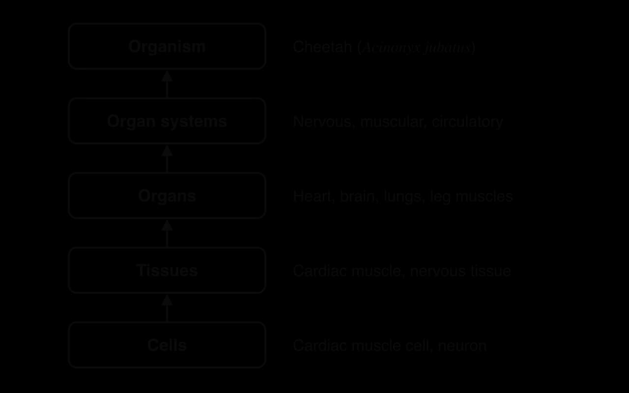
The body of a multicellular organism is organised as a hierarchy of subsystems, each built from the level beneath it. This hierarchy is one of the levels of organisation that run through the whole of biology, from molecules to ecosystems.
Cells are the smallest units of life. In a multicellular organism most cells are specialised for one function, such as a cardiac muscle cell that contracts or a neuron that carries impulses.
A tissue is a group of similar cells that carry out the same function. Cardiac muscle, nervous tissue and the epithelium lining the gut are tissues.
An organ is a structure made of two or more tissues that work together to perform a function. The heart contains cardiac muscle, nervous tissue, connective tissue and blood vessels.
An organ system is a group of organs that cooperate to perform a major function. The circulatory system (heart, arteries, veins and capillaries) transports materials around the body.
The organism is the whole living individual, made of several organ systems working together.
Each level depends on the level below it, but it also has abilities that the level below does not have. A single cardiac muscle cell can contract, but only the whole heart, with its chambers and valves, can pump blood in one direction. An organ such as the heart is therefore more than a collection of cells, and an organ system is more than a collection of organs.
A property of a whole system that its separate parts do not have is called an emergent property. Emergent properties arise from the interactions between the parts, so they cannot be predicted by studying each part on its own. Consciousness, the beating of a heart and the ability to hunt are all emergent properties.
The cheetah (Acinonyx jubatus) is the fastest land animal. Wild cheetahs fitted with GPS collars in Botswana reached top speeds of 25.9 m s⁻¹ (about 93 km h⁻¹) during hunts. No single organ makes this possible. Speed depends on the integration of several organ systems:
the skeletal and muscular systems, with a flexible spine that lengthens each stride and large leg muscles that contract with great power;
the nervous system, which coordinates the timing of muscle contraction in all four legs and keeps the animal balanced at high speed, while the eyes track the prey and the brain predicts its movements;
the respiratory system, with large nasal passages and lungs that take in oxygen rapidly;
the circulatory system, with a large heart that delivers oxygen and glucose to the muscles and removes carbon dioxide and heat.
A cheetah can only keep up a sprint for a short distance, usually a few hundred metres, before it must slow down and recover. Its success as a predator is therefore an emergent property of the whole organism, and its limits are set by the same interacting systems. This is a good example of form and function at the level of the organism.
In animals, organs are integrated in three ways. The nervous system sends electrical impulses along neurons. The endocrine system releases hormones into the blood. The blood itself transports materials and energy between organs. These three methods are linked: nerves control some endocrine glands, hormones affect the activity of neurons, and the blood carries hormones to their targets.
The nervous system is made of the central nervous system (CNS: the brain and spinal cord) and the peripheral nerves that connect it to every part of the body. Messages travel as nerve impulses along neurons. At the end of a neuron the message is passed on by a chemical, a neurotransmitter, which crosses a narrow gap called a synapse (the mechanism is covered in C2.2 Neural signalling).
The endocrine system is made of glands that secrete hormones into the blood. The blood carries a hormone to every part of the body, but only target cells with the right receptor respond to it. The two systems differ in speed, duration, targeting and the type of signal, as the table shows.
Feature | Nervous system | Endocrine system |
|---|---|---|
Type of signal | Electrical impulses along neurons, with chemical transmission at synapses | Chemical (hormones) carried in the blood |
Route | Along nerve fibres to specific cells | Through the blood to all parts of the body |
Speed of transmission | Very fast: impulses travel at up to about 100 m s⁻¹ | Slower: seconds to hours, depending on blood flow and the hormone |
Duration of effect | Usually short: stops soon after impulses stop | Usually longer: lasts until the hormone is broken down or removed |
Targeting | Precise: a neuron acts only on the cells it synapses with | Widespread: any cell with receptors for the hormone responds |
Typical uses | Rapid responses such as reflexes and movement | Long-term processes such as growth, puberty, metabolism and reproduction |
The two systems are suited to different tasks. A pain reflex must happen within a fraction of a second and must affect only certain muscles, so it is controlled by nerves. Growth during childhood takes years and affects almost every tissue, so it is controlled by hormones. Some responses use both: in a sudden emergency, nerves act within milliseconds and the hormone epinephrine then keeps the body prepared for several minutes.
The blood integrates organs by carrying materials between them. It transports oxygen from the lungs to respiring tissues and carbon dioxide back, glucose and amino acids from the small intestine to the liver and the rest of the body, urea from the liver to the kidneys, and hormones from endocrine glands to their target cells.
Blood also transports energy in the form of heat. Active organs such as the liver and contracting skeletal muscles produce heat as a by-product of respiration. The blood absorbs this heat and carries it to cooler parts of the body, and to the skin, where it can be lost to the surroundings. Water has a high specific heat capacity, so blood plasma can carry a large amount of heat with only a small rise in temperature.
The brain receives information from sensory receptors all over the body, combines it, compares it with stored memories, and sends instructions to muscles and glands. It contains about 86 billion neurons, each of which may form synapses with thousands of other neurons. Processing in the brain happens in these networks of neurons, where signals from many inputs are added together and passed on.
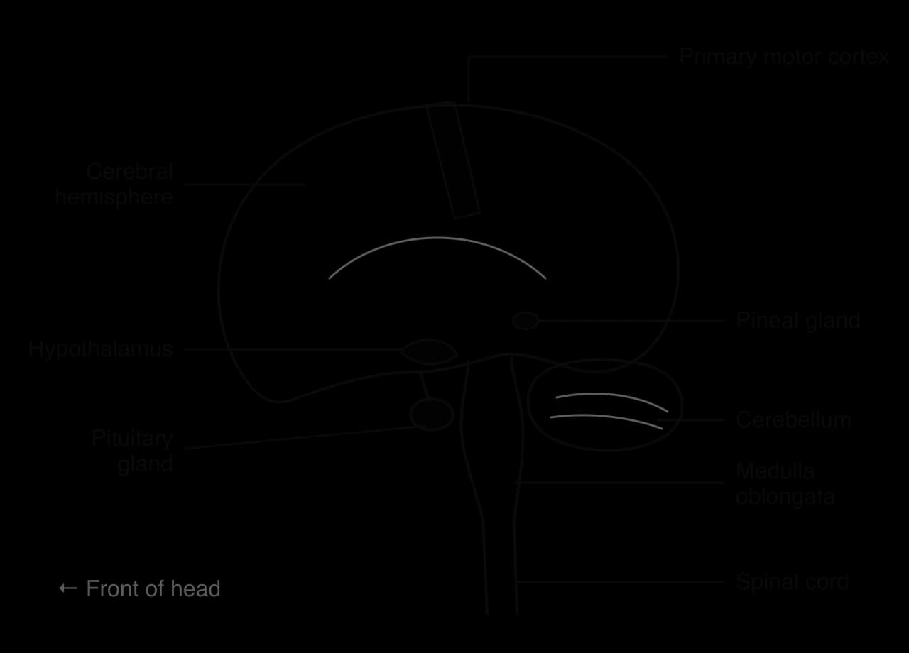
The largest part of the human brain is the cerebrum, made of the left and right cerebral hemispheres. Their outer layer, the cerebral cortex, is folded, which increases its surface area and the number of neurons it can hold. The cortex processes information from the senses, initiates voluntary movement and is responsible for thought, language, learning and memory. Below the cerebrum are the cerebellum, which coordinates movement, and the brainstem, whose lowest part, the medulla oblongata, controls the heart rate and breathing.
Very few decisions depend on one sense alone. Catching a ball uses information from the eyes (position and speed of the ball), the inner ear (balance and the position of the head) and stretch receptors in muscles and joints (the position of the arms). The brain combines these inputs, predicts where the ball will be, and sends a coordinated set of instructions to many muscles at once.
Learning is the acquisition of new knowledge or skills through experience, and memory is the ability to store that information and recall it later. Both depend on changes in the brain. When two neurons are repeatedly active together, the synapses between them become stronger, so a signal crosses them more easily in future. New synapses can also form and unused ones can be lost. A memory is stored as a particular pattern of strengthened connections in a network of neurons.
Different kinds of memory depend on different parts of the brain. Memories of facts and events (declarative memory, such as remembering a friend's birthday) need the hippocampus, a curved structure deep in each cerebral hemisphere, to be formed. Memories of skills (procedural memory, such as riding a bicycle) depend on other regions, including the cerebellum and the motor areas of the cortex. The strongest evidence for this division comes from people with damage to particular brain regions.
Henry Molaison, known in research papers as patient H.M. until his death in 2008, had severe epilepsy that drugs could not control. In 1953, at the age of 27, he had an operation in which the neurosurgeon William Scoville removed parts of the medial temporal lobe from both cerebral hemispheres, including most of each hippocampus. The operation reduced his seizures, but it left him unable to form new long-term memories of facts and events.
After the operation, H.M. could hold a conversation and remember a number for a short time by repeating it, but within minutes of being distracted he forgot what had happened. He met the same researchers for decades but did not recognise them. His older memories from childhood remained, as did his intelligence, language and personality. This showed that the hippocampus is needed to convert short-term memories into long-term memories, but not to store all old memories or for thinking in general.
The psychologist Brenda Milner then tested whether H.M. could learn a skill. He was asked to trace the outline of a star while seeing his hand and the paper only in a mirror, which reverses the direction of every movement. Over 30 trials spread across 3 days, his tracing improved steadily, with fewer errors each time, just as it does in people without brain damage. Yet at the start of each day he said he had never done the task before.
Kind of memory or ability | Example tested | H.M. after 1953 | What this shows |
|---|---|---|---|
New memories of facts and events | Recognising people met after the operation | Lost | The hippocampus is needed to form new declarative memories |
Old memories of facts and events | Events from his childhood | Mostly kept | Long-term memories are stored elsewhere in the cortex |
Short-term memory | Repeating a number for a few seconds | Kept | Short-term memory does not depend on the hippocampus |
Learning a motor skill | Mirror tracing of a star | Kept: improved over 3 days | Skill learning uses other regions, such as the cerebellum and motor cortex |
Sources: Scoville and Milner, Journal of Neurology, Neurosurgery and Psychiatry, 1957; Milner, 1962.
H.M.'s case changed the understanding of memory. Before it, many researchers thought that memory was spread evenly through the cortex. The evidence from one carefully studied person showed instead that particular brain regions carry out particular parts of learning and memory. The case also raises questions of research ethics. H.M. took part in studies for more than 50 years, although his amnesia meant that he could not remember the studies he had agreed to, and after his death his brain was cut into more than 2,000 thin sections that were photographed and made available to researchers.
Some processes in the body are conscious: the person is aware of them and can usually control them. Deciding to pick up a pen, reading a sentence and choosing words to speak are conscious processes, and they are coordinated by the cerebral hemispheres. Other processes are unconscious: they happen without awareness or deliberate control. The heartbeat, the narrowing of the pupils in bright light and the withdrawal of a hand from a hot object are unconscious.
Conscious processes | Unconscious processes | |
|---|---|---|
Awareness | The person is aware of them | Happen without awareness (the person may notice them afterwards) |
Control | Voluntary: can be started or stopped deliberately | Involuntary: cannot normally be stopped by choice |
Integrating centre | Cerebral hemispheres (cerebral cortex) | Spinal cord (reflexes), brainstem (heart rate, breathing), enteric nervous system (gut) |
Examples | Walking to the door, writing, speaking | Pain reflex, knee-jerk reflex, peristalsis, heart rate |
The spinal cord is a cylinder of nervous tissue about 45 cm long that runs from the base of the brain down the back, protected by the vertebrae. It carries impulses between the brain and the rest of the body, but it is also an integrating centre in its own right. Many reflexes are coordinated entirely within the spinal cord, so the response happens before any signal reaches the brain.
In a transverse section, the spinal cord has a butterfly-shaped central region of grey matter surrounded by white matter. Grey matter contains the cell bodies of neurons, their dendrites and synapses, so this is where integration takes place. White matter consists mainly of myelinated axons running up and down the cord, and the fatty myelin gives it its pale colour.
Information enters the nervous system through sensory receptors. A receptor is a cell, or the ending of a neuron, that detects a particular kind of stimulus and converts it into nerve impulses. Receptors are classified by the stimulus they detect:
Type of receptor | Stimulus detected | Example |
|---|---|---|
Photoreceptors | Light | Rod and cone cells in the retina of the eye |
Chemoreceptors | Chemicals | Taste buds, olfactory cells in the nose, chemoreceptors that monitor blood CO₂ and pH |
Mechanoreceptors | Pressure, touch, stretch, sound, movement | Touch receptors in the skin, hair cells in the ear, baroreceptors, stretch receptors in muscles |
Thermoreceptors | Temperature | Receptors for heat and cold in the skin and hypothalamus |
Pain receptors (nociceptors) | Damaging stimuli: high temperature, acid, crushing | Free nerve endings in the skin |
A sensory neuron carries impulses from a receptor to the CNS. Sensory neurons enter the spinal cord through the dorsal root, and their cell bodies lie just outside the cord in a swelling called the dorsal root ganglion. Some sensory information is processed in the spinal cord. Other information travels up the cord, or along cranial nerves from the eyes, ears, nose and tongue, to the cerebral hemispheres, where it becomes a conscious sensation such as sight, sound or pain.
Skeletal muscles only contract when they are stimulated by a motor neuron. A motor neuron has its cell body in the CNS (in the grey matter of the spinal cord or in the brainstem) and a long axon that runs out to a muscle. Where the axon ends on a muscle fibre it forms a neuromuscular junction: an impulse arriving there causes release of acetylcholine, which stimulates the muscle fibre to contract. The muscle is the effector, the part of the body that carries out the response.
Voluntary movements begin in the primary motor cortex, a strip of cortex that runs across the top of each cerebral hemisphere, from one side of the head to the other. Different parts of the strip control different parts of the body, and the areas for the hands, lips and tongue are especially large because these parts make fine, precise movements. Neurons in the motor cortex send impulses down the spinal cord to motor neurons, which then carry them to the skeletal muscles. Most of these pathways cross over in the brainstem, so the left hemisphere controls the right side of the body and the right hemisphere the left side.
A nerve is a bundle of many nerve fibres, held together and protected by connective tissue. Each nerve fibre is the long axon (or dendron) of a single neuron, so a nerve contains parts of hundreds or thousands of different neurons. The cell bodies of these neurons are not in the nerve: they are in the CNS or in ganglia.
Nerves can carry information in one direction or in both. The optic nerve, from the eye to the brain, contains only sensory fibres. Some nerves to muscles contain mainly motor fibres. Most spinal nerves are mixed nerves that contain both sensory and motor fibres, so impulses travel towards the CNS in some fibres and away from it in others at the same time.
A transverse section (TS) is a section cut across a structure at right angles to its length. In a TS of a nerve, the fibres are cut across, so each one appears as a small circle. The diagram of a nerve in transverse section shows the main features.
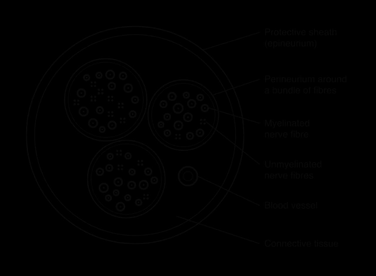
Protective sheath (epineurium): the tough outer layer of connective tissue that surrounds the whole nerve.
Bundles of fibres (fascicles): groups of nerve fibres, each surrounded by a thinner layer of connective tissue (the perineurium).
Myelinated nerve fibres: axons surrounded by a myelin sheath, which appears as a thick ring (darkly stained in many micrographs) around a pale central axon. Myelinated fibres conduct impulses quickly.
Unmyelinated nerve fibres: thinner axons without a myelin sheath, often lying in small groups. They conduct impulses more slowly; many carry pain and temperature information.
Blood vessels: small arteries, veins and capillaries in the connective tissue supply the fibres with oxygen and glucose.
A reflex is a rapid, automatic response to a stimulus that does not involve conscious thought. Reflexes protect the body from harm because they are faster than conscious responses. The pathway that impulses follow in a reflex is a reflex arc.
The pain withdrawal reflex is a typical example. When a finger touches a very hot object, the arm muscles contract and pull the hand away within a fraction of a second. The pain is felt only after the hand has started to move, because the reflex is coordinated in the spinal cord and the brain is informed afterwards. The reflex is an involuntary response by skeletal muscle, the same type of muscle that is used in voluntary movements.
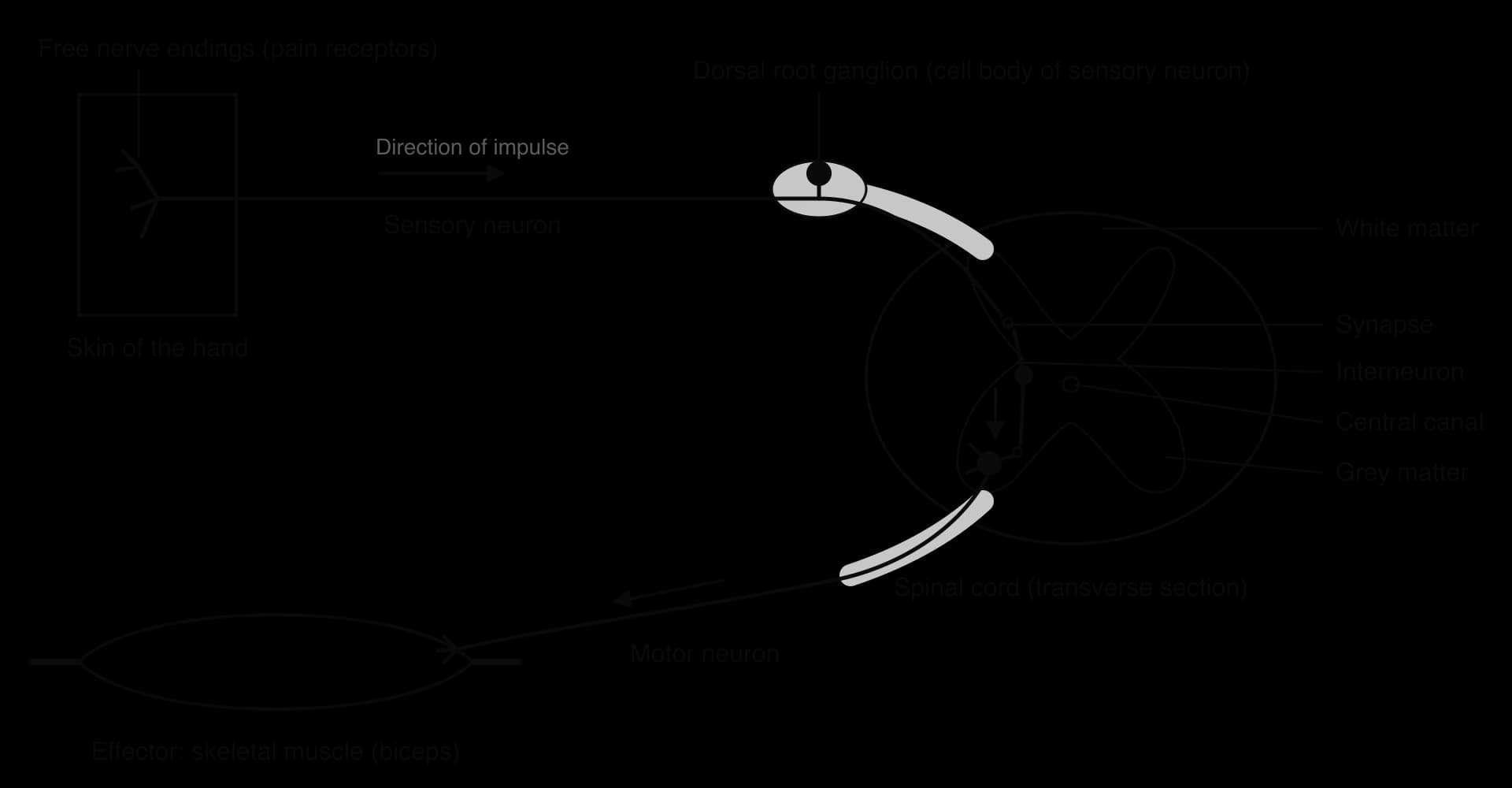
Stimulus and receptor. Heat damages the skin of the hand. Free nerve endings of sensory neurons in the skin act as pain receptors. They contain ion channels that open in response to high temperature, which depolarises the nerve ending and starts nerve impulses.
Sensory neuron. The impulses travel along the sensory neuron to the spinal cord. They pass the cell body in the dorsal root ganglion and enter the grey matter of the spinal cord through the dorsal root.
Interneuron. In the grey matter, the sensory neuron synapses with an interneuron (also called a relay neuron). The interneuron is the integrating step: it passes the signal on to a motor neuron, and branches of the interneuron also carry impulses up the spinal cord to the brain.
Motor neuron. The interneuron synapses with a motor neuron, whose cell body is in the grey matter. Impulses travel out of the spinal cord through the ventral root and along the motor neuron to the arm.
Effector and response. At the neuromuscular junctions, the motor neuron stimulates the biceps muscle to contract. The arm bends at the elbow and the hand is pulled away from the hot object.
Because only three neurons and two synapses are involved, and the pathway does not pass through the brain, the response is very fast. The brain does receive information about the event through other neurons in the spinal cord. That is why pain is felt a moment later, and why a person can later remember the event and learn to avoid hot objects.
The cerebellum sits at the back of the brain, below the cerebral hemispheres. Although it makes up only about a tenth of the volume of the brain, it contains more than half of the brain's neurons. It does not start movements; the primary motor cortex does that. Instead, the cerebellum makes movements smooth, accurate and well timed.
The cerebellum receives information from the motor cortex about the movement that is planned, and information from the inner ear, the eyes and stretch receptors in muscles and joints about the movement that is actually happening. It compares the two and sends corrections, so that the right muscles contract with the right force at the right moment. Its main roles are:
timing and coordination of contractions in many muscles at once, as in walking, running or playing a musical instrument;
balance and posture, using information from the inner ear about the position and movement of the head;
fine motor control, for precise movements such as writing or threading a needle;
learning motor skills, so that with practice a movement such as riding a bicycle becomes automatic.
Damage to the cerebellum, for example by a stroke, does not cause paralysis, because the motor cortex can still start movements. Instead the person moves clumsily, may walk with a wide, unsteady gait, and finds it hard to touch a target accurately. Alcohol affects the cerebellum strongly, which is why a person who is drunk may lose balance and slur their speech.
The central and peripheral nervous systems, sensory and motor neurons, and the way the brain and spinal cord integrate information are summarised in the video.
 YouTube
Many processes in the body follow a cycle of about 24 hours, even when a person is kept in constant light or darkness. These cycles are circadian rhythms (from the Latin circa diem, "about a day"). Body temperature, alertness, the secretion of several hormones and the pattern of sleep and waking all follow circadian rhythms.
The master clock of the body is the suprachiasmatic nucleus (SCN), a small group of about 20,000 neurons in the hypothalamus. Its neurons produce a rhythm of activity of about 24 hours by themselves, through a cycle of gene expression in which certain proteins build up and then switch off their own genes. Light resets this clock every day: special light-sensitive ganglion cells in the retina send impulses along the optic nerve directly to the SCN.
The SCN controls the pineal gland, a small endocrine gland near the centre of the brain. The pineal gland secretes the hormone melatonin into the blood. When light reaches the retina, the SCN inhibits melatonin secretion, so its concentration is very low during the day. After dark, the inhibition stops and melatonin secretion rises, reaching a peak in the middle of the night before falling again towards morning.
The graph of blood melatonin shows this diurnal pattern. Melatonin acts on receptors in the SCN and in other tissues. It lowers body temperature slightly and increases sleepiness, which helps the body prepare for sleep at night. Its fall in the morning helps the body to wake. In this way melatonin links the clock in the brain to the timing of sleep in the whole body.
Genes that build circadian clocks were first identified in the fruit fly Drosophila melanogaster. Jeffrey Hall, Michael Rosbash and Michael Young received the Nobel Prize in Physiology or Medicine in 2017 for discovering how the period gene and its protein produce a daily rhythm of activity inside cells. Similar clock genes have since been found in humans and most other animals, which shows that the mechanism is very ancient.
After a long flight across several time zones, the SCN is still running on the time of the place of departure. Melatonin is secreted at the wrong time of day, and the person feels sleepy during the day and awake at night. The clock is gradually reset by daylight at the new location, typically by about one hour per day. Melatonin tablets taken at the local bedtime are sometimes used to reduce jet lag, and bright light at the right time of day helps reset the clock. People who work night shifts have a similar problem, because their sleep is out of step with their melatonin rhythm.
The two adrenal glands sit on top of the kidneys. Their inner region, the adrenal medulla, secretes the hormone epinephrine (also called adrenaline). Epinephrine is released within seconds when the body faces a threat or prepares for intense physical activity, as in the "fight or flight" response. Secretion is triggered by impulses from the sympathetic nervous system, so this is an example of the nervous and endocrine systems working together.
Epinephrine travels in the blood to almost every organ, and many cell types have receptors for it. Its effects are widespread, but all of them have the same overall result: more oxygen and glucose reach the skeletal muscles so that they can contract intensely for longer.
Target | Effect of epinephrine | How this helps vigorous muscle contraction |
|---|---|---|
Heart | Increases heart rate and the force of contraction, so stroke volume rises | More blood, and so more oxygen and glucose, reaches the muscles each minute |
Airways (bronchioles) | Smooth muscle relaxes, so the airways dilate | Air flows into the alveoli more easily |
Breathing | Ventilation rate and depth increase | More oxygen is absorbed and more CO₂ removed |
Arterioles to skeletal muscles | Vasodilation | More blood flows to the muscles that are working |
Arterioles to the gut and skin | Vasoconstriction | Blood is diverted away from organs that are not needed for immediate activity |
Liver | Glycogen is broken down to glucose, which is released into the blood | Blood glucose rises, giving muscles more substrate for respiration |
Eyes | Pupils dilate | More light enters the eye, improving vision |
The changes described in the table also show why epinephrine is used in medicine. In anaphylaxis, a severe allergic reaction, the airways narrow and blood vessels widen so much that blood pressure falls dangerously. An epinephrine auto-injector (for example an EpiPen) delivers a dose of epinephrine into the thigh muscle. The hormone dilates the airways and constricts blood vessels, raising blood pressure, within minutes.
The hypothalamus is a small region at the base of the brain, just above the pituitary gland. It is the main link between the nervous system and the endocrine system. Like the rest of the brain it receives impulses from other neurons, and it also has a rich blood supply, which allows it to monitor the blood directly: it contains receptors that detect blood temperature, solute concentration and the levels of several hormones.
The pituitary gland is a pea-sized gland that hangs from the hypothalamus on a short stalk. It is often called the "master gland" because many of its hormones control other endocrine glands, such as the thyroid gland, the adrenal glands, the ovaries and the testes. The pituitary has two lobes, and the hypothalamus controls each of them in a different way.

Anterior pituitary. Neurosecretory cells in the hypothalamus secrete releasing hormones into a short portal blood vessel that runs down the stalk to the anterior lobe. These releasing hormones stimulate (or inhibit) endocrine cells in the anterior pituitary, which then secrete their own hormones into the blood. Examples are follicle-stimulating hormone (FSH) and luteinising hormone (LH), which control the ovaries and testes; thyroid-stimulating hormone (TSH), which stimulates the thyroid gland; and growth hormone (GH).
Posterior pituitary. Other neurosecretory cells in the hypothalamus make the hormones antidiuretic hormone (ADH) and oxytocin. These hormones travel down the axons of the neurosecretory cells, through the stalk, and are stored in the posterior lobe. When impulses arrive, the hormones are released into the blood.
The hypothalamus–pituitary system is controlled by feedback. For example, when the concentration of a hormone from the thyroid gland is high, the hypothalamus and anterior pituitary secrete less TSH, so less thyroid hormone is produced. In this way the hypothalamus integrates information from the nervous system and from the blood, and adjusts the activity of glands throughout the body.
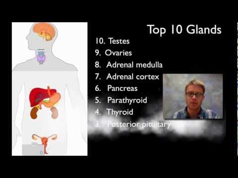
The way the hypothalamus controls the pituitary, and the feedback loops that keep hormone concentrations stable, are summarised in the video.
 YouTube
Many conditions in the body are kept close to a set value by negative feedback. A receptor detects a change away from the set value, a coordinator processes the information, and effectors produce a response that reverses the change. As conditions return to normal, the stimulus is reduced and so is the response. Negative feedback is the basis of homeostasis (D3.3).
Positive feedback works the other way: a change triggers a response that increases the change still further. Positive feedback does not keep conditions stable. It is used where a process must be completed quickly once it has started, such as the contractions of the uterus during childbirth or, in plants, the ripening of fruit (an HL example).
The heart muscle contracts without any stimulus from nerves. Each beat starts in the sinoatrial node (SA node), a small group of specialised cardiac muscle cells in the wall of the right atrium, which acts as the pacemaker. A heartbeat that starts in the muscle itself is described as myogenic. A heart removed from the body for a transplant continues to beat for a time, which shows that the brain is not needed to start each beat.
Although the beat is myogenic, its rate is adjusted by the brain to match the needs of the body. At rest the SA node of an adult produces about 60–80 beats per minute; during intense exercise the heart rate can rise to 180 beats per minute or more. The brain also adjusts the force of each contraction, and so the stroke volume (the volume of blood pumped by the left ventricle in one beat).
Two types of receptor provide the information that the brain uses:
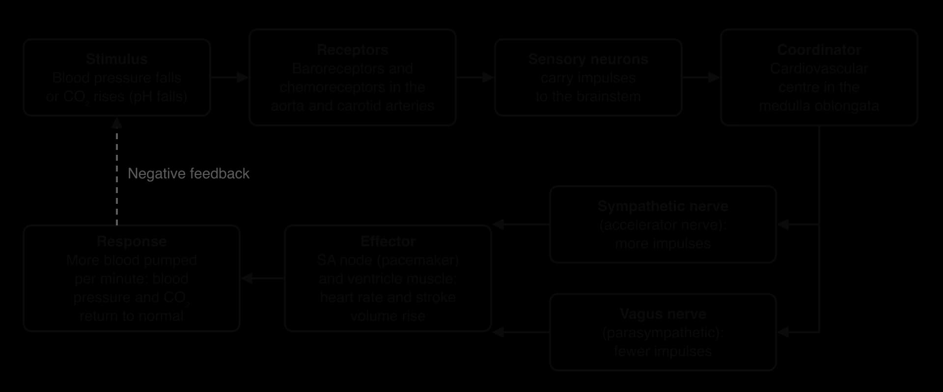
Baroreceptors are stretch receptors in the walls of the aorta (the aortic arch) and the carotid arteries (the carotid sinuses), which supply the head. They detect blood pressure: when pressure rises the artery walls stretch more and the baroreceptors send more impulses.
Chemoreceptors in the carotid bodies and aortic bodies, and in the medulla itself, detect the chemical composition of the blood: its pH and its concentrations of carbon dioxide and oxygen.
Impulses from these receptors travel along sensory neurons to the cardiovascular centre in the medulla oblongata. The cardiovascular centre integrates this information and sends impulses along two nerves to the SA node:
the sympathetic nerve (accelerator nerve), whose impulses increase the heart rate and the force of contraction;
the vagus nerve (part of the parasympathetic system), whose impulses decrease the heart rate.
The heart rate at any moment depends on the balance between impulses in these two nerves. Epinephrine in the blood also raises the heart rate by acting directly on the SA node.
The rate and depth of breathing are also controlled by negative feedback. The coordinator is the respiratory centre in the medulla oblongata, which sends rhythmic bursts of impulses along nerves to the diaphragm and the intercostal muscles. Each burst causes these muscles to contract, producing an inspiration; when the impulses stop, the muscles relax and expiration follows.
The main stimulus that changes the ventilation rate is the concentration of carbon dioxide in the blood. CO₂ reacts with water to form carbonic acid, which releases hydrogen ions:
A rise in blood CO₂ therefore lowers the pH of the blood. Chemoreceptors in the medulla detect the fall in pH of the fluid around them, and chemoreceptors in the carotid and aortic bodies detect the changes in the blood. The carotid and aortic bodies also respond to a large fall in oxygen concentration, but in healthy people at sea level this is rarely the stimulus.
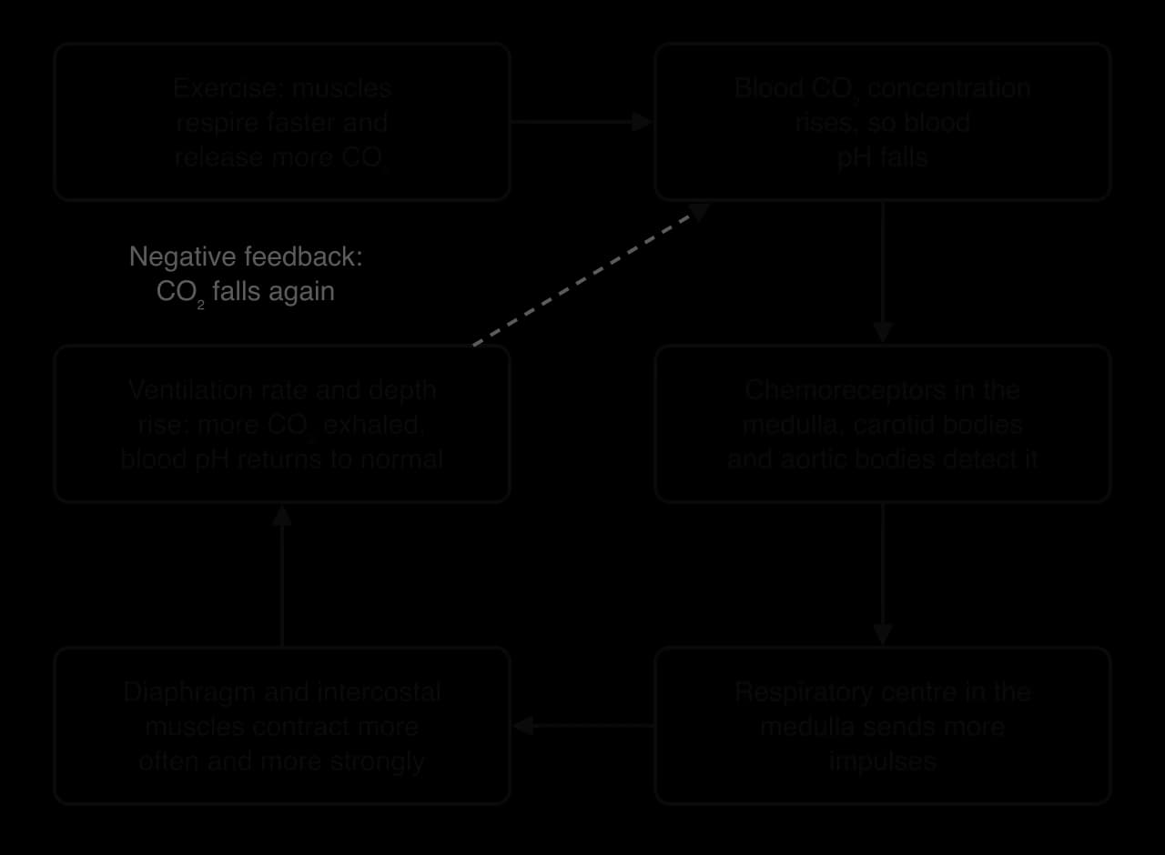
During exercise, muscle cells respire faster and release more CO₂ into the blood.
The CO₂ concentration of the blood rises, so its pH falls.
Chemoreceptors detect the fall in pH and send impulses to the respiratory centre in the medulla.
The respiratory centre sends impulses more often, and more strongly, to the diaphragm and intercostal muscles.
The muscles contract more frequently and more forcefully, so the ventilation rate and the depth of breathing (tidal volume) increase.
More CO₂ is exhaled, so blood CO₂ falls and pH rises back towards normal. The chemoreceptors then send fewer impulses, and breathing slows again.
Food is moved along the gut by peristalsis, a wave of muscle contraction that passes along the wall of the oesophagus, stomach and intestines. The wall of the gut contains two layers of smooth muscle: an inner layer of circular muscle, whose fibres run around the gut, and an outer layer of longitudinal muscle, whose fibres run along it.
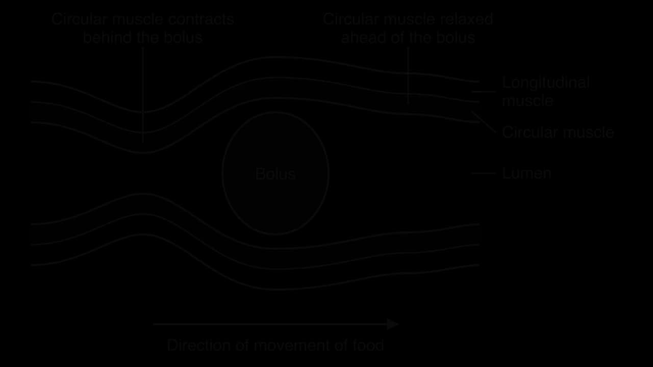
Behind a mass of food (a bolus), the circular muscle contracts, which narrows the gut and squeezes the food forwards. Just ahead of the bolus, the circular muscle relaxes and the longitudinal muscle contracts, which shortens and widens that section so the food can move into it. This wave of contraction and relaxation passes along the gut and pushes food in one direction only.
Only the two ends of the digestive system are under voluntary control by the CNS. Swallowing is started voluntarily: the tongue, controlled by the brain, pushes food to the back of the mouth, and from that point a swallowing reflex coordinated by the brainstem takes over. At the other end, egestion of faeces is controlled by a ring of skeletal muscle (the external anal sphincter), which can be relaxed or contracted by choice.
Between swallowing and egestion, peristalsis is involuntary. It is controlled by the enteric nervous system (ENS), a network of hundreds of millions of neurons in the wall of the gut, from the oesophagus to the anus. The ENS includes sensory neurons that detect stretching of the gut wall by food, interneurons, and motor neurons that stimulate the circular and longitudinal muscle. When food stretches the wall, the ENS causes contraction behind the bolus and relaxation in front of it.
The ENS can work even when the nerves that connect it to the brain are cut, which is why it is sometimes called the "second brain". The CNS can still adjust its activity: impulses from the brain speed up or slow down digestion, which is why stress can affect the gut. The importance of the ENS is shown by Hirschsprung disease, a condition present at birth in about 1 in 5,000 babies, in which ENS neurons are missing from the last part of the large intestine. Peristalsis cannot happen in that region, so faeces cannot pass, and the affected section must be removed by surgery.
Plants cannot move from place to place, but they can change the direction in which they grow. A tropism is a growth response in which the direction of growth depends on the direction of the stimulus. Growth towards the stimulus is a positive tropism, and growth away from it is a negative tropism.
The two tropisms most often studied are responses to light and to gravity:
Phototropism is a growth response to light from one direction. Shoots show positive phototropism: they grow towards the light. Roots of many species show weak negative phototropism.
Gravitropism is a growth response to gravity. Roots show positive gravitropism (they grow downwards), and shoots show negative gravitropism (they grow upwards).
Together these responses allow a germinating seed, whichever way up it lands in the soil, to send its root down towards water and minerals and its shoot up towards light.
Many experiments on phototropism use the coleoptiles of grass seedlings such as oat (Avena sativa) and canary grass (Phalaris canariensis). A coleoptile is the protective sheath that covers the first leaf as it grows up through the soil. It is a simple, straight, fast-growing cylinder, so its bending is easy to see and measure.
In 1880 Charles Darwin and his son Francis reported that canary grass coleoptiles bend towards light from one side only if their tip is exposed. When the tip was covered with an opaque cap, or cut off, there was no bending, even though the bending happens in the region below the tip. They concluded that the tip detects the light and sends an "influence" down to the growing region. In the 1920s Frits Went placed oat coleoptile tips on small blocks of agar and then put the blocks, without the tips, on one side of cut coleoptiles kept in the dark. The coleoptiles bent away from the side with the block, which showed that the influence is a chemical that diffuses out of the tip. This chemical was named auxin. These experiments are good examples of hypotheses tested by controlled experiments, each step narrowing down the explanation.
Tropic responses can be recorded qualitatively and quantitatively. Qualitative observations describe what happens without numbers, for example "the shoots bent towards the window". They are usually recorded as labelled diagrams drawn at intervals, showing the shape and direction of growth of each seedling. Quantitative data are numerical. The usual measurement for a tropic response is the angle of curvature: the angle between the original direction of growth and the direction in which the tip is now growing.
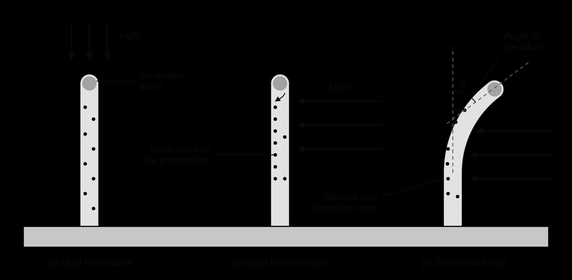
A typical investigation of phototropism uses seedlings of cress (Lepidium sativum), oat or mustard grown in the dark until they are a few centimetres tall. They are then placed in a box with a slit on one side, so that light reaches them from one direction only. After a set time, each seedling is photographed from the side against a grid, and the angle of curvature is measured on the photograph with a protractor or with image software. A control group is grown with light from above or on a slowly rotating turntable. Gravitropism is investigated in a similar way, using a clinostat: a slowly rotating disc that turns the seedlings so that gravity acts on all sides equally, as a control for seedlings that are kept still.
Measurements of curvature are only useful if their quality is known. Three different ideas are involved:
Precision describes how close repeated measurements are to each other, and how fine the scale of the instrument is. A protractor read to the nearest 1° is more precise than an estimate to the nearest 10°.
Accuracy describes how close a measurement is to the true value. Measuring from a photograph taken at an angle, or from the base rather than the original direction of growth, gives inaccurate angles even if they are read precisely.
Reliability describes whether the results can be repeated. Seedlings vary, so several seedlings (for example 10 or more) are measured for each condition and a mean is calculated. If repeated trials give similar means, the results are reliable.
Positive phototropism is a directional growth response to lateral light, that is, light from one side. Its consequence is that the leaves are moved into a position where they absorb more light for photosynthesis. Seedlings growing in the shade of other plants can bend towards gaps, and house plants on a windowsill gradually turn towards the window.
The response happens in three stages:
Detection. Blue-light receptor proteins called phototropins, in the cells near the shoot tip, detect the direction of the light.
Redistribution of auxin. Auxin made in the tip is transported towards the shaded side of the shoot, so its concentration becomes higher on the shaded side than on the lit side.
Differential growth. Cells on the shaded side elongate more than cells on the lit side, so the shoot bends towards the light.
A phytohormone (plant hormone) is a signalling chemical produced in small amounts in one part of a plant that controls growth, development or a response to a stimulus, either in the same tissue or elsewhere in the plant. Phytohormones are carried from cell to cell, in the xylem or in the phloem. Unlike animal hormones, they are not made in special glands, and many cells can both produce them and respond to them.
Plants use a variety of chemicals as phytohormones. Five main groups are well known, and each has several effects that depend on its concentration, the tissue and the other phytohormones present.
Phytohormone | Main site of production | Examples of effects |
|---|---|---|
Auxin (IAA) | Shoot tips, young leaves | Cell elongation in shoots, phototropism and gravitropism, apical dominance, formation of roots |
Cytokinins | Root tips | Cell division, growth of side shoots, delay of leaf ageing |
Gibberellins | Young leaves, seeds | Stem elongation, seed germination, flowering in some species |
Ethylene (ethene) | Ripening fruit, ageing tissues | Fruit ripening, leaf and fruit fall |
Abscisic acid (ABA) | Leaves, roots in dry soil | Closing of stomata in drought, seed dormancy |
The effects of phytohormones overlap and interact, so plant growth is integrated by the balance between them. This is the plant equivalent of the way several hormones together control human metabolism.
The main natural auxin is indole-3-acetic acid (IAA). It is a weak acid, so it exists in two forms: the uncharged form, IAAH, and the negatively charged ion, IAA⁻. The form that predominates depends on pH.
In the cell wall, the pH is about 5.5. At this pH a large proportion of auxin is in the uncharged form, IAAH. Being uncharged and fairly lipid-soluble, IAAH diffuses across the plasma membrane into the cell.
In the cytoplasm, the pH is about 7. Here IAAH loses an H⁺ ion and becomes IAA⁻. The charged ion cannot diffuse back out through the phospholipid bilayer, so auxin is trapped inside the cell.
IAA⁻ can only leave the cell through auxin efflux carriers, membrane proteins called PIN proteins. These are often located on one side of the cell only, for example the lower (basal) side.
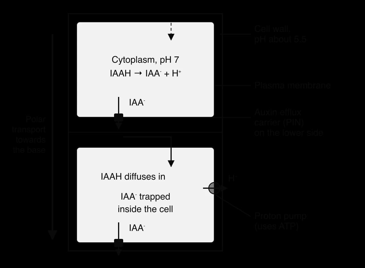
When every cell in a tissue places its efflux carriers on the same side, auxin is passed from cell to cell in one direction, like a bucket chain. This directional movement is called polar transport, and it moves auxin from the shoot tip down towards the roots. Proton pumps in the plasma membrane keep the wall acidic and the cytoplasm neutral, which maintains the pH difference that makes the system work, so polar transport uses energy from ATP.
Cells can change the position of their efflux carriers in response to stimuli. In a shoot lit from one side, cells near the tip move PIN proteins to the side of the cell that faces away from the light. Auxin is then pumped sideways towards the shaded side of the shoot, building up a concentration gradient across it. By controlling the position of efflux carriers, a plant concentrates auxin in particular places, such as the shaded side of a stem, the lower side of a horizontal root or the sites where new leaves will form.
Plant cells grow mainly by taking in water and elongating. The cell wall resists this expansion, because its cellulose microfibrils are held together by cross-links of other polysaccharides. For a cell to elongate, these cross-links must loosen so that turgor pressure can stretch the wall.
Auxin promotes elongation by stimulating cells to pump H⁺ ions into the cell wall. Auxin binds to receptors in the cell, which activates proton pumps in the plasma membrane and increases the number of pumps. The pumps use ATP to move H⁺ from the cytoplasm into the cell wall, and the pH of the wall falls further, to about 4.5–5. The low pH activates proteins in the wall called expansins, which break hydrogen bonds between the cellulose microfibrils and the cross-linking polysaccharides. The microfibrils can then slide apart, and the pressure of the cell contents stretches the wall. This mechanism is called acid growth.
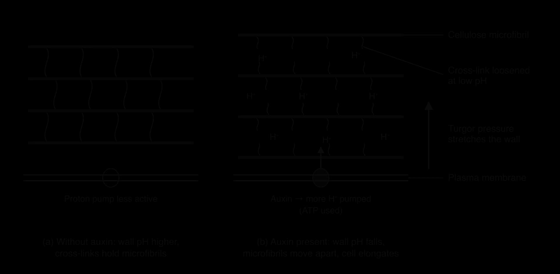
Acid growth explains phototropism. Auxin concentration is higher on the shaded side of a shoot, so the cells there pump more H⁺ into their walls, their walls loosen more, and they elongate faster than cells on the lit side. A concentration gradient of auxin across the shoot therefore produces a difference in growth rate, which bends the shoot towards the light. The effect of auxin depends on concentration: concentrations that promote growth in shoots inhibit elongation in roots, which are far more sensitive.
A plant needs a balance between its shoot system, which makes sugars by photosynthesis, and its root system, which absorbs water and minerals. Two phytohormones, made at opposite ends of the plant, help to keep this balance. Shoot tips produce auxin, which is transported down the stem to the roots. Root tips produce cytokinins, which are carried up to the shoots in the xylem.
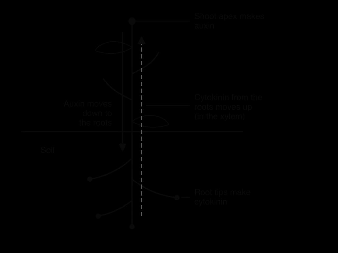
Auxin and roots. Auxin arriving in the roots stimulates the formation of new lateral roots. A large, actively growing shoot sends more auxin down, so the root system grows to match it.
Auxin and side shoots. Auxin from the shoot tip inhibits the growth of side buds lower down the stem, so the main shoot grows strongly. This is apical dominance. When the tip is removed, as in pruning, the auxin supply falls and the side buds start to grow, which makes the plant bushier.
Cytokinin and shoots. Cytokinin arriving in the shoot promotes cell division and the growth of side buds, opposing apical dominance. A large, healthy root system sends more cytokinin up, so the shoot can branch and grow.
The effects depend on the ratio of the two hormones. This was shown clearly in plant tissue culture in the 1950s, when Folke Skoog and Carlos Miller grew undifferentiated tobacco tissue (callus) on media with different amounts of auxin and cytokinin. A high ratio of auxin to cytokinin caused roots to form, a high ratio of cytokinin to auxin caused shoots to form, and equal amounts kept the callus growing without forming either. Gardeners use the same principle when they dip cuttings in rooting powder, which contains a synthetic auxin.
Ripening makes a fruit attractive to animals that eat it and disperse its seeds. During ripening the fruit softens as cell walls are broken down, starch is converted to sugars, acids decrease, the colour changes (for example from green to yellow in bananas as chlorophyll is broken down) and scented compounds are released. In many fruits, such as bananas, tomatoes, apples and avocados, ripening is triggered and coordinated by the gas ethylene (ethene, C₂H₄).
Ethylene stimulates ripening, and ripening tissue produces more ethylene. A small rise in ethylene therefore causes some ripening, which releases more ethylene, which causes faster ripening. This is positive feedback. Because ethylene is a gas, it also diffuses out of the fruit and into neighbouring fruits, where it triggers the same cycle.
The graph of ethylene production shows the typical shape of a process controlled by positive feedback: a slow start followed by a steep, accelerating rise. Positive feedback has two consequences for the plant:
Ripening is rapid: once it has started in a fruit, it is completed quickly instead of dragging on.
Ripening is synchronised: all the parts of one fruit, and the fruits on one plant, ripen at about the same time. Seed-dispersing animals are therefore attracted to a plant with many ripe fruits at once, which increases the number of seeds dispersed.
People make use of this positive feedback. Bananas are harvested and shipped green and hard, so they are not damaged in transport, and are then ripened in sealed rooms filled with ethylene gas before sale. At home, an unripe avocado ripens faster in a paper bag with a ripe banana, and one rotting apple can make the rest of the apples in a box ripen and spoil faster. In the opposite direction, apples in long-term storage are treated with 1-methylcyclopropene (1-MCP), which blocks ethylene receptors and delays ripening for months; it was approved for use on apples in the United States in 2002.
Integration of body systems is assessed across the papers. Paper 1A multiple-choice questions often ask which part of the brain controls a function, which receptor detects a stimulus, or which row of a table correctly compares nervous and hormonal signalling. Paper 1B data questions use heart rate, ventilation or hormone data and, at HL, measurements of curvature in seedlings. Paper 2 short-answer questions ask candidates to label a reflex arc or a section of a nerve, and extended-response questions ask them to explain feedback control of heart rate or ventilation, or (HL) phototropism. The command terms used most are state, outline, distinguish, explain and evaluate.
The practice question uses the following data. Heart rate and ventilation rate were measured in a student at rest and after 5 minutes of running.
Measurement | At rest | After running |
|---|---|---|
Heart rate / beats min⁻¹ | 68 | 150 |
Ventilation rate / breaths min⁻¹ | 14 | 35 |
Drawing questions on integration are marked strictly. A diagram of a reflex arc or a nerve earns marks only when the labelled structures are clearly identifiable, and when labels are attached by straight lines that end exactly on the structure.
The two drawings on integration that appear most often are the pain reflex arc and the transverse section of a nerve. Both are easier to reproduce accurately after they have been drawn several times from memory and checked against a labelled diagram.
I can explain why integration is needed and give the cheetah as an example of an emergent property.
I can compare the nervous and endocrine systems in speed, duration, targeting and type of signal.
I can outline the roles of the cerebral hemispheres, primary motor cortex, cerebellum, medulla oblongata and spinal cord.
I can use the case of Henry Molaison to explain the brain's role in learning and memory.
I can draw and label a transverse section of a nerve and explain the difference between a nerve and a neuron.
I can describe the pain reflex arc from free nerve ending to effector.
I can explain how melatonin, epinephrine and the hypothalamus and pituitary gland integrate body functions.
I can explain the feedback control of heart rate and ventilation rate.
I can outline how the CNS and the enteric nervous system control swallowing, peristalsis and egestion.
I can measure angles of curvature in seedlings and evaluate precision, accuracy and reliability. (HL)
I can explain phototropism using auxin efflux carriers and acid growth. (HL)
I can explain auxin–cytokinin interaction and positive feedback in fruit ripening. (HL)
[7 max]
Distinguish between nervous and hormonal signalling in animals.
4 marks
a. nervous signals are electrical (impulses) with chemical synapses but hormonal signals are chemical only;
b. nerve impulses travel along neurons but hormones are carried in the blood;
c. nervous responses are rapid but hormonal responses are (usually) slower;
d. nervous effects are short-lived but hormonal effects (often) last longer;
e. nervous signals go to specific effectors / a localized target but hormones reach all cells / any cell with receptors / are widespread;
f. nervous effectors are muscles and glands but hormones can act on many cell types / tissues;
[4 max]
Each point must give both sides of the difference. Accept answers in a table.
One additional mark is available for the construction of the answer.
The nervous system processes information through the interaction of many neurons.
Outline why consciousness is regarded as an emergent property of the brain.
4 marks
Explain how excitatory and inhibitory neurotransmitters together determine whether a postsynaptic neuron transmits an impulse.
7 marks
Describe the pain reflex arc that withdraws the hand from a sharp object.
4 marks
A peregrine falcon (Falco peregrinus) can dive at more than 300 km h-1 and seize a flying bird in mid-air. Why is this hunting ability described as an emergent property?