Explain how neurons generate resting and action potentials, how impulse speed varies and how synapses pass signals between cells; at HL, the ion channels, propagation, summation, pain and consciousness.
Each is in the Biology glossary, with every other term in the subject.
A neurotransmitter (ACh) released at many synapses, including neuromuscular junctions, that binds to receptors that open Na⁺ channels; it is broken down by acetylcholinesterase.
An enzyme in the synaptic cleft that hydrolyses acetylcholine into choline and acetate, ending its effect on the postsynaptic membrane.
A rapid, temporary reversal of the membrane potential of a neuron, from about −70 mV to about +30 mV and back, caused by the movement of Na⁺ and K⁺ ions across the membrane.
The property of an action potential that it either occurs fully, with the same size, once the threshold potential is reached, or does not occur at all.
The single long nerve fibre of a neuron that carries impulses away from the cell body to the axon terminals.
The part of a neuron that contains the nucleus and most of the cytoplasm, from which the nerve fibres (dendrites and axon) project.
The square of the correlation coefficient (R²), a value from 0 to 1 giving the proportion of the variation in the dependent variable explained by its linear relationship with the independent variable.
The state of awareness of oneself and one’s surroundings; an emergent property arising from the interactions of very large numbers of neurons in the brain.
The nervous system carries information around the body as electrical signals in nerve cells. These signals let an animal detect a change, process it and respond to it within a fraction of a second, far faster than hormones can.
Neural signalling has two parts. Along a nerve cell, the signal is electrical: a brief change in the charge across the plasma membrane that travels from one end of the cell to the other. Between nerve cells, the signal is chemical: a neurotransmitter released by one cell changes the membrane of the next.
42 exam-style questions on this subtopic, written in the style of the IB papers. The first has its markscheme open. Counted on
The table shows estimates of the number of motor neurons supplying three human skeletal muscles and the number of muscle fibres in each muscle.
Muscle | Number of motor neurons | Number of muscle fibres | Mean number of fibres per motor unit |
|---|---|---|---|
muscle that moves the eyeball | 2 970 | 27 000 | 9 |
muscle that moves the index finger | 120 | 40 800 | 340 |
calf muscle that raises the heel | 580 | 1 040 000 |
Calculate the mean number of fibres per motor unit in the calf muscle.
1 mark
1 793 (accept 1 790 to 1 800);
Working: 1 040 000 ÷ 580.
State the name of the junction between a motor neuron and a muscle fibre.
1 mark
The official deck for this part of the syllabus.
How does the speed of neural signalling compare with hormonal signalling?
State the two parts of neural signalling and the form the signal takes in each.
With a free account, Augment remembers where you stopped and what you have practiced.
One of the multiple short, branched nerve fibres of a neuron that receive signals and carry impulses towards the cell body.
A reduction in the potential difference across a membrane, making the inside less negative, as when Na⁺ ions diffuse into a neuron.
A local depolarisation of the postsynaptic membrane, caused by a neurotransmitter opening channels that let positive ions such as Na⁺ into the cell, which makes an action potential more likely.
A change in membrane potential that makes the inside more negative than the resting potential, as after an action potential or during an inhibitory postsynaptic potential.
A hyperpolarisation of the postsynaptic membrane, caused by an inhibitory neurotransmitter such as GABA opening Cl⁻ or K⁺ channels, which makes an action potential less likely.
The movement of ions, mainly Na⁺ diffusing along the inside of an axon, from a depolarised region to the adjacent resting region, which depolarises that region to the threshold potential.
The difference in electrical charge (potential difference) across a plasma membrane, measured in millivolts, with the inside usually negative compared with the outside.
An insulating layer around an axon, made of many layers of plasma membrane wrapped round by Schwann cells, with small gaps at intervals, that increases the speed of nerve impulses.
An insecticide that binds to nicotinic acetylcholine receptors in insect synapses and is not broken down by acetylcholinesterase, so it blocks synaptic transmission.
An elongated extension of a neuron, either an axon or a dendrite, along which electrical impulses are conducted.
An action potential propagated along a nerve fibre, carrying a signal from one end of a neuron to the other.
A synapse between a motor neuron and a skeletal muscle fibre, at which acetylcholine is released to stimulate contraction.
A cell of the nervous system, made of a cell body with a nucleus and elongated nerve fibres, that carries electrical impulses.
A signalling chemical released by a presynaptic neuron that diffuses across the synaptic cleft and binds to receptors on the postsynaptic membrane.
A short gap in the myelin sheath of an axon where the membrane is exposed and voltage-gated channels are concentrated, so action potentials can occur.
An instrument that displays changes in voltage against time; used to record resting and action potentials from electrodes inside and outside a neuron.
The neuron (or effector cell) on the receiving side of a synapse, whose membrane carries receptors for the neurotransmitter.
The neuron that releases neurotransmitter into the synaptic cleft at a synapse.
The short time after an action potential, about 1–2 ms, during which a region of membrane cannot produce another action potential, because its voltage-gated sodium channels are inactivated.
The return of the membrane potential towards the negative resting potential after depolarisation, caused by voltage-gated potassium channels opening and K⁺ diffusing out of the neuron.
The potential difference across the plasma membrane of a neuron that is not conducting an impulse, about −70 mV, with the inside negative compared with the outside.
The removal of a neurotransmitter from the synaptic cleft by transporter proteins that carry it back into the presynaptic neuron for reuse.
The conduction of a nerve impulse along a myelinated axon in which action potentials occur only at the nodes of Ranvier, so the impulse jumps from node to node.
The combining of excitatory and inhibitory postsynaptic potentials arriving at a postsynaptic neuron, which produces an action potential only if their combined effect reaches the threshold potential.
A junction between a neuron and another neuron or an effector cell, across which a signal is passed by a neurotransmitter diffusing across a narrow gap, the synaptic cleft.
The narrow gap, about 20 nm wide, between the presynaptic and postsynaptic membranes at a synapse, across which neurotransmitter diffuses.
The membrane potential, about −50 mV, that must be reached for enough voltage-gated sodium channels to open to start an action potential.
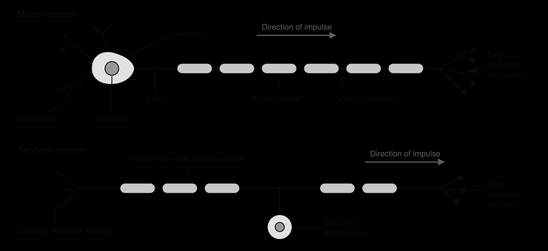
The cells of the nervous system that carry signals are neurons. Each neuron has a cell body, which contains the nucleus and most of the cytoplasm, and elongated nerve fibres that project from it. The fibres can be very long: the axon of a motor neuron that runs from the spinal cord to a muscle in the foot is about 1 m long, although the cell body is only about 0.1 mm across.
There are two kinds of nerve fibre:
Dendrites are multiple, short, branched fibres. A typical dendrite receives signals from other neurons and carries impulses towards the cell body.
The axon is a single, long fibre. The axon carries impulses away from the cell body to the axon terminals, where it passes the signal on to other neurons or to effectors such as muscle fibres.
Electrical impulses are conducted along these fibres. In many neurons the axon is wrapped in a myelin sheath, made of many layers of plasma membrane from Schwann cells (in nerves outside the brain and spinal cord). The sheath has small gaps, about 1 mm apart, where the axon membrane is exposed. Myelin increases the speed of the impulse.
Neurons differ in shape according to their function, which is a clear case of form fitting function. A motor neuron carries impulses from the central nervous system (the brain and spinal cord) to an effector, such as a muscle. Its cell body lies inside the spinal cord, with many dendrites around it and one long myelinated axon that ends in branches on muscle fibres. A sensory neuron carries impulses from a receptor to the central nervous system. Its fibre starts at sensory receptor endings, often in the skin, and runs all the way to the spinal cord, with the cell body on a short side branch near the spinal cord. Interneurons within the central nervous system connect sensory and motor neurons and each other.
All cells have a difference in electrical charge across their plasma membrane: the inside is negative compared with the outside. This difference is the membrane potential, measured in millivolts (mV). A membrane with a potential across it is described as polarised. In a neuron that is not carrying an impulse, the membrane potential is called the resting potential and is about −70 mV.
The resting potential depends on concentration gradients of sodium ions (Na⁺) and potassium ions (K⁺), which are set up by the sodium–potassium pump. This pump protein in the plasma membrane uses energy from ATP to move ions against their concentration gradients, a form of active transport. Each cycle of the pump moves 3 Na⁺ out of the cell and 2 K⁺ into it, using one ATP.
Running continuously, the pumps build up a high concentration of Na⁺ outside the neuron and a high concentration of K⁺ inside it. A large part of the ATP made by brain cells is used to drive these pumps.
Ion | Concentration inside / mmol dm⁻³ | Concentration outside / mmol dm⁻³ |
|---|---|---|
Na⁺ | 5–15 | 145 |
K⁺ | 140 | 5 |
Source: typical values for a mammalian neuron, Purves et al., Neuroscience (table of intracellular and extracellular ion concentrations). Exact values vary between cell types.
Three things together make the inside of the neuron negative:
Unequal pumping. The pump moves three positive charges out for every two it moves in, so each cycle leaves the inside slightly more negative.
Potassium leaks out more than sodium leaks in. The membrane contains K⁺ leak channels that are always open, and far fewer open Na⁺ channels. K⁺ ions diffuse out down their concentration gradient much faster than Na⁺ ions diffuse in, so positive charge is lost from the cell. This is the largest effect.
Negatively charged proteins stay inside. The cytoplasm contains many large proteins and other organic molecules with negative charges. They cannot cross the membrane, so they remain inside and add to its negative charge.
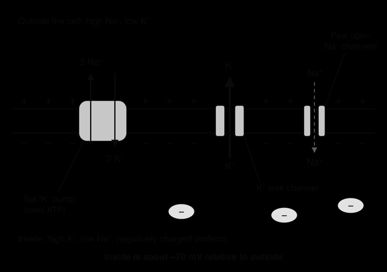
The resting potential is a steady state maintained by continuous pumping. Ions leak across the membrane all the time, and the pumps keep working to restore the gradients. If the pumps are stopped, for example by a poison that blocks ATP production, the gradients slowly run down and the neuron can no longer carry impulses.
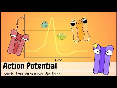
A nerve impulse is an action potential that travels along a nerve fibre. An action potential is a rapid, temporary reversal of the membrane potential in one part of the fibre: the inside of the membrane briefly becomes positive and then returns to negative. The whole sequence at one point on the membrane lasts about 1–2 ms.
The changes in charge are caused by the movement of positively charged ions across the membrane:
Channels for Na⁺ open, and Na⁺ ions diffuse into the fibre down their concentration gradient. The inside becomes less negative and then positive, reaching about +30 mV. This reduction in the potential difference across the membrane is called depolarisation.
The Na⁺ channels close and channels for K⁺ open. K⁺ ions diffuse out of the fibre, and the inside becomes negative again.
The sodium–potassium pump continues to work in the background, keeping the concentration gradients ready for further impulses.
A nerve impulse is therefore electrical, because it involves the movement of charged particles. The charged particles are positive ions crossing the membrane at each point along the fibre; electrons do not flow along the fibre as they do in a wire. Each action potential causes an action potential in the next section of the fibre, so the impulse is propagated along the fibre from one end to the other.
 YouTube
Nerve impulses travel at different speeds in different fibres. The two main factors are the diameter of the nerve fibre and whether it has a myelin sheath.
Impulses travel faster along wide fibres than along narrow ones, because a wide fibre has less resistance to the flow of ions along its inside. Squid such as Loligo depend on this effect. Their escape response, a sudden jet of water that shoots the squid backwards away from a predator, is controlled by giant axons up to about 1 mm in diameter that conduct impulses at about 25 m s⁻¹ without any myelin. Many non-myelinated fibres in other animals, including humans, are less than 1 µm wide and conduct at about 0.5–2 m s⁻¹.
Giant axons were also important in the history of physiology. Alan Hodgkin and Andrew Huxley used the squid giant axon in the 1940s and early 1950s because it was wide enough to insert an electrode inside it. Their recordings and measurements of ion movements explained how action potentials are generated, and they shared the Nobel Prize in Physiology or Medicine in 1963. The development of electrodes fine enough for the job is an example of new technology making new discoveries possible.
Myelin increases the speed of impulses much more than an increase in diameter does. Myelin is an electrical insulator, so ions can cross the axon membrane only at the gaps in the sheath, and the impulse moves along the axon from gap to gap instead of passing along every part of the membrane. The fastest human nerve fibres are myelinated and only 13–20 µm wide, yet they conduct at up to 120 m s⁻¹, several times faster than a squid giant axon fifty times wider. Myelination allows vertebrates to have many fast fibres packed into a small space, which is necessary in a large nervous system.
Type of fibre | Myelinated? | Diameter / µm | Conduction speed / m s⁻¹ | Example of function |
|---|---|---|---|---|
Aα | Yes | 13–20 | 80–120 | Motor neurons to skeletal muscle |
Aβ | Yes | 6–12 | 33–75 | Touch and pressure |
Aδ | Yes (thinly) | 1–5 | 3–30 | Sharp pain and cold |
C | No | 0.2–1.5 | 0.5–2 | Dull, burning pain and warmth |
Source: typical ranges in the Erlanger–Gasser classification of human peripheral nerve fibres, as summarised in Wikipedia, "Axon" (accessed 2026).
The data on fibre types show a clear pattern: wider fibres conduct faster. When two variables change together in this way they are correlated. In a positive correlation both increase together (conduction speed and fibre diameter). In a negative correlation one decreases as the other increases: the time an impulse takes to travel 1 m decreases as fibre diameter increases.
The strength of a linear correlation is measured by the correlation coefficient, r, which ranges from −1 to +1. A value close to +1 or −1 shows a strong linear relationship; a value close to 0 shows little or no linear relationship. Squaring r gives the coefficient of determination, R², which ranges from 0 to 1. R² gives the proportion of the variation in the dependent variable that can be explained by its linear relationship with the independent variable. An R² of 0.81, for example, means that 81% of the variation in conduction speed is explained by variation in diameter, and 19% by other factors.
Multiple sclerosis (MS) shows what happens when the myelin sheath is damaged. MS is an autoimmune disease: the immune system attacks and destroys the myelin around axons in the brain and spinal cord, leaving patches of damaged tissue called plaques or lesions. The axons themselves may survive at first, but without their insulation they conduct impulses slowly or fail to conduct them at all.

The symptoms depend on which fibres are affected. Damage to the optic nerve causes blurred vision; damage in the spinal cord and brain causes weakness, numbness, problems with balance and coordination, and fatigue. In the common relapsing–remitting form, attacks of symptoms are followed by periods of partial recovery, as inflammation settles and some myelin is repaired. Over years, repeated damage can lead to the loss of axons and permanent disability.
Measure | Value |
|---|---|
People living with MS worldwide, 2013 estimate | 2.3 million |
People living with MS worldwide, 2020 estimate | 2.8 million |
Global prevalence, 2020 | 35.9 per 100,000 people |
Rate of new diagnoses, 2020 | About one every 5 minutes |
Source: Atlas of MS, 3rd edition (Walton et al., Multiple Sclerosis Journal 26: 1816–1821, 2020), MS International Federation.
The rise from 2.3 million to 2.8 million between the two estimates does not necessarily mean that MS has become more common. Part of the increase reflects better diagnosis and more complete counting in more countries, and part reflects people with MS living longer. This is a common problem when comparing data collected at different times by different methods.
MS links several themes of biology. It connects structure to function at the level of the cell (the role of myelin in conduction speed), the immune system at the level of the organism (C3.2 Defence against disease) and the evidence-based interpretation of global health data.
A synapse is a junction between a neuron and another cell. Synapses link neurons to other neurons, and neurons to effector cells such as muscle fibres and gland cells. At most synapses the two cells are not in contact but separated by a narrow gap, the synaptic cleft, about 20 nm wide. The signal crosses the cleft as a chemical, a neurotransmitter. These are chemical synapses, usually called simply synapses.
The neuron that releases the neurotransmitter is the presynaptic neuron, and the cell that receives it is the postsynaptic neuron (or effector cell). The structure of a synapse makes signals pass in only one direction:
Only the presynaptic neuron contains vesicles of neurotransmitter and the channels that trigger their release.
Only the postsynaptic membrane has receptors for the neurotransmitter.
One-way transmission keeps signals flowing in a definite direction through the nervous system, from receptors through the central nervous system to effectors. Synapses are also where signals are combined, strengthened or weakened, so they are the sites where most processing of information in the nervous system takes place.
The end of a presynaptic axon is swollen into a synaptic knob, which contains mitochondria and many small vesicles filled with neurotransmitter. The neurotransmitter is released when an action potential arrives:
An action potential reaches the end of the presynaptic neuron and depolarises the presynaptic membrane.
Depolarisation opens voltage-gated calcium ion channels in the presynaptic membrane. Calcium ions (Ca²⁺), which are much more concentrated outside the neuron, diffuse in.
Inside the neuron, Ca²⁺ acts as a signalling chemical. It causes the vesicles of neurotransmitter to move to the presynaptic membrane and fuse with it.
The vesicles release their neurotransmitter into the synaptic cleft by exocytosis.
The amount of neurotransmitter released depends on how much Ca²⁺ enters, which depends on how many action potentials arrive. A rapid series of impulses releases more neurotransmitter than a single impulse. The Ca²⁺ is then pumped back out of the synaptic knob, ready for the next impulse.
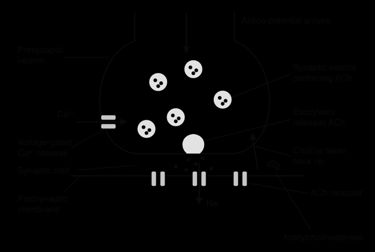
After release, the neurotransmitter diffuses across the synaptic cleft and binds to specific receptors on the postsynaptic membrane. These are transmembrane receptors. The best-studied example is acetylcholine (ACh), which is secreted by presynaptic neurons at many synapses, including the neuromuscular junctions between motor neurons and skeletal muscle fibres.
Acetylcholine diffuses across the synaptic cleft and binds to acetylcholine receptors on the postsynaptic membrane.
These receptors are ligand-gated ion channels: binding of acetylcholine opens them.
Na⁺ ions diffuse through the open channels into the postsynaptic cell, down their concentration gradient.
The entry of positive charge depolarises the postsynaptic membrane. This local depolarisation is an excitatory postsynaptic potential (EPSP). If it is large enough, it starts an action potential in the postsynaptic neuron or muscle fibre.
If acetylcholine stayed bound to its receptors, the postsynaptic cell would be stimulated continuously and a muscle would stay contracted. The signal is ended quickly by acetylcholinesterase, an enzyme in the synaptic cleft that breaks acetylcholine down into choline and acetate by hydrolysis. Choline is reabsorbed into the presynaptic neuron, where it is combined with an acetyl group again to make new acetylcholine, which is loaded into vesicles. Acetylcholinesterase is one of the fastest enzymes known, so each molecule of acetylcholine acts for only a very short time.
At a neuromuscular junction, one impulse in the motor neuron releases enough acetylcholine to produce an action potential in the muscle fibre almost every time, which then causes it to contract (B3.3 Muscle and motility). At synapses between neurons, a single EPSP is usually too small on its own, and the postsynaptic neuron responds only when several arrive together.
 YouTube
The channels that open and close during an action potential are voltage-gated channels: they change shape and open or close in response to changes in the membrane potential. A nerve fibre contains two kinds, voltage-gated Na⁺ channels and voltage-gated K⁺ channels, scattered along its membrane.
An action potential starts only if the membrane is first depolarised from −70 mV to about −50 mV. This value is the threshold potential. A small depolarisation that does not reach threshold opens only a few Na⁺ channels, and the membrane returns to rest. Once the threshold is reached, enough voltage-gated Na⁺ channels open for the inflow of Na⁺ to depolarise the membrane further, which opens more Na⁺ channels. This positive feedback makes depolarisation very rapid and self-sustaining.
Depolarisation. At threshold, voltage-gated Na⁺ channels open. Na⁺ diffuses into the axon, down both its concentration gradient and the electrical gradient (the inside is negative). The inside becomes positive, reaching about +30 to +40 mV.
Repolarisation. About 1 ms after opening, the Na⁺ channels close and become inactivated: they cannot reopen for a short time. Voltage-gated K⁺ channels, which open more slowly in response to the depolarisation, are now open. K⁺ diffuses out of the axon, down its concentration gradient, and the inside becomes negative again. This return towards the resting potential is repolarisation.
Hyperpolarisation. The K⁺ channels close slowly, so slightly too much K⁺ leaves and the membrane potential briefly falls below the resting potential, to about −80 mV. This is hyperpolarisation, also called the undershoot.
Return to rest. The K⁺ channels close, and the resting potential of −70 mV is restored by K⁺ leak channels and the sodium–potassium pump.

Only a tiny fraction of the Na⁺ and K⁺ ions in the axon cross the membrane during one action potential, so the concentration gradients hardly change. A neuron can fire thousands of action potentials before the gradients would run down, and the sodium–potassium pump restores them continuously.
Evidence that voltage-gated Na⁺ channels are essential comes from tetrodotoxin, a poison found in pufferfish. It binds to and blocks voltage-gated Na⁺ channels. Axons treated with tetrodotoxin can no longer produce action potentials, and poisoning causes paralysis, including of the muscles used in breathing.
Once threshold is reached, every action potential in a given neuron reaches the same peak and has the same shape, whatever the strength of the stimulus. A stimulus that does not reach threshold produces no action potential at all. Action potentials are therefore all-or-nothing. The intensity of a stimulus is coded by the frequency of action potentials (how many pass per second) and by the number of neurons that fire, not by the size of each one.
During an action potential, the inside of the depolarised region of the axon is positive and the inside of the neighbouring resting regions is negative. Na⁺ ions that have entered the depolarised region diffuse along the inside of the axon towards the neighbouring regions, attracted by their negative charge. Outside the axon, positive ions move in the opposite direction, towards the depolarised region. These movements of charge are called local currents.
The local currents reduce the potential difference across the membrane in the region ahead. When that region reaches the threshold potential of about −50 mV, its voltage-gated Na⁺ channels open and it generates its own action potential. This in turn depolarises the next region, and so on along the axon. The action potential is therefore regenerated at every point, so it arrives at the far end of the axon at full size.
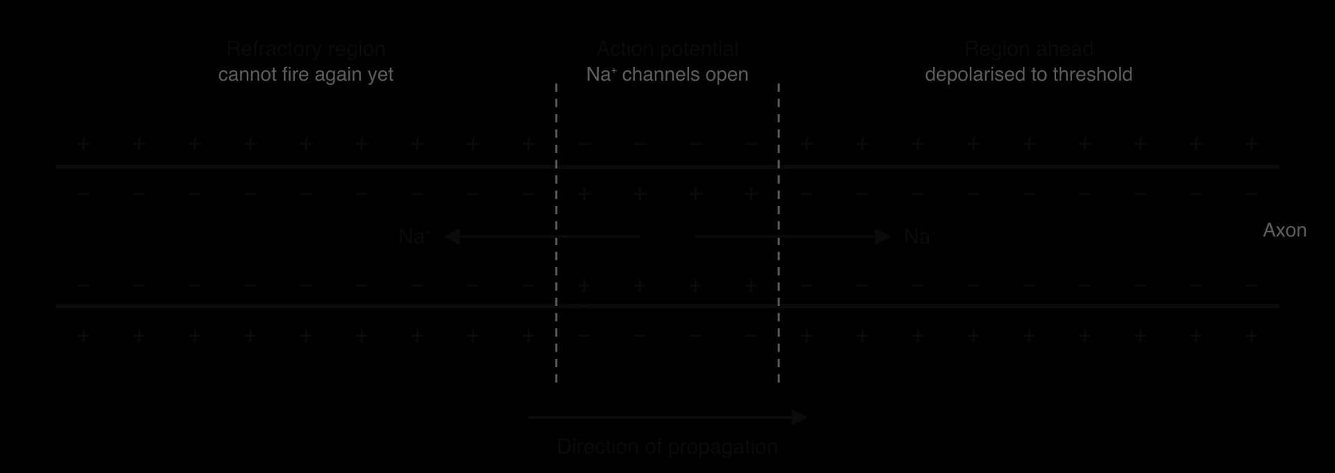
Local currents spread in both directions from an active region, yet the impulse travels in only one direction. After an action potential, there is a short refractory period of about 1–2 ms during which that region of membrane cannot produce another action potential. The voltage-gated Na⁺ channels are inactivated and cannot reopen until the membrane has repolarised, and the K⁺ channels are still open. The region the impulse has just left is therefore refractory, and only the region ahead can be depolarised to threshold.
The refractory period has two consequences. It makes impulses travel in one direction along an axon, and it limits how often a neuron can fire, so action potentials are always separate events and cannot merge.
The membrane potential of a neuron is measured with a microelectrode inside the axon and a reference electrode outside it. The potential difference between them is displayed against time on an oscilloscope (today usually a computer). The resulting trace has a characteristic shape.
Each part of the trace corresponds to an event at the membrane:
Part of the trace | Membrane potential | Events at the membrane |
|---|---|---|
Resting potential | Steady at about −70 mV | Voltage-gated channels closed; K⁺ leak and Na⁺/K⁺ pump maintain the potential |
Rise to threshold | −70 to −50 mV | Local currents (or a stimulus) depolarise the membrane slightly |
Depolarisation | Steep rise from −50 to about +40 mV | Voltage-gated Na⁺ channels open; Na⁺ diffuses in |
Repolarisation | Steep fall from +40 mV towards −70 mV | Na⁺ channels inactivated; voltage-gated K⁺ channels open; K⁺ diffuses out |
Hyperpolarisation | Dips to about −80 mV | K⁺ channels slow to close; extra K⁺ leaves |
Return to rest | Back to −70 mV | K⁺ channels close; resting potential restored |
A trace over a longer time shows a series of separate action potentials. Counting them over a measured time gives the frequency of impulses, which shows the intensity of the stimulus.
In a myelinated axon, the myelin sheath insulates the membrane, so ions cannot cross it except at the gaps between Schwann cells. These gaps, about 1 µm long and spaced about 1 mm apart, are the nodes of Ranvier. Voltage-gated Na⁺ and K⁺ channels and sodium–potassium pumps are concentrated at the nodes, and there are very few under the myelin.
An action potential at one node produces local currents that flow along the inside of the axon, under the myelin, to the next node. The next node is depolarised to threshold and generates its own action potential. The impulse therefore jumps from node to node, a process called saltatory conduction (from the Latin saltare, to jump).
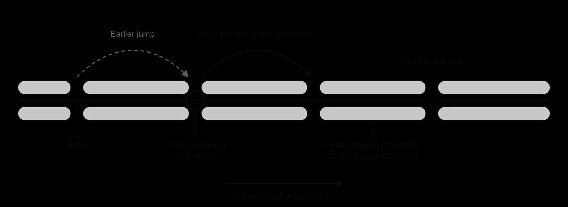
Saltatory conduction is faster than continuous conduction because the slow step, opening channels and moving ions across the membrane, happens only at the nodes, not along the whole length of the axon. Myelinated fibres can conduct at up to about 120 m s⁻¹, compared with 0.5–2 m s⁻¹ in thin non-myelinated fibres. It is also more efficient: far fewer ions cross the membrane per impulse, so the sodium–potassium pumps use less ATP to restore the gradients.
In multiple sclerosis, loss of myelin exposes stretches of axon membrane that have few voltage-gated Na⁺ channels. Local currents from the last node leak out through the damaged region and may not depolarise the next node to threshold, so conduction slows or fails completely, which explains the large effect of demyelination on nerve function.
Exogenous chemicals are substances that come from outside the body. Many of them act at synapses, because synaptic transmission depends on neurotransmitters binding to specific receptors and being removed quickly. A chemical that binds to a receptor, blocks an enzyme or blocks a transporter can increase or decrease transmission.
Neonicotinoids are insecticides with a shape similar to nicotine. They bind to the nicotinic acetylcholine receptors on postsynaptic membranes in the central nervous system of insects. Neonicotinoids are not broken down by acetylcholinesterase, so they stay bound to the receptors far longer than acetylcholine.
At first, the bound neonicotinoid opens the receptor channels, so postsynaptic neurons are overstimulated.
With the receptors occupied and unable to respond normally, acetylcholine released by presynaptic neurons can no longer bind and transmit signals.
Synaptic transmission is blocked, causing paralysis and death of the insect.
Neonicotinoids bind much more strongly to insect acetylcholine receptors than to those of mammals, which made them attractive as crop pesticides with low toxicity to humans. However, they are taken up into all parts of treated plants, including pollen and nectar, so bees and other pollinators are exposed. After a scientific review concluded that three widely used neonicotinoids, imidacloprid, clothianidin and thiamethoxam, posed a risk to bees, the European Union banned their outdoor use in 2018. The case shows how understanding a mechanism at the level of one receptor informs decisions that affect whole ecosystems.
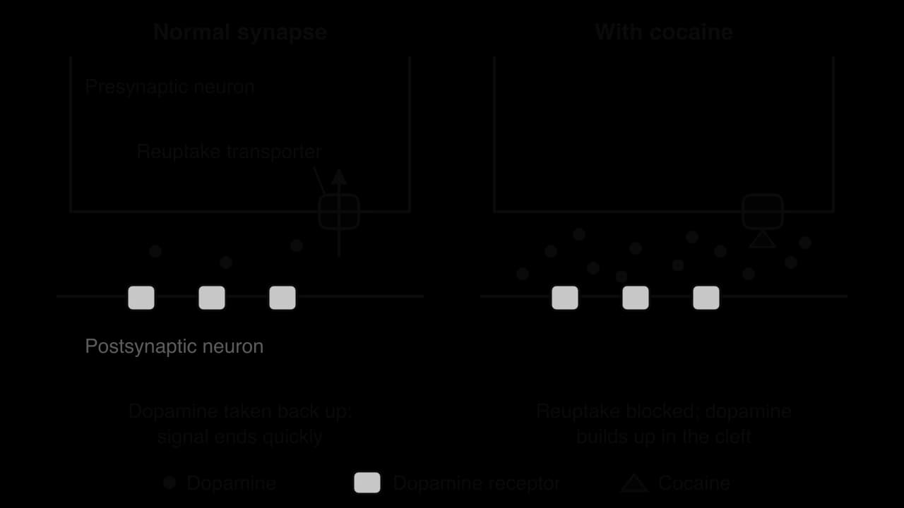
At many synapses, the neurotransmitter is removed from the cleft by reuptake: transporter proteins in the presynaptic membrane pump it back into the presynaptic neuron, where it is reused. Dopamine is removed in this way at synapses in the brain’s reward pathway, a group of neurons whose activity produces feelings of pleasure and reinforces behaviour.
Cocaine binds to the dopamine reuptake transporters and blocks them. Dopamine is still released normally, but it is not taken back up, so it stays in the synaptic cleft and keeps binding to receptors on the postsynaptic neurons. Transmission in the reward pathway is increased and prolonged, which produces the euphoria of cocaine use. With repeated use, the brain adapts, for example by reducing the number of dopamine receptors, so more of the drug is needed for the same effect, and normal sources of pleasure produce a weaker response. These changes contribute to addiction.
Some neurotransmitters inhibit the postsynaptic cell: they make an action potential less likely. The main inhibitory neurotransmitter in the brain is GABA (gamma-aminobutyric acid). When GABA binds to its receptors on the postsynaptic membrane, it opens channels for chloride ions (Cl⁻), which diffuse into the cell. At other inhibitory synapses, channels open that let K⁺ diffuse out. Either way, the inside of the postsynaptic cell becomes more negative than the resting potential: the membrane is hyperpolarised, for example to about −75 mV.
This hyperpolarisation is an inhibitory postsynaptic potential (IPSP). It moves the membrane potential further away from the threshold, so a larger depolarisation is needed to produce an action potential.
A single neuron in the brain may receive synapses from thousands of presynaptic neurons, some excitatory and some inhibitory. Individual EPSPs and IPSPs are small, a few millivolts each, and they spread over the cell body to the start of the axon. The postsynaptic neuron adds them together, a process called summation. An action potential is generated in its axon only if the combined effect brings the membrane to the threshold potential.
Temporal summation: one presynaptic neuron fires several times in quick succession, so each EPSP begins before the previous one has faded, and they add together.
Spatial summation: several different presynaptic neurons release neurotransmitter at the same time at different synapses on the same postsynaptic neuron, and their effects add together.
Excitatory and inhibitory inputs partly cancel each other. If a neuron receives EPSPs at some synapses and IPSPs at others at the same time, the IPSPs reduce the net depolarisation, and the threshold may not be reached even though the excitatory inputs alone would have reached it. The result is all-or-nothing: the postsynaptic neuron either fires an action potential or it does not. Summation allows a neuron to integrate information from many sources, which is the basis of information processing in the nervous system.
Pain is detected by neurons with free nerve endings in the skin and other tissues. These endings have no special receptor cell around them: the end of the sensory neuron itself is the receptor. Its membrane contains channel proteins for positively charged ions that open in response to harmful stimuli:
temperatures above about 43 °C
acid (a high concentration of H⁺ ions), for example from damaged tissue
certain chemicals, such as capsaicin, the substance that makes chilli peppers taste hot.
When the channels open, positive ions (Na⁺ and Ca²⁺) diffuse into the nerve ending and depolarise it. If the depolarisation reaches the threshold potential, action potentials are generated and travel along the sensory neuron to the spinal cord and then to the brain. Pain is perceived only when the impulses reach the sensory areas of the cerebral cortex; the nerve ending detects the stimulus, but the brain creates the sensation. The fibres that carry pain are thin Aδ fibres (fast, sharp pain) and unmyelinated C fibres (slower, burning pain).
The receptor responsible for heat and capsaicin was identified in 1997 by David Julius and his colleagues, who searched for the gene that made cells sensitive to capsaicin. The receptor, TRPV1, is a cation channel that opens at temperatures above about 43 °C, in acid conditions and when capsaicin binds. Capsaicin therefore feels hot because it opens the same channels as heat. Julius shared the Nobel Prize in Physiology or Medicine in 2021 with Ardem Patapoutian for discoveries of receptors for temperature and touch.
The human brain contains about 86 billion neurons, and each one forms synapses with thousands of others. Each neuron on its own does only simple things: it sums its inputs and either fires an action potential or does not. The brain as a whole, however, is conscious, which means it is aware of itself and its surroundings, has thoughts and feelings, and makes decisions.
Consciousness is an example of an emergent property, a property of a whole system that arises from the interactions of its parts and that none of the parts has on its own. No single neuron is conscious, and consciousness cannot be found by studying neurons one at a time. It emerges from the patterns of activity across vast networks of interconnected neurons.
Emergent properties are found at every level of organisation: the properties of a cell emerge from its molecules, the functions of an organ from its tissues, and the stability of an ecosystem from the interactions of its species. Consciousness also marks a limit of the reductionist approach in science, which explains a system by studying its parts in isolation: understanding the action potential in full detail does not, by itself, explain awareness.
Questions on neural signalling appear in every part of the assessment. Paper 1A tests the parts of a neuron, the ions involved in the resting and action potentials, and the events at a synapse. Paper 1B and Paper 2 Section A use data such as conduction speeds against fibre diameter (with correlation coefficients), oscilloscope traces (HL) and summation graphs (HL). Paper 2 Section B often asks for an extended explanation of synaptic transmission, of the resting potential or, at HL, of the generation and propagation of an action potential. Common command terms are outline, explain, describe, annotate, calculate and deduce.
The following question is based on these data for three types of human nerve fibre.
Fibre | Myelinated | Diameter / µm | Speed / m s⁻¹ |
|---|---|---|---|
Aα | Yes | 13–20 | 80–120 |
Aδ | Yes | 1–5 | 3–30 |
C | No | 0.2–1.5 | 0.5–2 |
The question totals 12 marks. Parts (d) and (e) are HL only; parts (a) to (c) could appear on either paper.
Neural signalling links to C2.1 Chemical signalling (ligand-gated receptors and the acetylcholine receptor), B2.1 Membranes and membrane transport (pumps and channels) and C3.1 Integration of body systems (reflex arcs and the brain).
I can identify the cell body, nucleus, dendrites, axon and myelin sheath on diagrams of motor and sensory neurons.
I can explain how the sodium–potassium pump, K⁺ leakage and negatively charged proteins generate a resting potential of about −70 mV.
I can explain why a nerve impulse is electrical and describe it as an action potential propagated along a fibre.
I can compare conduction speeds in squid giant axons, non-myelinated fibres and myelinated fibres, and interpret r and R² for these data.
I can describe how Ca²⁺ triggers release of neurotransmitter by exocytosis and why synapses work in one direction.
I can explain how acetylcholine generates an excitatory postsynaptic potential and how acetylcholinesterase ends the signal.
I can explain depolarisation and repolarisation in terms of voltage-gated channels and the threshold potential. (HL)
I can explain propagation by local currents and the role of the refractory period, and annotate an oscilloscope trace. (HL)
I can explain saltatory conduction at nodes of Ranvier. (HL)
I can explain the effects of neonicotinoids and cocaine on synaptic transmission. (HL)
I can explain inhibitory postsynaptic potentials and spatial and temporal summation. (HL)
I can outline how free nerve endings detect pain and explain consciousness as an emergent property. (HL)
neuromuscular junction;
Accept motor end plate.
Suggest, using the data, why the muscle that moves the eyeball has small motor units.
2 marks
a. each motor unit contains only about 9 fibres, so recruiting one more unit adds very little force;
b. allows very fine / precise control of movement;
c. eye movements must be precise to keep an image focused on the fovea / track objects;
[2 max]
Explain how the force of contraction of the calf muscle can be varied.
2 marks
a. all fibres in a motor unit contract together (when the neuron fires);
b. more motor units are recruited / activated for greater force;
c. motor neurons can send impulses at a higher frequency for greater force;
[2 max]
Describe the events at a neuromuscular junction that lead to depolarization of the muscle fibre membrane.
3 marks
a. action potential / nerve impulse arrives at the axon terminal (of the motor neuron);
b. calcium ions diffuse into the axon terminal (through channels);
c. vesicles release acetylcholine (into the gap / synaptic cleft) by exocytosis;
d. acetylcholine diffuses across the gap;
e. binds to receptors on the muscle fibre membrane;
f. sodium ions enter the muscle fibre, depolarizing its membrane;
g. acetylcholine is broken down by acetylcholinesterase (to end the signal);
[3 max]
Movement in animals depends on the coordinated action of nerves, muscles and bones.
Outline how an action potential is propagated along an axon.
4 marks
Explain how skeletal muscle fibres contract, using the sliding filament model.
7 marks
Describe the structure of the human hip joint.
4 marks
The graph shows the increase in permeability of the plasma membrane of a neuron to Na+ and K+, above the resting level, during one action potential at a point on its axon.
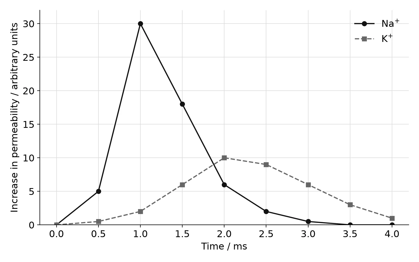
State the time at which the permeability of the membrane to Na+ is highest.
1 mark
Compare and contrast the changes in permeability to Na+ and to K+.
2 marks
Explain the changes in permeability to Na+ between 0 and 2.0 ms.
3 marks
Outline the role of voltage-gated potassium channels during the action potential.
2 marks
Many action potentials in neurons are started at synapses where acetylcholine is the neurotransmitter. Outline how nicotinic acetylcholine receptors cause depolarization of the postsynaptic membrane.
2 marks