Notes on C2.1 Chemical signalling
Cells constantly receive chemical signals from other cells and from their surroundings. Chemical signalling allows a multicellular organism to coordinate the activities of trillions of cells, and allows even single-celled bacteria to act together as a group.
Every chemical signal works in the same basic way. A signalling molecule released by one cell binds to a receptor on or in another cell, and that binding changes what happens inside the receiving cell. The differences between signals lie in what the molecules are, how far they travel, where their receptors sit and what sequence of events the binding sets off.
Receptors and the ligands that bind to them
A receptor is a protein with a binding site for one particular signalling chemical. The signalling chemical is called the ligand. Each receptor binds only its own ligand, or a small group of closely related molecules, because the binding site has a shape and a pattern of chemical groups that are complementary to the ligand.
Binding is held by weak interactions: hydrogen bonds, ionic attractions between charged groups and hydrophobic interactions between non-polar groups. These form only when the ligand and the binding site fit together closely, which is the source of the specificity. Because the bonds are weak, binding is reversible. When the concentration of ligand around the cell falls, ligands leave their receptors and the signal stops.
Binding causes a conformational change
When the ligand binds, the receptor changes shape. This conformational change is what passes the signal on. In a receptor that spans the plasma membrane, the change in shape on the outside of the cell is transmitted through the protein to the part inside the cell, which can then interact with other molecules. The ligand itself does not need to enter the cell.
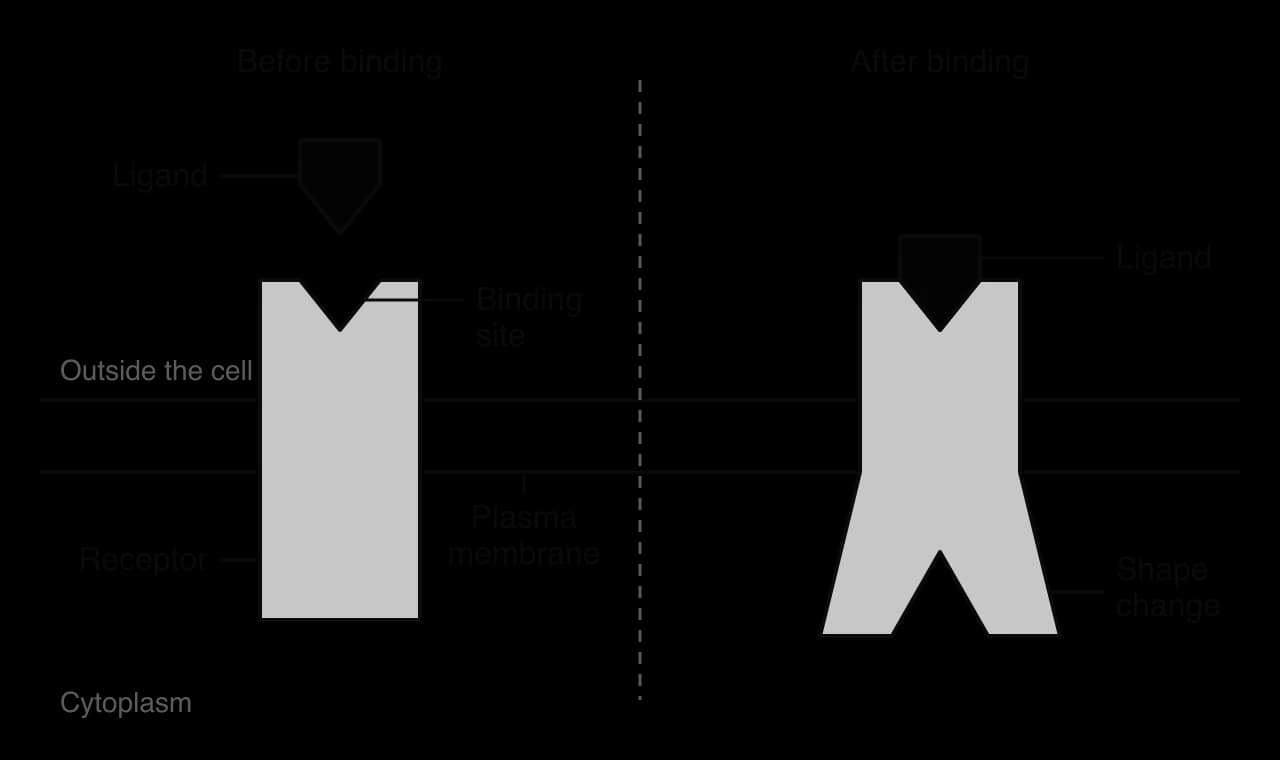
The same principle explains why the same signal can have different effects in different cells. A cell responds to a signal only if it makes the receptor for it, and what follows depends on the proteins that receptor interacts with in that particular cell type. A cell without the receptor is unaffected, even if the ligand is present in high concentration around it.
Quorum sensing in bacteria
Chemical signalling also occurs in single-celled organisms. Many bacteria release signalling molecules and detect the concentration of those molecules around them. Because each cell releases a small amount, the concentration of the signal rises as the number of cells in a confined space rises. Bacteria use this to sense their own population density and to switch on certain genes only when the population is large enough for those genes to be useful. This is called quorum sensing, from "quorum", the minimum number of members needed for a meeting to make decisions.
Bioluminescence in Vibrio fischeri
The best-studied example is the marine bacterium Vibrio fischeri. It produces light by bioluminescence, a reaction catalysed by the enzyme luciferase. Producing light uses a large amount of energy, and a single cell gives out too little light to have any effect. V. fischeri therefore produces light only when many cells are present together.
Each V. fischeri cell makes an autoinducer, a small signalling molecule (an acyl-homoserine lactone) produced by an enzyme called LuxI. The autoinducer is small and lipid-soluble, so it diffuses freely in and out of the cell across the plasma membrane. Inside the cell it can bind to a receptor protein called LuxR, which is found in the cytoplasm.
At low population density, autoinducer diffuses away from the cells into the water. Its concentration stays low, few LuxR receptors have autoinducer bound, and the light-producing genes are not transcribed.
As the population grows in a confined space, every cell releases autoinducer, so its concentration around and inside the cells rises.
Above a threshold concentration, most LuxR receptors have autoinducer bound. The LuxR–autoinducer complex binds to the promoter of the lux genes and switches on their transcription.
The lux genes code for luciferase and the enzymes that make its substrate, so the cells emit light. They also include the gene for LuxI, so more autoinducer is made, which reinforces the response.
Because every cell in the population detects the same autoinducer concentration, the whole population switches on its light genes at about the same time.

This is a clear example of a gene expression change caused by a signal from outside the cell. The response is all or nothing at the level of the population: the colony is either dark or glowing, with only a short transition between the two states.
The graph of light emission against cell density has an S-shape. Below the threshold the cells are dark even though they are dividing. Once the autoinducer concentration passes the threshold, light output rises steeply, because each cell that switches on also makes more LuxI and therefore more autoinducer.
The Hawaiian bobtail squid
In the open ocean V. fischeri lives at low density and does not glow. It does glow inside the light organ of the Hawaiian bobtail squid (Euprymna scolopes), a small squid that hunts at night in shallow water off Hawaii. The squid supplies the bacteria with nutrients in its light organ, where they grow to a very high density and produce light. The light shines downwards from the underside of the squid and matches the moonlight coming from above, so the squid casts no shadow that a predator below could see. This camouflage is called counter-illumination.

The partnership is a mutualism: the bacteria gain food and a protected habitat, and the squid gains camouflage. Each morning the squid expels most of the bacteria into the sea and the remaining cells grow back to high density by the next night. Quorum sensing ensures that the bacteria spend energy on light only when they are in the light organ, where it benefits them.
Quorum sensing is widespread among bacteria. Vibrio cholerae, the bacterium that causes cholera, uses it to control its virulence genes, and many other pathogens switch on toxin production only once their population in a host is large. Research into blocking autoinducer receptors is exploring ways to disarm pathogens without killing them.
 YouTube
Functional categories of signalling chemicals in animals
Animals use many signalling chemicals. They can be grouped by what they do and where they act. Four functional categories are hormones, neurotransmitters, cytokines and calcium ions. The categories describe the role of a molecule, not its chemical structure, so chemically similar molecules can belong to different categories.
Hormones
Hormones are signalling chemicals secreted by endocrine glands directly into the blood. The blood carries them to all parts of the body, but only target cells with the right receptors respond. Examples are insulin from the pancreas, adrenaline from the adrenal glands and oestradiol from the ovaries. Hormones can act on cells far from the gland that made them, and their effects often last minutes, hours or days.
Neurotransmitters
Neurotransmitters are signalling chemicals released by a neuron at a synapse, the junction between a neuron and another cell. They diffuse across the narrow gap between the two cells and bind to receptors on the membrane of the receiving cell. Examples are acetylcholine, dopamine and GABA. Neurotransmitters act only on the cell immediately across the synapse, and their effect is very fast and very brief, because they are broken down or taken back up within milliseconds. C2.2 Neural signalling describes synapses in detail.
Cytokines
Cytokines are small proteins secreted by many kinds of cell, especially cells of the immune system. Most act locally, on the cell that released them or on nearby cells, and they regulate cell growth, cell division and immune responses. Examples are the interleukins, by which helper T-lymphocytes stimulate B-lymphocytes to divide during an immune response, and the interferons released by virus-infected cells, which make neighbouring cells more resistant to the virus.
Calcium ions
Calcium ions (Ca²⁺) act as signals inside cells. The concentration of Ca²⁺ in the cytoplasm is normally kept very low by pumps that move Ca²⁺ out of the cell or into stores such as the endoplasmic reticulum. When channels in the plasma membrane or in these stores open, Ca²⁺ floods into the cytoplasm and binds to proteins that change their activity. Three examples are:
At a synapse, depolarisation of the end of a neuron opens Ca²⁺ channels, and the entry of Ca²⁺ triggers release of neurotransmitter (C2.2).
In muscle fibres, Ca²⁺ released from the sarcoplasmic reticulum binds to proteins on the actin filaments and allows contraction (B3.3).
In an egg cell at fertilisation, a wave of Ca²⁺ release triggers the changes that prevent a second sperm from entering (D3.1).
Calcium ions often act as a second messenger, carrying a signal from a receptor at the plasma membrane to targets inside the cell as part of a signal transduction pathway.
Feature | Hormones | Neurotransmitters | Cytokines | Calcium ions |
|---|---|---|---|---|
Released by | Endocrine glands | Neurons, at synapses | Many cells, especially immune cells | Stores inside the cell, or enter through membrane channels |
Route to target | Carried in the blood | Diffuse across the synaptic gap | Diffuse through tissue fluid to nearby cells | Diffuse through the cytoplasm |
Distance of action | Long (any tissue the blood reaches) | Very short (about 20 nm) | Short (same or nearby cells) | Within one cell |
Speed and duration | Seconds to days | Milliseconds | Minutes to hours | Milliseconds to seconds |
Example | Insulin, adrenaline, oestradiol | Acetylcholine, dopamine, GABA | Interleukins, interferons | Ca²⁺ triggering neurotransmitter release |
Signalling systems like these have evolved many times, in bacteria, plants, fungi and animals, using different molecules. This is a pattern of unity and diversity: the underlying logic of a ligand binding a specific receptor is shared by all of them, while the molecules themselves vary widely.
Chemical diversity of hormones and neurotransmitters
Hormones and neurotransmitters belong to several different chemical groups, and the chemical nature of each signal affects how it is made, how it travels and how it acts.
Chemical groups of hormones
Amines are small molecules made from a single amino acid. Adrenaline and noradrenaline are made from tyrosine; thyroxine is made from tyrosine with iodine added; melatonin is made from tryptophan.
Proteins and peptides are chains of amino acids. Insulin (51 amino acids), glucagon, antidiuretic hormone (ADH) and oxytocin are examples. Follicle-stimulating hormone and luteinising hormone are glycoproteins, with carbohydrate attached.
Steroids are lipids made from cholesterol, with four fused carbon rings. Oestradiol, progesterone, testosterone, cortisol and aldosterone are steroid hormones.
Chemical groups of neurotransmitters
Amino acids: glutamate, the main excitatory neurotransmitter in the brain; GABA (gamma-aminobutyric acid) and glycine, which are inhibitory.
Peptides: short chains of amino acids such as the endorphins, which reduce the sensation of pain, and substance P, which is released by pain-sensing neurons.
Amines: dopamine, noradrenaline and serotonin, all made from single amino acids.
Nitric oxide (NO): a gas, made on demand from the amino acid arginine. It diffuses through membranes in all directions and lasts only a few seconds.
Acetylcholine is in a group of its own: it is an ester formed from choline and acetate.
Why so many different chemicals are used
Signalling only works if each receptor can tell its own signal apart from every other molecule around it. A large number of chemically different signals, each with a distinctive shape, allows many separate messages to be sent through the same blood or the same tissue fluid without being confused. The variety of molecules also gives signals different properties that suit their roles:
Peptide and protein hormones are hydrophilic, so they dissolve in plasma and travel freely, but they cannot cross the plasma membrane. They act through receptors on the cell surface and often produce rapid responses.
Steroid hormones are hydrophobic. They travel in the blood bound to carrier proteins, cross the plasma membrane by simple diffusion and act on receptors inside the cell, usually by changing gene expression. Their effects are slower to start and last longer.
Small neurotransmitters such as amino acids and amines can be made quickly, stored in vesicles in large numbers, released in milliseconds and removed rapidly, which suits fast, short signals.
Nitric oxide cannot be stored in vesicles at all; it is made only when needed and spreads to all nearby cells, which suits it to signals that affect a small region of tissue.
The same molecule can also serve in more than one category. Noradrenaline is a neurotransmitter released by neurons of the sympathetic nervous system and also a hormone secreted by the adrenal glands. Its role depends on where it is released and how far it travels.
Localised and distant effects of signalling molecules
Signalling molecules differ in how far they travel before they act. Neurotransmitters and hormones are the two extremes, and comparing them shows how the distance travelled affects the whole character of a signal.
A neurotransmitter is released from the end of one neuron into a synaptic cleft only about 20 nm wide. It diffuses across this gap in a fraction of a millisecond and binds to receptors on the one cell on the other side. Its effect is therefore localised to a single target cell, very fast, and over within milliseconds because enzymes or transporters remove the neurotransmitter from the cleft.
A hormone is secreted into the blood and carried around the whole body, reaching every tissue within about a minute. Any cell with the right receptor responds, so a single hormone can have widespread effects on many organs at once. Adrenaline released from the adrenal glands, for example, increases heart rate, widens the airways and causes the liver to release glucose. Hormones remain in the blood for minutes to hours before they are broken down, so their effects last longer.
Feature | Neurotransmitter | Hormone |
|---|---|---|
Released from | Presynaptic neuron | Endocrine gland |
Transported by | Diffusion across the synaptic cleft only | The blood, to the whole body |
Distance to target | About 20 nm | Up to the length of the body |
Target cells | Only the cell across the synapse | Every cell with the right receptor |
Speed of effect | Milliseconds | Seconds to hours |
Duration of effect | Milliseconds (rapid removal) | Minutes to days |
Many signals fall between these extremes. Cytokines, nitric oxide and many growth factors act on cells within a short distance of where they are released, without entering the blood. This is sometimes called paracrine signalling, while signalling by a cell to itself is autocrine signalling.
Transmembrane receptors and intracellular receptors
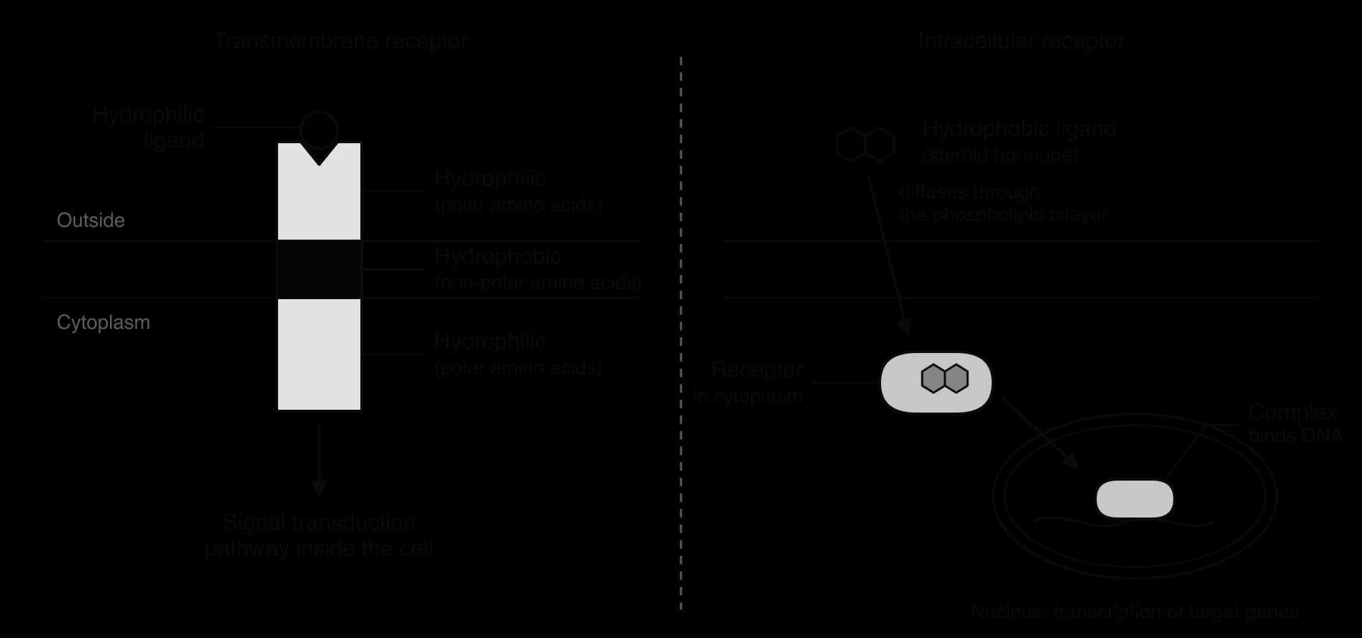
Receptors are found in two places. A transmembrane receptor spans the plasma membrane, with its binding site on the outside of the cell. An intracellular receptor is found in the cytoplasm or the nucleus. Which kind of receptor a signal uses depends on whether the signal can cross the plasma membrane.
Transmembrane receptors
Hydrophilic signals, such as peptide hormones, most amine hormones and most neurotransmitters, are charged or polar. They cannot pass through the hydrophobic core of the phospholipid bilayer, so they must act from outside the cell. Their receptors are integral proteins that pass through the membrane.
The distribution of amino acids in a transmembrane receptor matches the environment of each part of the protein.
The region that passes through the membrane is made of amino acids with hydrophobic (non-polar) R groups, usually arranged as one or more α-helices. These hold the receptor in the hydrophobic core of the bilayer.
The region outside the cell, which includes the binding site, and the region inside the cell are made largely of amino acids with hydrophilic (polar or charged) R groups, which interact with the water on each side of the membrane.
Intracellular receptors
Hydrophobic signals, mainly the steroid hormones and thyroxine, dissolve in the phospholipid bilayer and diffuse straight through it. Their receptors are proteins in the cytoplasm or the nucleus. These receptors do not need a hydrophobic region, because they are entirely surrounded by the aqueous cytoplasm or nucleoplasm; their surface is mostly hydrophilic. When the hormone binds, the hormone–receptor complex usually acts in the nucleus to change the transcription of particular genes.
Feature | Transmembrane receptor | Intracellular receptor |
|---|---|---|
Location | Spans the plasma membrane | In the cytoplasm or nucleus |
Ligands | Hydrophilic: peptide and protein hormones, most amines, neurotransmitters | Hydrophobic: steroid hormones, thyroxine |
Does the ligand enter the cell? | No | Yes, by diffusion through the phospholipid bilayer |
Amino acids | Hydrophobic band in the membrane; hydrophilic regions on both sides | Mostly hydrophilic surface |
How the signal is passed on | Signal transduction pathway in the cytoplasm | Hormone–receptor complex binds DNA and regulates transcription |
Speed of response | Usually rapid (seconds to minutes) | Slower (hours), but long-lasting |
Signal transduction pathways
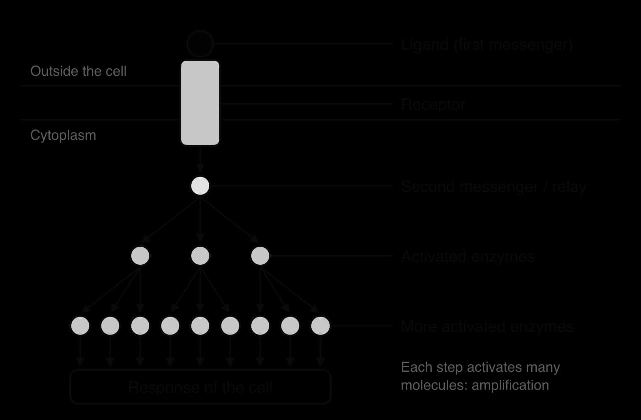
Binding of a ligand to a transmembrane receptor takes place outside the cell, but the response happens inside it. The sequence of events that carries the signal from the receptor to the part of the cell that responds is a signal transduction pathway. "Transduction" means converting a signal from one form into another: here, from the binding of an external ligand into chemical changes in the cytoplasm.
Most pathways have the same parts:
First messenger. The ligand (a hormone or neurotransmitter) binds to the receptor. It is called the first messenger because it carries the signal to the cell. In most pathways it never enters the cell.
Receptor activation. The receptor changes shape and becomes active.
Relay and second messenger. The active receptor activates other proteins in or on the membrane, which may produce a small molecule that diffuses through the cytoplasm. This second messenger carries the signal to targets inside the cell. Cyclic AMP and Ca²⁺ are common second messengers.
Cascade. The signal is passed along a chain of proteins, often enzymes called protein kinases that activate one another by adding phosphate groups.
Response. The final proteins in the chain change the cell’s activity: an ion channel opens and the membrane potential changes, an enzyme is activated or inhibited, or a transcription factor alters gene expression.
Amplification and control
Each active enzyme in a cascade can activate many molecules of the next enzyme before it is switched off. The signal is therefore amplified at every step, so a very low concentration of hormone in the blood, binding to a few hundred receptors, can change the activity of millions of molecules inside the cell. Cascades also allow a signal to branch, so one ligand can trigger several responses at once, and they allow the pathway to be regulated at several points.
Every step must also be reversible, so that the response ends when the ligand is no longer present. Enzymes called phosphatases remove the phosphate groups added by kinases, and other enzymes break down second messengers. Without these off-switches, a cell would keep responding long after the signal had gone.
Acetylcholine receptors and changes in membrane potential
The simplest transmembrane receptors are ion channels that open when a ligand binds. They are called ligand-gated ion channels. Receptors for neurotransmitters are often of this type, because they produce a response in less than a millisecond.
The best-known example is the nicotinic acetylcholine receptor, found in the postsynaptic membrane at synapses between motor neurons and muscle fibres, and at many synapses in the nervous system. It is made of five protein subunits arranged in a ring around a central pore that passes through the membrane. Two of the subunits have a binding site for acetylcholine (ACh) on the outside of the membrane.
With no acetylcholine bound, the pore is closed and ions cannot pass.
When acetylcholine is released at the synapse, two ACh molecules bind to the two binding sites.
Binding causes a conformational change that opens the pore.
Positively charged ions, mainly sodium ions (Na⁺), diffuse through the pore into the cell, down their concentration gradient and attracted by the negative charge inside the cell. A smaller number of K⁺ ions move out.
The net inflow of positive charge makes the inside of the cell less negative. This change in membrane potential is a depolarisation, and if it is large enough it starts an action potential in the muscle fibre or neuron.

In this kind of receptor there is no second messenger and no cascade: the change in shape of the receptor directly causes the response. This is why transmission at synapses is so fast. The channel closes again as soon as acetylcholine leaves the binding sites, and acetylcholine is broken down within milliseconds by the enzyme acetylcholinesterase in the synaptic cleft. C2.2 Neural signalling describes how the depolarisation leads to an action potential.
G protein-coupled receptors
The largest family of transmembrane receptors in animals is the G protein-coupled receptors (GPCRs). Each is a single polypeptide that crosses the plasma membrane seven times, as seven hydrophobic α-helices. The ligand binds on the outside of the cell or among the helices within the membrane. On the inside, the receptor interacts with a G protein.
How a G protein is switched on and off
A G protein is a protein attached to the inner surface of the plasma membrane, made of three subunits called α, β and γ. Its name comes from the guanine nucleotides it binds: GDP (guanosine diphosphate) and GTP (guanosine triphosphate). It acts as a molecular switch that is on when GTP is bound and off when GDP is bound.
In the inactive state, the α subunit has GDP bound and the three subunits are together.
A ligand binds to the GPCR, which changes shape. The active receptor binds the G protein.
The receptor causes the α subunit to release its GDP. GTP, which is more abundant in the cytoplasm, binds in its place.
With GTP bound, the α subunit separates from the β and γ subunits. The α–GTP subunit moves along the membrane and activates or inhibits a target protein, such as an enzyme or an ion channel.
The α subunit slowly hydrolyses its GTP to GDP. With GDP bound it becomes inactive, leaves its target and rejoins the β and γ subunits, ready to be activated again.
Because the α subunit switches itself off by hydrolysing GTP, the signal stops soon after the ligand leaves the receptor. While a ligand stays bound, one receptor can activate many G proteins one after another, which is the first step of amplification.
The variety of G protein-coupled receptors
There are about 800 GPCR genes in the human genome. Different GPCRs respond to hormones such as adrenaline and glucagon, to neurotransmitters such as dopamine and serotonin, to odour molecules in the nose and to light in the eye, where the GPCR rhodopsin absorbs photons. About a third of all approved medicines act on GPCRs, including beta blockers, antihistamines and opioid painkillers. The sharing of one receptor design across so many different signals is another example of unity within diversity: the same seven-helix structure has been adapted, by evolution, to bind hundreds of different ligands.
How adrenaline acts through a G protein and cyclic AMP
Adrenaline is an amine hormone secreted by the adrenal glands in response to stress, exercise or danger. It prepares the body for intense activity, and one of its most important effects is to raise the concentration of glucose in the blood by causing the liver to break down glycogen. Adrenaline is hydrophilic, so it acts through a GPCR on liver and muscle cells.
Adrenaline (the first messenger) binds to its GPCR, the adrenergic receptor, on the outside of a liver cell. The receptor changes shape.
The receptor activates a G protein: GDP on the α subunit is replaced by GTP, and the α–GTP subunit separates from the β and γ subunits.
The α–GTP subunit binds to and activates adenylyl cyclase, an enzyme in the plasma membrane.
Adenylyl cyclase converts ATP into cyclic AMP (cAMP). Each active enzyme makes many cAMP molecules, which diffuse through the cytoplasm as the second messenger.
Cyclic AMP binds to and activates protein kinase A (PKA).
Protein kinase A phosphorylates and activates another enzyme, phosphorylase kinase, which in turn phosphorylates and activates glycogen phosphorylase.
Glycogen phosphorylase breaks glycogen down into glucose phosphate, which liver cells convert to glucose and release into the blood. Protein kinase A also inhibits the enzyme that makes glycogen, so glucose is not stored again.
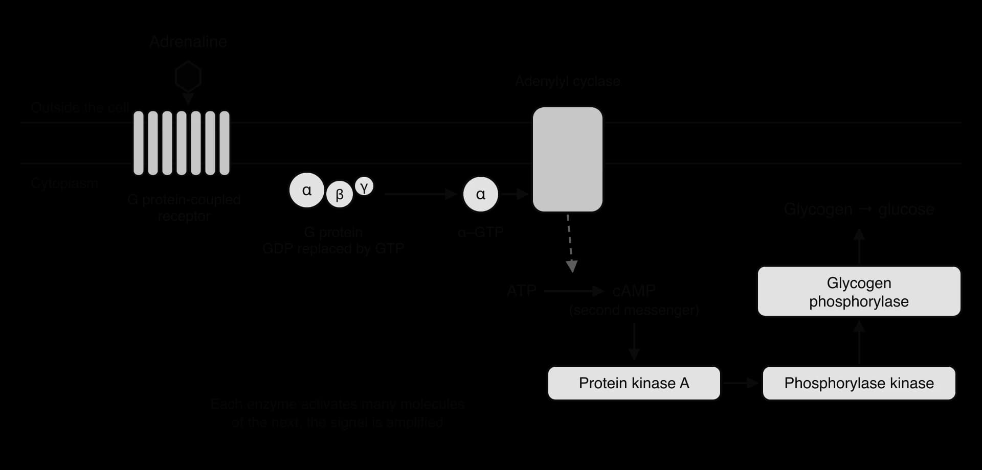
The pathway amplifies the signal enormously. One adrenaline–receptor complex activates many G proteins, each adenylyl cyclase makes many cAMP molecules, and each kinase phosphorylates many molecules of the next enzyme. A tiny concentration of adrenaline in the blood can therefore cause the release of large amounts of glucose within seconds. The response is switched off when adrenaline leaves the receptor: G proteins hydrolyse their GTP, and an enzyme called phosphodiesterase converts cAMP to an inactive form.
Cyclic AMP was the first second messenger to be discovered. Earl Sutherland showed in the 1950s that adrenaline caused liver tissue to make a heat-stable small molecule, later identified as cAMP, which could activate glycogen breakdown even when added to broken cells without any adrenaline present. The discovery that hormones work through second messengers inside the cell earned him the Nobel Prize in Physiology or Medicine in 1971.
 YouTube
Receptors with tyrosine kinase activity: the insulin receptor
A third type of transmembrane receptor is itself an enzyme. Its region inside the cell is a tyrosine kinase, an enzyme that adds phosphate groups to the amino acid tyrosine in proteins. The insulin receptor is a well-studied example.
Insulin is a protein hormone secreted by β cells in the pancreas when the concentration of glucose in the blood rises. Its main targets are muscle cells and adipose (fat) cells, which take up glucose in response, and liver cells, which store glucose as glycogen. The insulin receptor is made of two identical halves, each with a part outside the cell that binds insulin, a single section that crosses the membrane and a tyrosine kinase region in the cytoplasm.
Insulin binds to the parts of the receptor outside the cell, causing a conformational change that brings the two kinase regions inside the cell close together.
Each kinase region adds phosphate groups to tyrosine residues on the other half of the receptor. This is autophosphorylation, and it activates the receptor fully.
The phosphorylated tyrosines act as docking sites. Relay proteins bind to them and are themselves phosphorylated, starting a cascade of kinases inside the cell.
The cascade causes vesicles in the cytoplasm whose membranes contain the glucose transporter GLUT4 to move to the plasma membrane and fuse with it.
GLUT4 is now in the plasma membrane, so glucose enters the cell by facilitated diffusion down its concentration gradient. The rate of glucose uptake rises many times.
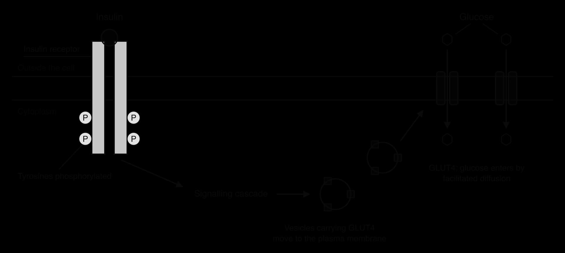
When the insulin concentration falls, the receptors stop signalling and GLUT4 is removed from the plasma membrane by endocytosis back into vesicles. The number of transporters in the membrane, and therefore the rate of glucose uptake, is controlled by the hormone. In type 2 diabetes, target cells respond poorly to insulin at or after the receptor, a condition called insulin resistance, so fewer GLUT4 transporters reach the membrane and blood glucose stays high (D3.3 Homeostasis).
Feature | Acetylcholine receptor | G protein-coupled receptor | Insulin receptor |
|---|---|---|---|
Type | Ligand-gated ion channel | Activates a G protein | Tyrosine kinase |
Example ligand | Acetylcholine | Adrenaline | Insulin |
First change after binding | Pore opens | G protein exchanges GDP for GTP | Tyrosines on the receptor are phosphorylated |
Second messenger | None | Cyclic AMP | None; a cascade of relay proteins and kinases |
Response | Na⁺ enters; membrane depolarised | Glycogen broken down to glucose | GLUT4 moves to the membrane; glucose uptake rises |
Speed | Under a millisecond | Seconds | Minutes |
Intracellular receptors that affect gene expression
A steroid hormone acts in a different way from adrenaline and insulin. Oestradiol, progesterone and testosterone are all made from cholesterol in the gonads, and all have the four-ring structure of a steroid. Being hydrophobic, they are carried in the blood bound to carrier proteins, and they pass through the plasma membrane of every cell they reach, although only target cells contain the matching receptor.
The steroid hormone diffuses through the phospholipid bilayer into the cytoplasm.
It binds to a specific receptor protein in the cytoplasm or nucleus. Oestradiol binds the oestrogen receptor, progesterone the progesterone receptor and testosterone the androgen receptor.
Binding causes a conformational change in the receptor. Receptors in the cytoplasm release the proteins that held them inactive, and the hormone–receptor complex moves into the nucleus through a nuclear pore.
The hormone–receptor complex binds to specific DNA sequences, called hormone response elements, in the promoter regions of particular genes.
The complex acts as a transcription factor: it promotes (or in some genes reduces) the transcription of those genes, so the cell makes more of the proteins they code for.
Because the response requires transcription and translation of new proteins, steroid hormones act more slowly than hormones that work through second messengers, usually over hours. Their effects also last longer, because the new proteins remain after the hormone has gone. Testosterone, for example, binds androgen receptors in muscle cells and increases transcription of genes for muscle proteins, which contributes to the larger muscle mass in males after puberty. The same hormone acting on different cells switches on different genes, because the genes that are accessible to the receptor complex differ between cell types.
Case study: tamoxifen and the oestrogen receptor in breast cancer
Breast cancer shows why the type of receptor in a cell determines how it responds to a hormone. About four in every five invasive breast cancers are oestrogen-receptor positive (ER-positive): their cells contain oestrogen receptors in the nucleus. When oestradiol binds to these receptors, the complex acts as a transcription factor and switches on genes that stimulate cell division. In an ER-positive cancer, oestradiol in the blood therefore drives the growth of the tumour.
Tamoxifen is a drug with a shape similar to oestradiol. It binds to the binding site of the oestrogen receptor, so it competes with oestradiol. When tamoxifen is bound, the receptor takes up a different conformation that cannot activate transcription of the growth genes. Tamoxifen therefore blocks the effect of oestradiol on the cancer cells without changing the amount of oestradiol in the blood. Cancers whose cells lack oestrogen receptors (ER-negative) are not driven by oestradiol, so tamoxifen has little or no effect on them.
The strongest evidence for tamoxifen comes from combining the results of many randomised trials. A meta-analysis of 20 trials, including 10,645 women with ER-positive early breast cancer, compared women who took tamoxifen for about 5 years after surgery with women who did not. A rate ratio compares the rate of an event in the treated group with the rate in the untreated group: a value of 1 means no effect, and 0.5 means the rate was halved.
Outcome (ER-positive disease) | Rate ratio, tamoxifen vs no tamoxifen | Interpretation |
|---|---|---|
Recurrence of cancer, years 0–4 | 0.53 | Recurrence rate roughly halved |
Recurrence of cancer, years 5–9 | 0.68 | Recurrence rate about a third lower, after treatment had stopped |
Death from breast cancer, over 15 years | 0.70 | About 30% lower; 15-year breast cancer mortality about 24% vs 33% |
Any outcome in ER-negative disease | Close to 1 | Little or no effect |
Source: Early Breast Cancer Trialists' Collaborative Group, The Lancet 378: 771–784, 2011. Proportion of ER-positive cancers (79.4%): Breast Cancer Surveillance Consortium, cases 2005–2022.
The data support the mechanism. Tamoxifen works only where the target cells have the intracellular receptor it acts on, and its benefit continues after treatment stops, which suggests that blocking oestrogen-driven transcription for five years removed many cancer cells that would otherwise have grown. The case also shows how knowledge of a signalling pathway at the molecular level leads to treatments for whole patients, linking the level of molecules to the level of the organism.
Effects of oestradiol and progesterone on target cells
Oestradiol and progesterone are the main ovarian hormones of the menstrual cycle. Both are steroid hormones that act through intracellular receptors, and their effects depend on which target cells have the receptors. Two examples are oestradiol acting on cells in the brain and progesterone acting on the lining of the uterus.
Oestradiol and the GnRH-secreting cells of the hypothalamus
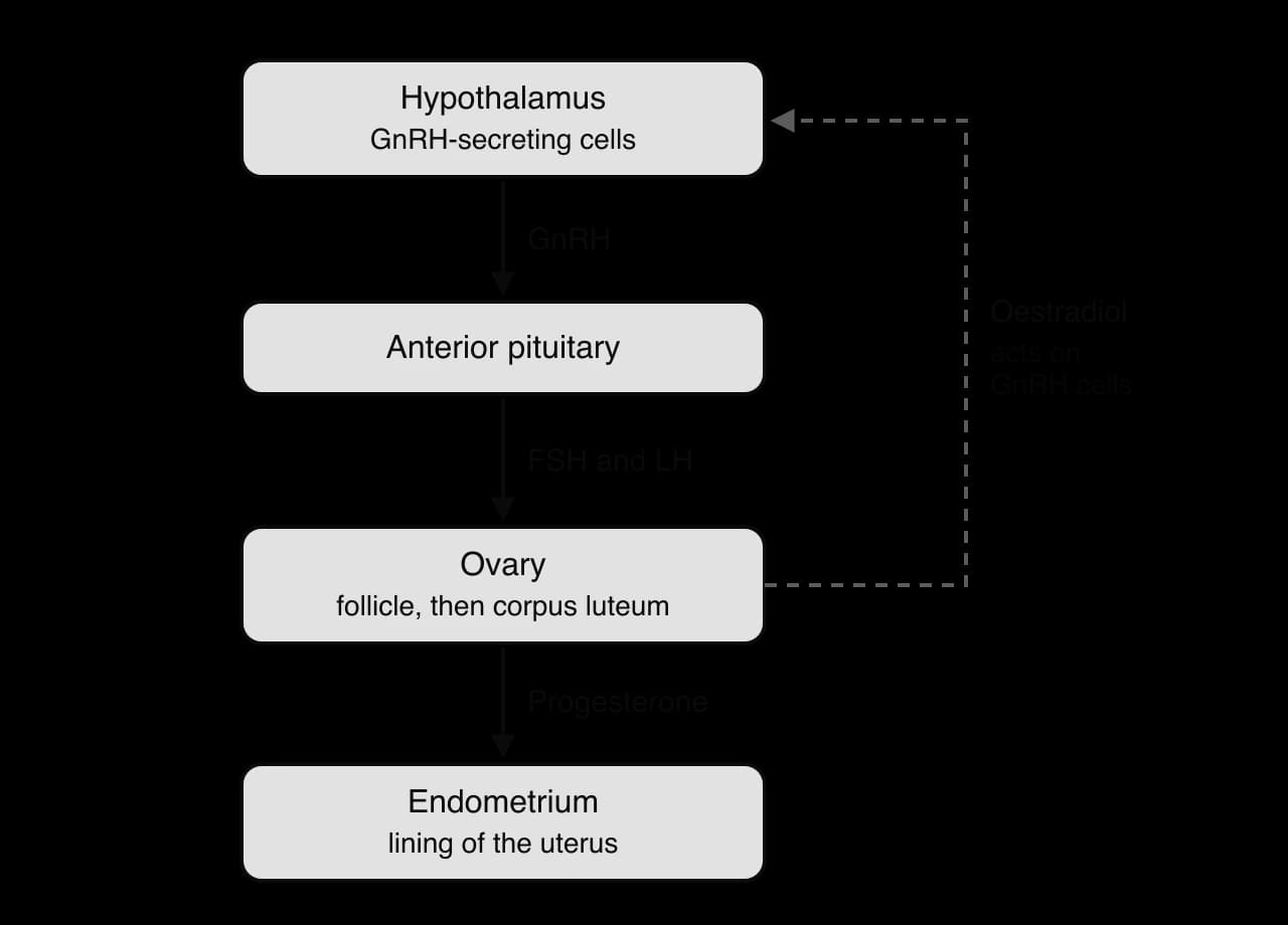
The hypothalamus, at the base of the brain, contains neurosecretory cells that release gonadotropin-releasing hormone (GnRH). GnRH travels a short distance in blood vessels to the anterior pituitary gland, where it stimulates the secretion of follicle-stimulating hormone (FSH) and luteinising hormone (LH). These two hormones act on the ovary, where FSH stimulates the growth of follicles, which secrete oestradiol.
Oestradiol from the growing follicles acts back on the hypothalamus, which contains cells with oestrogen receptors that regulate the GnRH-secreting cells. The effect depends on the concentration of oestradiol:
At low to moderate concentrations, during most of the cycle, oestradiol reduces the secretion of GnRH, and therefore of FSH and LH. This is negative feedback, and it prevents too many follicles developing at once.
When a large follicle produces a high concentration of oestradiol for about two days, late in the follicular phase, the effect reverses. Oestradiol now stimulates the GnRH-secreting cells, and GnRH and LH secretion rise sharply. This positive feedback causes the LH surge that triggers ovulation about a day later.
Progesterone and the endometrium
After ovulation, the remains of the follicle become the corpus luteum, which secretes large amounts of progesterone. Cells of the endometrium, the lining of the uterus, contain progesterone receptors. Progesterone diffuses into these cells and binds to its receptor, and the hormone–receptor complex acts as a transcription factor.
It switches on genes for growth factors and other proteins that make the endometrium thicken and develop a good blood supply.
It causes glands in the endometrium to secrete nutrients, preparing the lining to receive an embryo.
It maintains the lining for as long as the progesterone concentration stays high.
If no embryo implants, the corpus luteum breaks down after about 10–12 days and the progesterone concentration falls. Without progesterone the endometrium can no longer be maintained, and it breaks down and is shed during menstruation. If an embryo does implant, the corpus luteum keeps secreting progesterone, and the lining is maintained through pregnancy. The full sequence of the menstrual cycle is described in D3.1 Reproduction.
Regulation of signalling pathways by positive and negative feedback
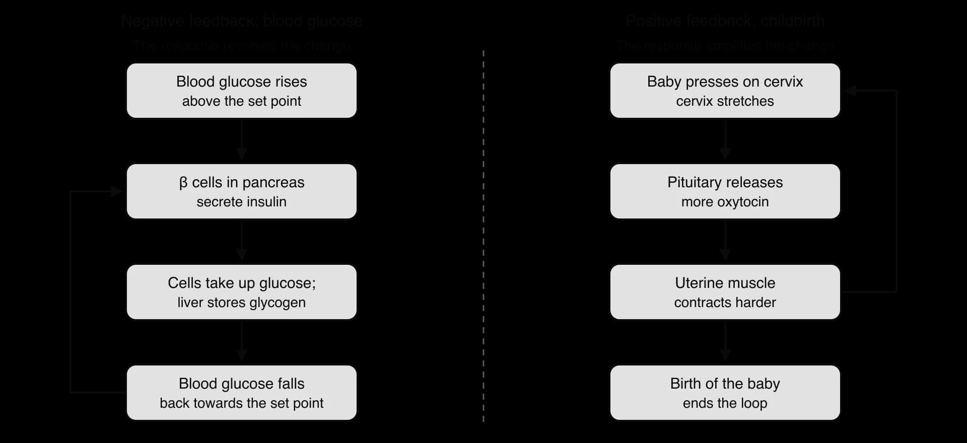
Many signalling pathways regulate themselves through feedback, in which the result of a process affects the process itself. In negative feedback the response reverses the change that triggered it. In positive feedback the response increases the change that triggered it.
Negative feedback
Negative feedback keeps a variable close to a set point and is the basis of homeostasis. When the variable moves away from the set point, the response moves it back, and the response then decreases as the variable returns to normal, which makes negative feedback systems stable.
Control of blood glucose by insulin is an example. After a meal, the blood glucose concentration rises. β cells in the pancreas detect the rise and secrete insulin. Insulin causes muscle and fat cells to take up glucose through GLUT4 and liver cells to convert glucose into glycogen. As the blood glucose concentration falls back towards the set point, the β cells secrete less insulin, so the response decreases as the change is corrected. The hormone thyroxine is controlled in a similar way: high thyroxine reduces the secretion of thyroid-stimulating hormone (TSH) by the pituitary, so less thyroxine is made.
Positive feedback
Positive feedback amplifies a change and drives a process rapidly to completion. It is unstable on its own, so it is useful only for events that must happen quickly and then stop, and every positive feedback loop needs an event that ends it.
Childbirth is an example. When labour begins, the baby’s head presses on the cervix and stretches it. Stretch receptors send impulses to the hypothalamus, and the posterior pituitary releases oxytocin. Oxytocin binds to receptors on the smooth muscle of the uterus and causes stronger contractions, which push the baby harder against the cervix and stretch it further, causing more oxytocin release. The contractions become stronger and more frequent until the baby is born, which removes the stretch and ends the loop. The LH surge before ovulation, triggered by high oestradiol, is a second example.
Feature | Negative feedback | Positive feedback |
|---|---|---|
Effect of the response | Reverses the original change | Increases the original change |
Result | Variable kept near a set point; stable | Process driven rapidly to completion; unstable |
How it ends | Response decreases as the variable returns to normal | An outside event breaks the loop |
Frequency in the body | Very common (most homeostasis) | Rare |
Examples | Blood glucose and insulin; thyroxine and TSH | Oxytocin in childbirth; LH surge before ovulation |
Feedback operates at every level of organisation. Inside a single cell, the product of a signalling pathway can inhibit an earlier step; in a whole organism, hormones regulate glands; in an ecosystem, predator and prey numbers regulate one another. In each case, interaction between the parts of a system keeps it stable or drives it to change.
Exam practice
Chemical signalling is assessed at HL only. Paper 1A questions test the receptor types, the categories of signalling chemicals and which hormones use which receptors. Paper 1B and Paper 2 Section A use data such as dose–response curves, hormone concentrations through the menstrual cycle or trials of drugs that act on receptors. Paper 2 Section B often asks for an extended account of one pathway (adrenaline and cAMP, insulin and GLUT4, or a steroid hormone acting on gene expression) or a comparison of two receptor types. The command terms that appear most often are outline, explain, compare and contrast, distinguish and suggest.
The question has 12 marks, a typical size for a structured question in Section A. Part (b) rewards differences written as pairs, and part (c) rewards the full sequence from diffusion through the membrane to the effect on the tissue.
Several of these pathways connect to other subtopics: synapses in C2.2 Neural signalling, blood glucose control in D3.3 Homeostasis and the menstrual cycle in D3.1 Reproduction.
Self-check
- To do:
I can explain what a ligand is and why binding to its receptor is specific and causes a conformational change.
- To do:
I can describe quorum sensing in Vibrio fischeri, including autoinducer, threshold concentration and bioluminescence.
- To do:
I can distinguish hormones, neurotransmitters, cytokines and calcium ions as signalling chemicals.
- To do:
I can give examples of amine, protein and steroid hormones and of amino acid, peptide, amine and gas neurotransmitters, and explain why chemically diverse signals are used.
- To do:
I can compare the localised effects of neurotransmitters with the distant effects of hormones.
- To do:
I can distinguish transmembrane and intracellular receptors by location, amino acid distribution and the ligands they bind.
- To do:
I can outline a signal transduction pathway, including first and second messengers and amplification.
- To do:
I can explain how the acetylcholine receptor changes the membrane potential.
- To do:
I can explain how adrenaline acts through a G protein, adenylyl cyclase, cAMP and protein kinase A.
- To do:
I can explain how the insulin receptor, a tyrosine kinase, increases glucose uptake through GLUT4.
- To do:
I can explain how steroid hormones regulate gene expression, including the effects of oestradiol on GnRH-secreting cells and of progesterone on the endometrium.