C1.1 Enzymes and metabolism
Explain how enzymes catalyse and control metabolism, interpret rate graphs and experiments, and (HL) explain inhibition, feedback control and how penicillin works.
- IB Biology
- Part of C Interaction and interdependence
- SL and HL
Key terms introduced here
Each is in the Biology glossary, with every other term in the subject.
- Activation energy
The energy that must be supplied to reactants to break or weaken their bonds so that a reaction can take place; enzymes lower it.
- Active site
The region of an enzyme, usually a pocket or groove on its surface, to which the substrate binds and where catalysis takes place.
- Allosteric site
A binding site on an enzyme, separate from the active site, to which a specific regulatory molecule binds reversibly, changing the shape of the enzyme and its active site.
- Anabolism
The metabolic reactions that build macromolecules from smaller molecules, mainly by condensation reactions; these reactions need an input of energy.
- Catabolism
The metabolic reactions that break down macromolecules into monomers by hydrolysis, or oxidise substrates into simpler molecules; many release energy.
- Catalyst
A substance that increases the rate of a chemical reaction without being changed or used up by the reaction.
- Collision theory
The theory that a reaction occurs only when particles collide with enough energy and in the correct orientation; the rate depends on the frequency of successful collisions.
- Competitive inhibition
Inhibition in which a molecule similar to the substrate binds reversibly to the active site, preventing substrate binding; it is overcome at high substrate concentrations.
- Controlled variable
A variable other than the independent variable that could affect the dependent variable and is kept constant so that the experiment is a fair test.
- Dependent variable
The variable that is measured in an investigation, because it is expected to change when the independent variable changes.
- Enzyme
A biological catalyst, usually a globular protein, that speeds up a specific chemical reaction in a living organism without being used up.
- Enzyme inhibitor
A substance that binds to an enzyme and reduces the rate of the reaction it catalyses; inhibitors may be competitive, non-competitive or irreversible.
- Enzyme specificity
The ability of an enzyme to catalyse only one reaction or a small group of similar reactions, because only a substrate with a complementary shape and chemistry can bind to its active site.
- Enzyme–substrate complex
The temporary structure formed when a substrate binds to the active site of an enzyme, before it is converted into products.
- Extracellular enzyme
An enzyme that is secreted from the cell that made it and catalyses reactions outside that cell, such as digestive enzymes in the gut.
- Feedback inhibition
Regulation of a metabolic pathway in which the end product inhibits an enzyme of an early step, usually the first, by binding to its allosteric site.
- Independent variable
The variable that is deliberately changed by the experimenter in an investigation, such as temperature in an enzyme experiment.
- Induced fit
A model of enzyme action in which binding of the substrate changes the shape of both the active site and the substrate, so they fit closely and bonds in the substrate are stressed.
- Intracellular enzyme
An enzyme that catalyses reactions inside the cell that produced it, in the cytoplasm or within an organelle, such as the enzymes of glycolysis.
- Lock-and-key model
An early model of enzyme action in which the active site is a rigid shape that exactly fits the substrate, as a key fits a lock; replaced by the induced-fit model.
- Mechanism-based inhibition
Irreversible inhibition in which an inhibitor in the active site is chemically changed by the enzyme and forms a covalent bond that permanently inactivates it, as penicillin does.
- Metabolic pathway
A sequence of enzyme-catalysed reactions in which the product of each reaction is the substrate of the next.
- Metabolism
The complex network of interdependent and interacting chemical reactions that take place in a living organism.
- Non-competitive inhibition
Inhibition in which the inhibitor binds reversibly to an allosteric site, changing the active site so the reaction cannot be catalysed; increasing substrate concentration does not overcome it.
- Optimum
The value of a factor, such as temperature or pH, at which the rate of an enzyme-catalysed reaction is highest.
- Rate of reaction
The change in the amount of substrate or product per unit time; for an enzyme it can also be estimated as 1/time for a fixed amount of change.
- Statin
A medicine that lowers blood cholesterol by competitively inhibiting HMG-CoA reductase, an enzyme of cholesterol synthesis in the liver.
- Substrate
The molecule on which an enzyme acts, which binds to the active site and is converted into products.
- Transpeptidase
A bacterial enzyme that forms peptide cross-links between peptidoglycan chains in the cell wall; the target of penicillin.
Notes on C1.1 Enzymes and metabolism
Every living cell carries out thousands of chemical reactions, and almost all of them are catalysed by enzymes. Enzymes determine which reactions take place in a cell, how fast they go and when they stop, so they control the whole of metabolism.
The study of enzymes connects several levels of organisation. The shape of a single protein molecule explains why an enzyme acts on only one substrate, the network of enzyme-catalysed reactions explains how a cell builds and breaks down its molecules, and the control of those reactions explains how an organism responds to changes in its needs.
Enzymes as biological catalysts
A chemical reaction converts one or more reactants into products. Many of the reactions that cells depend on, such as the breakdown of glucose or the joining of amino acids, happen extremely slowly on their own at body temperature. A catalyst increases the rate of such a reaction.
An enzyme is a catalyst made by a living organism. Almost all enzymes are proteins. At the end of a reaction an enzyme is chemically unchanged, so the same molecule can catalyse the reaction again and again. A single molecule of the enzyme catalase, for example, can break down millions of molecules of hydrogen peroxide every second. Because they are reused, enzymes are needed only in small amounts.
Why cells need fast reactions
A cell can only stay alive if it obtains energy, builds new molecules and removes waste faster than its structures decay. Without enzymes, most metabolic reactions would be far too slow for this. Some would take years, and a few would take millions of years, at the temperature and pH inside cells. Raising the temperature would speed them up, but cells cannot do this, because high temperatures damage their proteins and membranes.
Enzymes overcome this limitation, speeding up reactions by factors of millions or more under the mild conditions inside cells, around 37 °C and near-neutral pH in humans. This allows a cell to carry out the reactions it needs quickly enough to grow, respond to its environment and reproduce. Faster reactions also mean that a cell can switch a process on or off quickly by changing the activity of the enzyme that catalyses it.
Enzymes and metabolism
Metabolism is the complete set of chemical reactions in an organism. These reactions are not separate from each other. The product of one reaction is usually the reactant (substrate) of the next, so reactions are linked into chains and cycles, and many chains share the same molecules. Metabolism is therefore a complex network of interdependent and interacting reactions.
A sequence of reactions in which the product of each reaction is the substrate of the next, each catalysed by its own enzyme, is a metabolic pathway. Respiration, for example, is a pathway of dozens of steps that gradually releases energy from glucose. The pathways of a cell are linked where they share intermediates, in the same way that the roads of a city meet at junctions. A change in one pathway can therefore affect others.
Many enzymes for many reactions
Each enzyme catalyses only one reaction, or a small group of very similar reactions, because its shape fits only certain substrates. A cell that carries out thousands of different reactions therefore needs thousands of different enzymes. The human genome contains genes for several thousand enzymes, and each type of cell produces the particular set of enzymes needed for its functions.
Control of metabolism through enzymes
Because almost every reaction needs its own enzyme, a cell controls its metabolism by controlling its enzymes. A reaction happens at a significant rate only when the enzyme for it is present and active. Cells regulate metabolism in two main ways:
Which enzymes are made. Genes for enzymes are switched on and off, so the cell produces only the enzymes it needs at that time. Cells in the pancreas make digestive enzymes, for example, while cells in the liver make the enzymes that store and release glycogen.
How active the enzymes are. Existing enzymes can be activated or inhibited by other molecules, so their activity changes within seconds as conditions change.
This control through enzymes is an example of interaction and interdependence at the molecular level. The amount of each substance in a cell depends on the balance between the enzymes that make it and the enzymes that use it.
Anabolic and catabolic reactions
Metabolic reactions can be divided into two groups: those that build larger molecules from smaller ones, and those that break larger molecules into smaller ones.
Anabolism joins monomers together to form macromolecules. The monomers are usually joined by condensation reactions, in which a bond forms between two molecules and a molecule of water is released. Anabolic reactions need energy, which is usually supplied by ATP. Examples of anabolic processes include:
Protein synthesis: amino acids are joined by peptide bonds to form polypeptides on ribosomes.
Glycogen formation: glucose molecules are joined by glycosidic bonds to form glycogen, the storage polysaccharide of animals, in liver and muscle cells.
Photosynthesis: carbon dioxide is built into glucose and other carbohydrates in chloroplasts, using energy from light.
The synthesis of DNA and RNA from nucleotides, and of triglycerides from glycerol and fatty acids.
Catabolism breaks macromolecules down into monomers or into even simpler molecules. The breakdown of polymers into monomers happens by hydrolysis, in which water is used to split a bond. Catabolism also includes the oxidation of substrates such as glucose to release energy. Many catabolic reactions release energy. Examples include:
Digestion: starch, proteins and lipids in food are hydrolysed into sugars, amino acids, and fatty acids and glycerol, which can be absorbed.
Cell respiration: glucose and other substrates are oxidised to carbon dioxide and water, and the energy released is used to make ATP.
The hydrolysis of glycogen to glucose in the liver when blood glucose is low.
Feature | Anabolism | Catabolism |
|---|---|---|
What happens | Larger molecules built from smaller ones | Larger molecules broken into smaller ones |
Main type of reaction | Condensation (water released) | Hydrolysis (water used); oxidation |
Energy | Needs an input of energy (usually from ATP) | Many reactions release energy |
Examples | Protein synthesis, glycogen formation, photosynthesis | Digestion, cell respiration |
Enzyme structure and the active site
Enzymes are globular proteins. Each one is made of one or more polypeptide chains folded into a compact, roughly spherical three-dimensional shape. The sequence of amino acids in each chain determines how it folds, and the folded shape is held together by bonds between the R-groups of the amino acids: hydrogen bonds, ionic bonds, disulfide bonds and hydrophobic interactions. This folded shape is the tertiary structure of the protein, and in enzymes made of more than one chain the chains also fit together in a quaternary structure.
Somewhere on the surface of the enzyme is a small pocket or groove where the substrate binds and the reaction is catalysed. This region is the active site.
The substrate is the molecule that an enzyme acts on. The active site has a shape and chemical properties that are complementary to the substrate, so the substrate fits it closely. Only a few amino acids line the active site and take part directly in binding the substrate and catalysing the reaction, often fewer than ten out of several hundred in the whole enzyme. The diagram of an enzyme shows these few amino acids and the much larger folded chain around them.
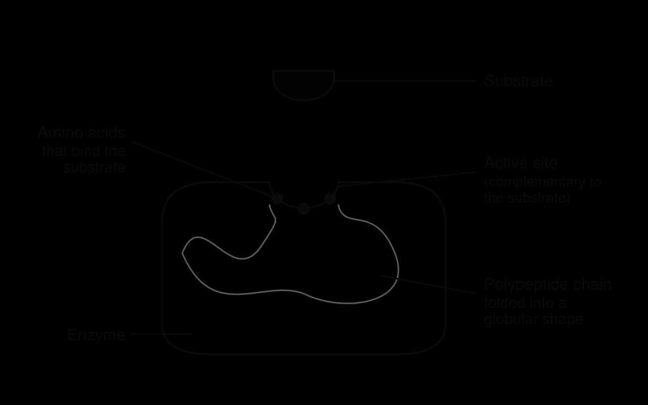
The rest of the enzyme molecule is essential for catalysis. The amino acids that line the active site are often far apart in the primary sequence of the polypeptide; they are brought together only when the whole chain folds. Interactions throughout the protein hold these amino acids in exactly the right positions, give the active site its shape, and allow small movements of the enzyme that help catalysis. A change anywhere in the structure, such as the substitution of a single amino acid far from the active site, can change the shape of the active site and reduce the activity of the enzyme, so the function of the active site depends on the form of the whole molecule.
Induced-fit binding of the substrate
For a reaction to be catalysed, the substrate binds to the active site, forming an enzyme–substrate complex. The substrate is held in the active site by temporary bonds, such as hydrogen bonds and ionic attractions, with the amino acids that line it. The reaction then takes place, the products are released, and the enzyme is free to bind another substrate molecule.
From lock and key to induced fit
An early model of enzyme action, proposed by Emil Fischer in 1894, was the lock-and-key model. It pictured the active site as a rigid shape into which only the correct substrate fits, like a key into a lock. The model explains specificity well, but it treats the enzyme and substrate as fixed shapes, which does not match later evidence that proteins are flexible.
The induced-fit model, proposed by Daniel Koshland in 1958, replaced it. In the induced-fit model, the active site is not exactly complementary to the substrate before binding. As the substrate approaches and binds, both the substrate and the enzyme change shape slightly, so the active site moulds itself around the substrate and the fit becomes closer. The change in the shape of the enzyme puts strain on particular bonds within the substrate, which weakens them and makes them easier to break or rearrange.
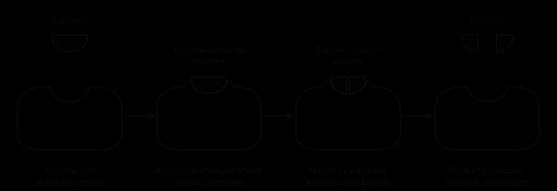
The change from the lock-and-key model to the induced-fit model is an example of how scientific models are refined when new evidence appears. Structures of enzymes determined by X-ray crystallography, with and without substrate bound, show that many enzymes really do change shape when the substrate binds. Hexokinase, the enzyme that adds a phosphate group to glucose at the start of glycolysis, closes around glucose like a pair of jaws.
Molecular motion and collisions
An enzyme can only catalyse a reaction when a substrate molecule enters its active site. In solution, molecules of enzyme and substrate move about randomly and continuously, colliding with water molecules and with each other. Collision theory explains that a reaction can only occur when the reacting particles collide.
Not every collision leads to binding. The substrate has to collide with the active site, not another part of the enzyme surface, and it has to approach in the correct orientation so that its groups line up with the amino acids of the active site. A collision that leads to binding and catalysis is a successful collision. The rate of reaction therefore depends on how often successful collisions happen, which in turn depends on how fast the molecules are moving and how many of them there are.
In most cases both the enzyme and the substrate are dissolved and move. In some cases only one of them moves:
Immobilised substrates. Some substrates are large or are part of a larger structure, such as a starch grain, a strand of DNA or the cellulose of a cell wall. The substrate is held still and the enzyme molecules move to it. DNA polymerase, for example, moves along a DNA template strand.
Immobilised enzymes. Many enzymes are fixed in membranes, such as the enzymes of the electron transport chain in the inner mitochondrial membrane and ATP synthase. Only the substrates move, diffusing to the enzymes.
Industry often uses immobilised enzymes deliberately, by attaching them to a surface or trapping them in beads of gel. Lactose-free milk is made by passing milk over immobilised lactase, which hydrolyses lactose into glucose and galactose. The enzyme stays in the reactor and can be reused, and it does not end up in the product.
Enzyme specificity and denaturation
Each enzyme acts on only one substrate or a small group of very similar substrates. This enzyme specificity comes from the active site. Its shape matches the shape of the substrate, and the chemical properties of the amino acids that line it (their charge, polarity or hydrophobic character) match the chemical groups of the substrate. Only a substrate with the right shape and the right chemical groups can bind and be held so that its bonds are strained.
Amylase, for example, hydrolyses the α-1,4 glycosidic bonds of starch but not the β-1,4 bonds of cellulose, even though both are polymers of glucose. Humans make amylase but no enzyme that digests cellulose, which is why cellulose passes through the human gut undigested.
Denaturation
The active site keeps its shape only while the bonds that hold the enzyme's tertiary structure are intact. Heat and extreme pH break these bonds. Denaturation is a change in the three-dimensional structure of a protein that destroys its function. In a denatured enzyme the active site has lost its shape and chemical properties, so the substrate can no longer bind and the reaction is no longer catalysed.
High temperature makes the atoms of the enzyme vibrate more strongly. The vibrations break hydrogen bonds and other weak interactions, so the polypeptide chain unfolds.
Extreme pH changes the charges on R-groups by adding or removing hydrogen ions. Ionic bonds and hydrogen bonds that depend on these charges are broken, and the shape of the active site changes. Charged amino acids in the active site may also lose the charges they need to bind the substrate.
Denaturation is usually permanent. Once the chain has unfolded it rarely refolds into the correct shape, even when conditions return to normal. A cooked egg white, in which the protein albumin has been denatured by heat, does not become clear again when it cools.
Factors affecting the rate of enzyme activity
The rate of an enzyme-catalysed reaction depends on how often substrate molecules collide successfully with active sites, and on how many active sites are working. Temperature, pH and substrate concentration all affect one or both of these. Each factor gives a characteristic graph of rate against the factor, and each graph can be explained using collision theory and denaturation.
 YouTube
Temperature
As temperature rises from a low value, the rate of reaction increases. Enzyme and substrate molecules gain kinetic energy and move faster, so they collide more often, and more of the collisions have enough energy to lead to a reaction. For many reactions in this range, the rate roughly doubles for every 10 °C rise.
At a certain temperature the rate reaches a maximum. This is the optimum temperature for the enzyme. Above the optimum, the rate falls steeply. The enzyme molecules vibrate so strongly that the bonds holding their tertiary structure break, the active sites change shape and more and more enzyme molecules are denatured. Fewer active sites can bind substrate, so the rate falls, even though molecules are still colliding frequently. When all the enzyme molecules are denatured, the rate falls to zero.
The graph of rate against temperature is not symmetrical. The rise up to the optimum is gradual, because collision frequency increases steadily. The fall after the optimum is steep, because denaturation happens quickly once the temperature is high enough to break the bonds of the tertiary structure.
Optimum temperature reflects the organism's environment
Most human enzymes have optimum temperatures close to 37 °C, the normal body temperature. Enzymes from organisms that live in other conditions have different optima. Thermus aquaticus, a bacterium first described in 1969 from hot springs in Yellowstone National Park, USA, makes a DNA polymerase (Taq polymerase) with an optimum of about 75–80 °C, and its proteins have extra bonds that stop them denaturing in the hot water. At the other extreme, the proteases added to biological washing powders, such as subtilisins from bacteria of the genus Bacillus, have been modified by changing particular amino acids so that they stay active and stable in cool washes, which saves the energy needed to heat the water.
pH
Each enzyme also has an optimum pH, at which its rate of reaction is highest. On either side of the optimum the rate falls. pH is a measure of the concentration of hydrogen ions (H⁺). A change in pH changes the charges on R-groups: excess H⁺ adds hydrogen ions to basic groups such as –NH₂, giving them a positive charge, while a shortage of H⁺ removes hydrogen ions from acidic groups such as –COOH, giving them a negative charge.
These changes affect the enzyme in two ways. Close to the optimum, small changes in the charges of amino acids in the active site make it harder for the substrate to bind, so the rate falls but the effect is reversible. Further from the optimum, the changed charges break the ionic bonds and hydrogen bonds that hold the tertiary structure, and the enzyme is denatured. The graph of rate against pH is therefore a curve with a peak at the optimum.
- Pepsin (stomach)
- Salivary amylase (mouth)
The optimum pH of an enzyme matches the place where it works. Pepsin, a protease that digests protein in the stomach, has an optimum of about pH 2, matching the hydrochloric acid secreted by the stomach wall. Salivary amylase works best at about pH 7 in the mouth and stops working when it reaches the stomach. Trypsin, a protease secreted by the pancreas into the small intestine, has an optimum of about pH 8.
Substrate concentration
At low substrate concentrations, increasing the concentration increases the rate of reaction in direct proportion. With more substrate molecules in the same volume, collisions with active sites happen more often. At this stage most active sites are empty most of the time, so substrate concentration is the limiting factor.
As the substrate concentration rises further, the rate increases less and less, and eventually it levels off at a maximum. At high substrate concentrations, every active site is occupied almost all the time. A substrate molecule can only bind when a product has been released, so adding more substrate cannot increase the rate. The enzyme is said to be saturated, and the number of enzyme molecules is now the limiting factor.
If the enzyme concentration is increased while substrate is in excess, the rate increases in proportion, because there are more active sites. This is why cells can increase the rate of a reaction by making more of the enzyme that catalyses it.
Sketch graphs as models
The graphs of rate against temperature, pH and substrate concentration are generalised sketch graphs. They are models: they show the typical shape of the relationship without giving real values. Results from an actual experiment never lie exactly on such a curve. Data points scatter because of random error, an optimum may be missed if the measurements are too far apart, and at high temperatures the rate depends on how long the enzyme was heated, because denaturation takes time. Comparing a sketch graph with real results is a way of evaluating both the model and the experiment.
Measuring the rate of enzyme-catalysed reactions
The rate of an enzyme-catalysed reaction is the change in the amount of substrate or product per unit time. It can be found by measuring how much product forms, or how much substrate disappears, over a period of time:
Typical units are cm³ s⁻¹ for a gas produced, or mol dm⁻³ s⁻¹ for a change in concentration. Sometimes the time taken for a fixed amount of change is measured instead, for example the time until all the starch has gone. Then the rate is taken as the reciprocal of the time, with units of s⁻¹:
Variables in an enzyme experiment
A fair experiment changes one variable at a time. The independent variable is the factor that is deliberately changed, such as temperature. The dependent variable is the factor that is measured, such as the volume of oxygen produced per minute. Every other factor that could affect the rate is a controlled variable, kept constant so that it cannot affect the results.
Catalase and hydrogen peroxide
Catalase is an enzyme found in most cells, which breaks down hydrogen peroxide, a toxic by-product of metabolism, into water and oxygen:
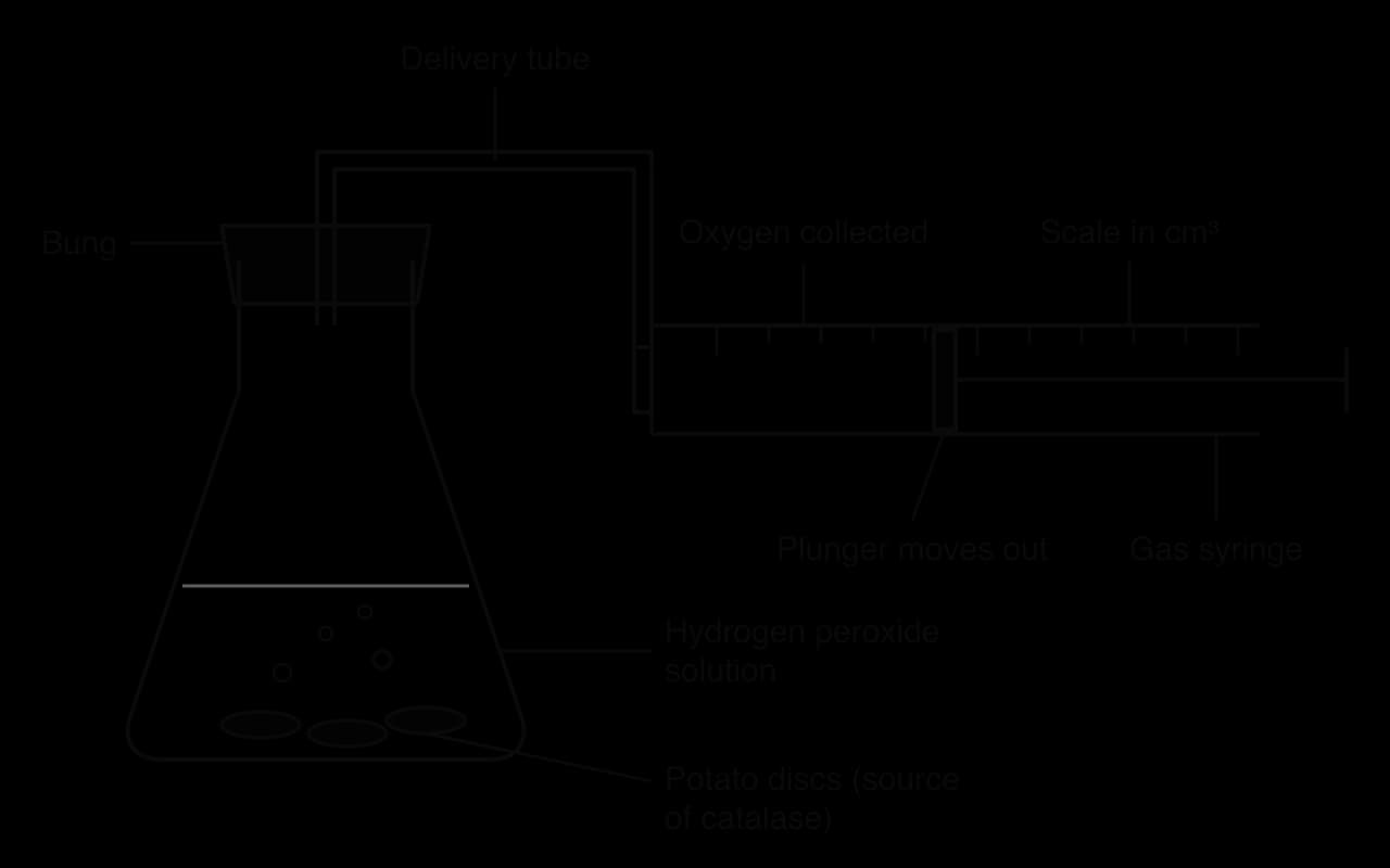
The rate can be measured by collecting the oxygen produced. Discs of potato tissue, or yeast suspension, are added to hydrogen peroxide solution in a flask, and the oxygen passes along a delivery tube into a gas syringe, as in the diagram of the apparatus. The volume of gas is read at regular intervals. Oxygen can also be collected by displacing water from an upturned measuring cylinder, or the rate can be estimated from the height of foam in a test tube or from the time taken for filter-paper discs soaked in catalase to rise through hydrogen peroxide.
Variable | In an investigation of the effect of temperature on catalase | How it is controlled or measured |
|---|---|---|
Independent | Temperature | Water baths at, for example, 10, 20, 30, 40, 50 and 60 °C, checked with a thermometer |
Dependent | Rate of oxygen production | Volume of oxygen in the gas syringe after a fixed time, for example 60 s |
Controlled | Concentration and volume of hydrogen peroxide | Same solution and measuring cylinder for every trial |
Controlled | Amount of enzyme | Same number of potato discs, cut with the same borer to the same thickness |
Controlled | pH | Same buffer solution in every trial |
Initial rate from a graph
Because the rate falls as substrate is used up, the fairest way to compare rates under different conditions is to measure the initial rate, the rate at the very start of the reaction, when substrate concentration is still known. On a graph of product against time, the initial rate is the gradient of a tangent drawn to the curve at time zero. Later, the curve levels off when all the substrate has been converted, and the rate at that point is zero.
- Oxygen produced
- Tangent at time 0
Amylase and starch
Amylase hydrolyses starch into maltose. The disappearance of starch can be followed with iodine solution, which turns from orange-brown to blue-black in the presence of starch. Drops of the reaction mixture are taken at regular intervals, for example every 10 s, and added to drops of iodine on a spotting tile. The time at which the iodine no longer turns blue-black shows when the starch has been fully hydrolysed, and the rate is calculated as 1/time. A colorimeter can measure the fading blue-black colour more precisely, giving quantitative data instead of a judgement by eye.
Other practicals follow the same logic. Lipase hydrolyses the fats in milk into fatty acids and glycerol, and the fatty acids lower the pH. The rate can be followed with a pH probe and data logger, or with an indicator such as phenolphthalein, which changes from pink to colourless as the pH falls.
Rates can also be determined from secondary data, such as a published table or graph. The method is the same: find the change in product or substrate and divide by the time over which it happened, or draw a tangent to a curve and calculate its gradient.
Enzymes and activation energy
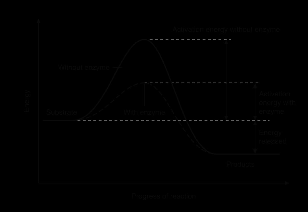
Even a reaction that releases energy overall does not start by itself. To react, the bonds in the substrate first have to be broken or weakened, and breaking bonds needs energy. Energy is released later in the reaction, when new bonds form in the products. The energy that has to be supplied to start the reaction is the activation energy.
In a test tube, activation energy can be supplied by heating. Cells cannot rely on this, so enzymes lower the activation energy instead. When the substrate binds to the active site, the induced fit stresses particular bonds, and amino acid side chains in the active site may donate or accept hydrogen ions or hold two substrates close together in the right orientation. All of these make the bonds easier to break, so less energy is needed to start the reaction. More substrate molecules have enough energy to react at body temperature, and the reaction goes faster.
An energy diagram shows the energy of the reactants and products as a reaction proceeds. The substrate starts at one energy level and the products end at another. The peak between them is the energy that has to be reached for the reaction to take place.
Case study: a reaction that would take 78 million years
Orotidine 5′-phosphate decarboxylase (OMP decarboxylase) catalyses the last step in making uridine monophosphate, the pyrimidine nucleotide from which the other pyrimidine nucleotides of RNA and DNA are made. The reaction removes a carboxyl group from the substrate, releasing CO₂. Every cell that makes its own nucleotides needs this reaction.
In 1995 the biochemists Anna Radzicka and Richard Wolfenden measured how fast the same reaction happens without the enzyme, using a similar compound in neutral water at 25 °C. The uncatalysed reaction is so slow that its rate had to be estimated from measurements made at high temperatures. Its half-time, the time for half of the substrate to react, is about 78 million years. With the enzyme, each active site converts about 39 substrate molecules every second.
Measure | Uncatalysed reaction (25 °C, neutral water) | Catalysed by OMP decarboxylase |
|---|---|---|
Rate constant | 2.8 × 10⁻¹⁶ s⁻¹ | 39 s⁻¹ (molecules converted per active site per second) |
Time scale | Half the substrate reacts in about 78 million years | One molecule converted in about 0.026 s |
Rate enhancement | 1 | About 1.4 × 10¹⁷ times faster |
Source: Radzicka and Wolfenden, Science, 1995.
The data show why life depends on enzymes. A cell that had to wait millions of years for a single step in nucleotide synthesis could never copy its DNA or make RNA. OMP decarboxylase achieves its enormous rate enhancement by binding its substrate very tightly and specifically: amino acids in its active site hold the substrate in a strained position and stabilise the unstable state halfway through the reaction, which lowers the activation energy. Such tight and precise binding makes OMP decarboxylase an extreme example of enzyme specificity.
Intracellular and extracellular enzymes (HL only)
Enzymes are made on ribosomes inside cells, but they do not all work there. An intracellular enzyme catalyses reactions inside the cell that produced it, in the cytoplasm or inside an organelle. An extracellular enzyme is secreted from the cell by exocytosis and catalyses reactions outside it.
Most enzymes are intracellular. The ten enzymes of glycolysis are dissolved in the cytoplasm, where glucose is broken down to pyruvate. The enzymes of the Krebs cycle are in the mitochondrial matrix, and those of DNA replication and transcription are in the nucleus. Keeping these enzymes inside compartments places them close to their substrates and to the other enzymes of the same pathway.
Extracellular enzymes are used where the substrate is too large to enter the cell or where the reaction would be harmful inside it. The clearest example is chemical digestion in the human gut. Cells of the salivary glands, stomach wall and pancreas make digestive enzymes such as amylase, pepsin, trypsin and lipase, package them into vesicles and release them by exocytosis into the lumen of the gut. There the enzymes hydrolyse food macromolecules, which are too large to be absorbed, into small molecules that can cross the gut wall. Saprotrophic fungi and bacteria also secrete extracellular enzymes onto dead matter (see B4.2 Ecological niches).
Feature | Intracellular enzymes | Extracellular enzymes |
|---|---|---|
Where they act | Inside the cell that made them | Outside the cell that made them |
How they leave the cell | They do not leave | Secreted in vesicles by exocytosis |
Examples | Enzymes of glycolysis (cytoplasm) and the Krebs cycle (mitochondrial matrix) | Amylase, pepsin, trypsin and lipase in the gut; enzymes secreted by saprotrophs |
Some extracellular enzymes are dangerous to the cells that make them. Pepsin and trypsin would digest the proteins of the cells that produce them, so they are made and secreted in an inactive form (pepsinogen and trypsinogen) and only activated in the lumen of the gut.
Heat generated by metabolism (HL only)
Every transfer of energy in metabolism is less than 100% efficient. When glucose is oxidised in cell respiration, only part of the energy released is captured in ATP; the rest is released as heat. More heat is released when ATP is used to power muscle contraction, active transport and anabolic reactions. Heat is therefore an unavoidable by-product of all metabolic reactions.
Most animals lose this heat to their surroundings, and their body temperature follows that of the environment. Mammals and birds are endotherms: they use the heat released by their own metabolism to keep their body temperature constant and higher than that of their surroundings, around 37 °C in most mammals and around 40 °C in most birds. Their metabolic rates are much higher than those of reptiles or fish of the same size, so they release far more heat.
A constant, raised body temperature keeps enzymes close to their optimum temperature, so metabolic reactions stay fast whatever the external temperature. Endotherms can therefore stay active at night and in cold climates. The cost is a large demand for food. When an endotherm is cold, it releases extra heat by shivering, by increasing its metabolic rate, and in many mammals by respiration in brown adipose tissue, where the energy is released directly as heat without making ATP (see D3.3 Homeostasis).
Linear and cyclical metabolic pathways (HL only)
Metabolic pathways take two basic forms. In a linear pathway, a starting substrate is converted through a chain of intermediates into a final product, each step catalysed by a different enzyme. Glycolysis is a linear pathway: in ten enzyme-catalysed steps in the cytoplasm, one glucose molecule (six carbon atoms) is converted into two molecules of pyruvate (three carbon atoms each).
In a cyclical pathway, the starting compound is regenerated at the end of each turn of the cycle, so it can react again. The Krebs cycle is a cyclical pathway. An acetyl group with two carbon atoms joins a four-carbon compound (oxaloacetate) to form a six-carbon compound (citrate). Through a series of reactions, two carbon atoms are released as CO₂ and oxaloacetate is regenerated, ready to accept the next acetyl group. The Calvin cycle in chloroplasts is also cyclical: carbon dioxide is fixed by combining with ribulose bisphosphate (RuBP), and RuBP is regenerated at the end of each turn (see C1.3 Photosynthesis).
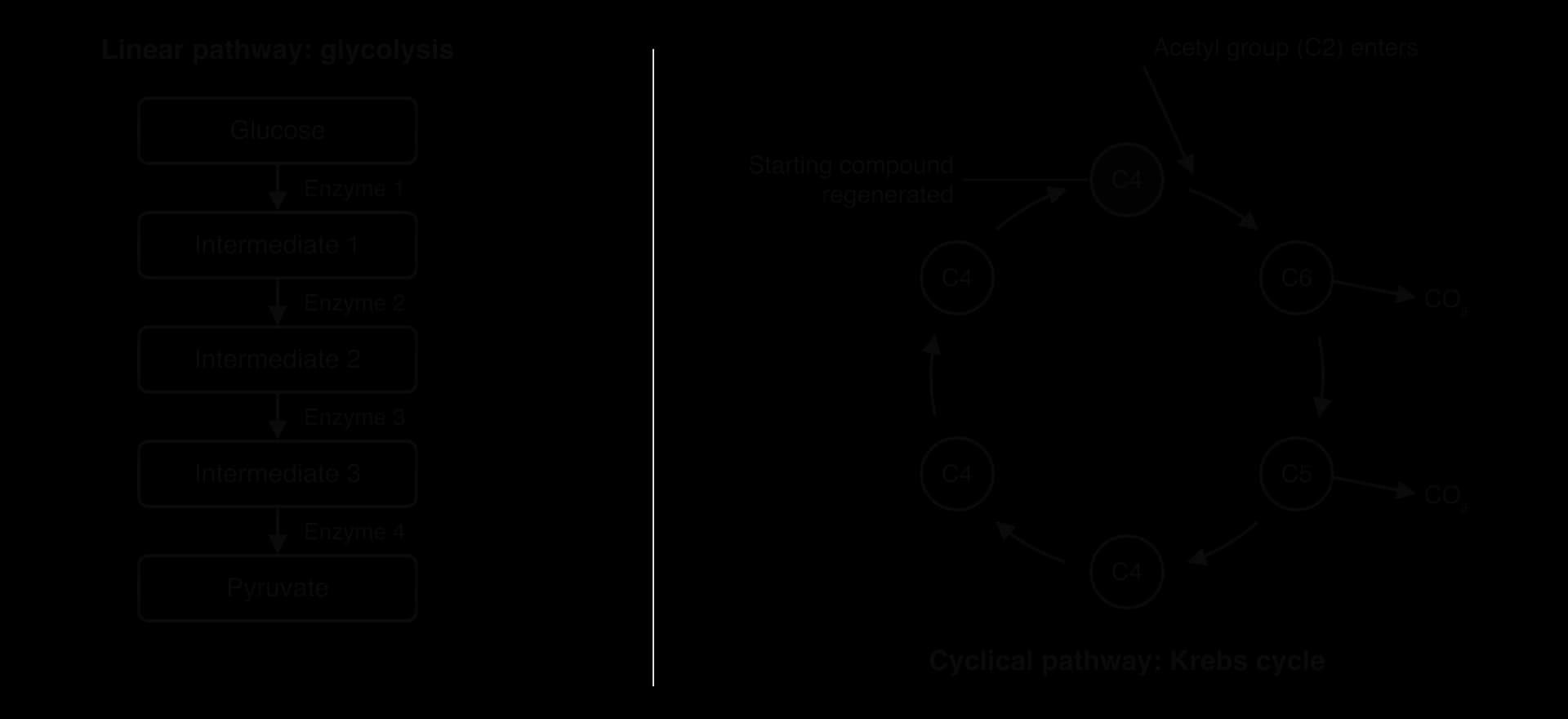
Feature | Linear pathway | Cyclical pathway |
|---|---|---|
Shape | Chain from a starting substrate to a final product | Ring of intermediates |
Starting compound | Used up | Regenerated at the end of each turn |
Examples | Glycolysis; the threonine to isoleucine pathway | Krebs cycle; Calvin cycle |
A cycle needs only a small amount of each intermediate, because the intermediates are recycled continuously. A cell can process a large flow of acetyl groups through the Krebs cycle with only a small, constant pool of oxaloacetate.
Enzyme inhibition (HL only)
An enzyme inhibitor is a substance that reduces the rate of an enzyme-catalysed reaction by binding to the enzyme. Cells use inhibitors to control their own metabolism, and many medicines and poisons work by inhibiting enzymes. Inhibitors are classified by where they bind and whether they bind reversibly.
Allosteric sites and non-competitive inhibition
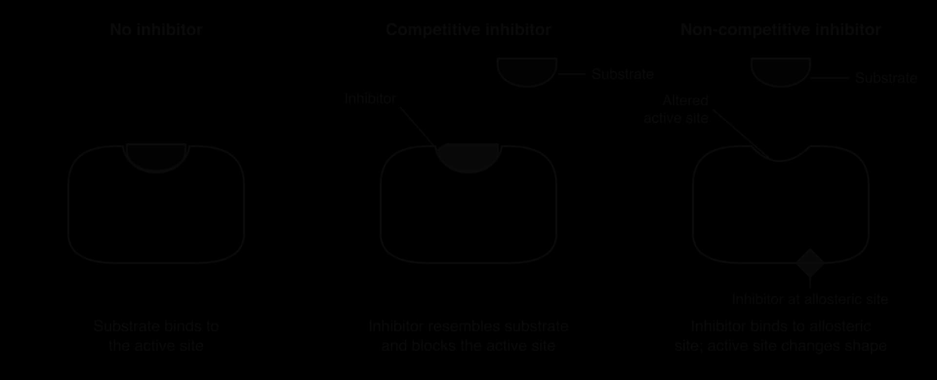
Many enzymes have, as well as the active site, a second binding site called an allosteric site. Like the active site, it binds only specific molecules. When a molecule binds to the allosteric site, it causes a change in the shape (a conformational change) of the whole enzyme, which spreads to the active site. Binding is reversible: the molecule attaches by weak bonds and can detach again.
In non-competitive inhibition, the inhibitor binds to an allosteric site. The conformational change alters the active site so that the substrate can no longer bind, or binds but cannot be converted into products. Because the inhibitor does not occupy the active site, it does not compete with the substrate. Adding more substrate cannot displace it, so the maximum rate of the reaction is reduced however much substrate is present. Each enzyme molecule with an inhibitor bound is effectively out of action, as if there were fewer enzyme molecules.
Competitive inhibition
In competitive inhibition, the inhibitor has a shape and chemical properties similar to those of the substrate. It binds reversibly to the active site, so it blocks the substrate from binding, but it is not converted into products. The inhibitor and the substrate compete for the same active sites, and the proportion of active sites occupied by each depends on their relative concentrations. Increasing the substrate concentration makes it more likely that a substrate molecule, not an inhibitor, enters each active site, so at very high substrate concentrations the effect of the inhibitor is overcome and the reaction approaches the same maximum rate as without the inhibitor.
Statins
Statins are medicines that lower the concentration of cholesterol in the blood, and they are a widely used example of competitive inhibition. In the liver, cholesterol is made by a long pathway. One of the early steps, the conversion of HMG-CoA to mevalonate, is catalysed by the enzyme HMG-CoA reductase, and this step controls the rate of the whole pathway. Part of the statin molecule closely resembles HMG-CoA, so statins bind to the active site of HMG-CoA reductase and compete with the substrate. Less mevalonate, and therefore less cholesterol, is made. The liver cells respond by taking up more cholesterol-carrying lipoproteins from the blood, which lowers blood cholesterol and reduces the risk of atherosclerosis and coronary heart disease.
The first statin, compactin (mevastatin), was isolated from the fungus Penicillium citrinum in 1973 by the Japanese biochemist Akira Endo, who was searching fungi for compounds that inhibit cholesterol synthesis. Lovastatin, from a different fungus, became the first statin approved for medical use, in 1987.
A second medical example is the treatment of methanol or ethylene glycol (antifreeze) poisoning. These alcohols are not very toxic themselves, but the enzyme alcohol dehydrogenase in the liver converts them into toxic products. The drug fomepizole is a competitive inhibitor of alcohol dehydrogenase: it occupies the active site, so the poison is converted only slowly and much of it is removed from the body unchanged.
 YouTube
Comparing competitive and non-competitive inhibition
The two types of inhibition can be distinguished by measuring the rate of reaction at a range of substrate concentrations, with and without the inhibitor. The graph of rate against substrate concentration shows the difference most clearly at high substrate concentrations.
- No inhibitor
- Competitive inhibitor
- Non-competitive inhibitor
Feature | Competitive inhibition | Non-competitive inhibition |
|---|---|---|
Shape of inhibitor | Similar to the substrate | Not similar to the substrate |
Where it binds | Active site | Allosteric site |
Effect on the enzyme | Blocks the active site; enzyme shape unchanged | Changes the shape of the active site |
Effect of increasing substrate concentration | Inhibition reduced; maximum rate eventually reached | Inhibition not reduced; maximum rate stays lower |
Example | Statins on HMG-CoA reductase; fomepizole on alcohol dehydrogenase | Isoleucine on threonine deaminase (feedback inhibition) |
Feedback inhibition of metabolic pathways (HL only)
Cells need to make the right amount of each substance: enough to meet demand, but not so much that resources are wasted. Many metabolic pathways are regulated by feedback inhibition, in which the end product of the pathway inhibits an enzyme near the start of it, usually the first enzyme, as a form of negative feedback.
The end product usually acts as a non-competitive inhibitor: it binds to the allosteric site of the first enzyme and changes the shape of its active site. When the concentration of the end product is high, more enzyme molecules have the end product bound, the first step slows and less end product is made. When the end product is used up and its concentration falls, it detaches from the allosteric sites, the enzyme becomes active again and production resumes, so the supply of the end product matches demand.
Threonine to isoleucine
Bacteria and plants make the amino acid isoleucine from another amino acid, threonine, in a pathway of five enzyme-catalysed steps. The first enzyme, threonine deaminase (also called threonine dehydratase), converts threonine to 2-oxobutanoate. Isoleucine, the end product, is an allosteric inhibitor of threonine deaminase.
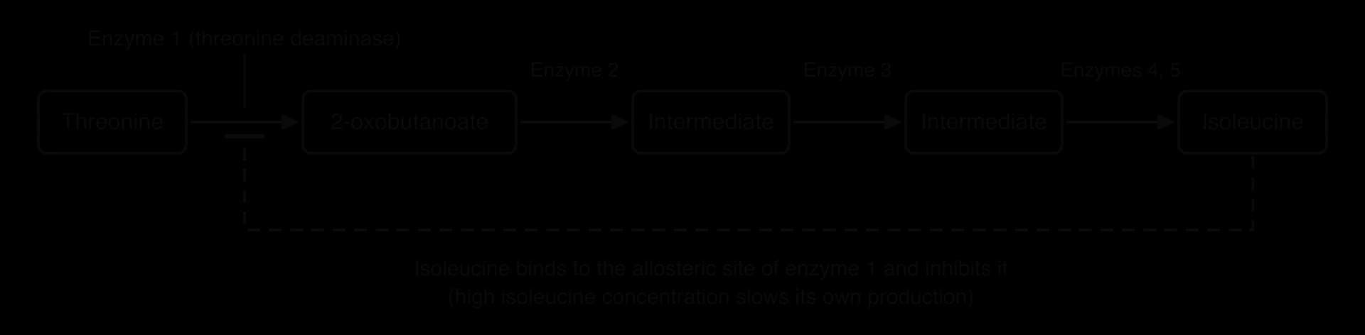
Inhibiting the first enzyme, not a later one, has two advantages. It stops the whole pathway at once, so none of the intermediates build up, and it saves threonine, which the cell also needs for making proteins. The pathway shows how an allosteric site allows a molecule that does not resemble the substrate at all to control an enzyme.
Mechanism-based inhibition (HL only)
Competitive and non-competitive inhibitors bind reversibly, by weak bonds, but some inhibitors bind irreversibly. In mechanism-based inhibition, the inhibitor enters the active site like a substrate, and the enzyme begins its normal catalytic mechanism on it. Instead of producing a product that is released, the reaction forms a covalent bond between the inhibitor and an amino acid in the active site. The active site is permanently changed and the enzyme molecule is inactivated for good. This is sometimes called suicide inhibition, because the enzyme takes part in its own inactivation.
Penicillin and transpeptidases
Penicillin, discovered by Alexander Fleming in 1928, was the first widely used antibiotic, and it is a mechanism-based inhibitor. Bacteria are surrounded by a cell wall made of peptidoglycan, which consists of long chains of sugars cross-linked by short peptides. The cross-links make the wall strong enough to stop the cell bursting by osmosis. They are formed by enzymes called transpeptidases, which join a peptide on one chain to a peptide on a neighbouring chain.
Penicillin has a four-membered ring, the β-lactam ring, which resembles the end of the peptide that is the normal substrate of transpeptidase. Penicillin therefore enters the active site, and an amino acid (serine) in the active site attacks the β-lactam ring as if it were the substrate. The ring opens and penicillin becomes covalently bonded to the serine. The bond is not broken, so the enzyme is permanently inactivated. Without working transpeptidases, a growing bacterium cannot build cross-links into new wall material. The wall weakens, water enters by osmosis, and the cell bursts. Human cells have no cell walls and no transpeptidases, which is why penicillin harms bacteria but not human cells.
Resistance by changed transpeptidases
Some bacteria are resistant to penicillin because they make transpeptidases with a changed active site. Meticillin-resistant Staphylococcus aureus (MRSA) carries a gene, mecA, that codes for an extra transpeptidase called PBP2a. The active site of PBP2a has a different shape, and β-lactam antibiotics such as penicillin and meticillin bind to it only weakly and slowly. When the bacterium is exposed to the antibiotic, its normal transpeptidases are inactivated, but PBP2a continues to form cross-links, so the wall is still built and the bacterium survives.
This is a change in the structure of the enzyme that is the target of the drug, and it spreads in bacterial populations by natural selection whenever antibiotics are used. Other bacteria resist penicillin in a different way, by making β-lactamase enzymes that hydrolyse the β-lactam ring before it reaches the transpeptidase. The spread of antibiotic resistance is discussed further in C3.2 Defence against disease.
Aspirin is another irreversible inhibitor. It transfers an acetyl group to a serine in the active site of cyclooxygenase, an enzyme that makes prostaglandins, the signalling molecules that cause pain and inflammation. The acetylated enzyme is permanently inactive, so the effect of aspirin lasts until the cell makes new enzyme molecules.
Exam practice
Enzymes are assessed throughout the papers. Paper 1A questions test definitions (active site, activation energy, denaturation), the shapes of the standard graphs and, at HL, the types of inhibition. Paper 1B uses data from enzyme practicals: identifying variables, calculating rates with units, and evaluating methods. Paper 2 asks for explanations of graphs, labelled sketch graphs, and extended answers on enzyme action, factors affecting rate and, at HL, the control of metabolism by inhibition. The command terms most often used are outline, explain, sketch, calculate, deduce and compare and contrast.
A common structure for a Section B question is a short description, a graph explanation and a longer explanation linking two ideas, such as specificity and denaturation, or inhibition and the control of a pathway.
Synoptic links also help with extended answers, because enzyme action connects protein structure to the control of whole pathways.
Self-check
- To do:
I can define enzyme and catalyst and explain why cells need enzymes.
- To do:
I can explain why metabolism needs many different enzymes and how cells control it through enzymes.
- To do:
I can distinguish anabolism from catabolism, with examples of each.
- To do:
I can describe the active site and explain specificity and the induced-fit model.
- To do:
I can explain how collisions between substrate and active site lead to catalysis.
- To do:
I can explain and sketch the effects of temperature, pH and substrate concentration on enzyme activity.
- To do:
I can calculate rates of reaction from experimental data, including initial rates from tangents.
- To do:
I can explain how enzymes lower activation energy and interpret an energy diagram.
- To do:
I can distinguish intracellular and extracellular enzymes and linear and cyclical pathways, and explain why metabolism releases heat. (HL)
- To do:
I can compare competitive and non-competitive inhibition, using statins as an example. (HL)
- To do:
I can explain feedback inhibition using the threonine to isoleucine pathway. (HL)
- To do:
I can explain mechanism-based inhibition by penicillin and resistance due to changed transpeptidases. (HL)
Questions on C1.1
43 exam-style questions on this subtopic, written in the style of the IB papers. The first has its markscheme open. Counted on
Question 1 · BIO-000371 mark
What is the main benefit to a cell of having enzymes that act as catalysts?
Explanation
Catalysts increase the rate of reactions without being used up, so metabolic reactions happen quickly enough to sustain life at the relatively low temperatures found in cells. Enzymes do not supply energy (B); they lower the activation energy. They do not change the amount of product at equilibrium (C), and they do not heat the cell (D).
The answer is A.
Question 2 · BIO-000381 mark
Which of these reactions are anabolic?
I. Formation of glycogen from glucose in liver cells
II. Digestion of starch to maltose by amylase in the mouth
III. Synthesis of a polypeptide at a ribosome
Answer it, then compare with the markscheme.Check this one with a free account
Question 3 · BIO-000391 mark
A mutation in the gene for an enzyme replaces one amino acid with another. The new amino acid is located far from the active site, yet the enzyme can no longer catalyse its reaction. What is the most likely explanation?
Answer it, then compare with the markscheme.Check this one with a free account
Try a quick-fire question
One of 1 short multiple-choice questions on C1.1. Pick an answer to see why it's right or wrong.
What happens to an enzyme far above its optimum temperature?
Flashcards for C1.1
The official deck for this part of the syllabus.
C1.1 Enzymes and metabolism · front
Outline why enzymes control the metabolism of a cell.
C1.1 Enzymes and metabolism · front
Define catalyst.
- C1.1 Enzymes and metabolism241 cards
Carry on with C1.1 in Augment
With a free account, Augment remembers where you stopped and what you have practiced.