Notes on B3.3 Muscle and motility
Muscle is a tissue that contracts, pulling on the structures it is attached to. Muscle contraction powers almost every movement of an animal, from the beating of the heart and the breathing movements of the ribs to running, flying and swimming.
The same molecular machinery, protein filaments that slide past one another using energy from ATP, is found in the muscles of insects, fish, birds and mammals. Its arrangement in muscles, skeletons and joints differs between animals, and these differences are adaptations to the way each animal lives.
Movement as a universal feature of living organisms
Every living organism moves in some way. Movement is one of the functions of life, alongside nutrition, growth, reproduction, excretion, response, homeostasis and metabolism. Some movement takes place inside the organism, and some moves the whole organism from one place to another.
Movement within the body and locomotion
Movement within the body includes the beating of the heart, the peristalsis that pushes food along the gut, the beating of cilia that sweep mucus out of the airways and the flow of cytoplasm inside cells. None of these moves the organism as a whole. Locomotion is movement of the whole organism from place to place, by walking, running, swimming, flying, crawling or burrowing.
An organism that can move itself from place to place is motile. An organism that stays fixed in one place for most of its life is sessile. Most animals are motile, while most plants, fungi and some animals are sessile. Sessile organisms still move parts of their bodies, and many have a motile stage in their life cycle that spreads the species to new places.
A sessile and a motile species compared
The common mussel (Mytilus edulis) lives on rocky shores and harbour walls. An adult mussel attaches itself to the rock with tough byssus threads that it secretes from its foot, and it stays in one place. It still moves internally: cilia on its gills beat to draw a current of water through the shell, from which it filters food particles, and muscles open and close the two valves of the shell. Mussel larvae are motile. They swim and drift in the plankton for several weeks before settling, so the species colonises new shores even though the adults cannot move.
The brown hare (Lepus europaeus) is a motile species of open grassland and farmland. Long, muscular hind legs allow it to run fast enough to escape predators such as foxes, and it moves around its range to feed on grasses and herbs. Like the mussel, it also has movements inside the body, such as the beating of its heart and the ventilation of its lungs.
Feature | Common mussel (Mytilus edulis) | Brown hare (Lepus europaeus) |
|---|---|---|
Adult lifestyle | Sessile: attached to rock by byssus threads | Motile: runs across grassland |
Movement within the body | Cilia on the gills draw water in; muscles close the shell | Heart beats; diaphragm and ribs ventilate the lungs; peristalsis in the gut |
Locomotion | Only in the larval stage (swimming and drifting) | Throughout life, by running with muscular hind legs |
Main benefit of its type of movement | Feeds without moving by filtering food from water | Finds food and escapes predators |
Plants are sessile, yet parts of a plant also move. Shoots bend towards light by phototropism, and the leaflets of the sensitive plant (Mimosa pudica) fold up within seconds of being touched. The universal presence of movement, achieved in very different ways by mussels, hares and plants, is an example of unity and diversity in living things.
Structure of skeletal muscle
Animals have three types of muscle tissue. Skeletal muscle is attached to bones and moves the skeleton; it is under voluntary control. Cardiac muscle forms the wall of the heart, and smooth muscle lines the gut and blood vessels. Skeletal muscle is also called striated muscle, because under the microscope it shows a regular pattern of light and dark stripes (striations). The sliding filament model explains how skeletal muscle contracts.
From whole muscle to filaments
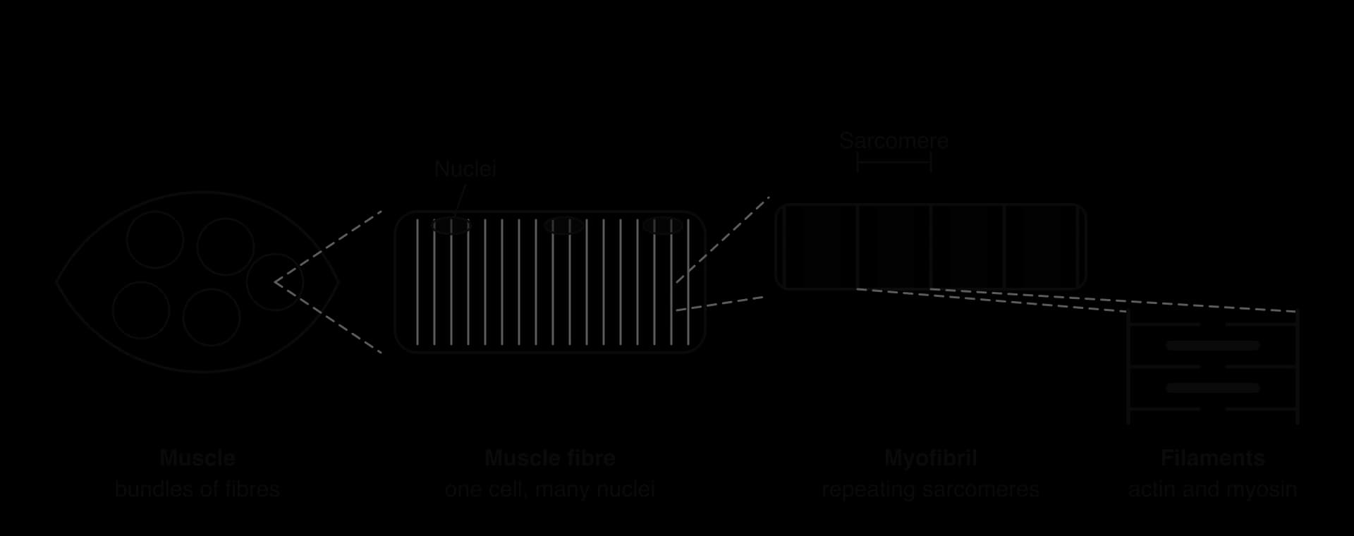
A skeletal muscle such as the biceps is made of bundles of long cells called muscle fibres, each one a striated muscle fibre. A single fibre can be several centimetres long. Each fibre forms when many embryonic cells fuse, so it contains many nuclei, lying just under its plasma membrane. The plasma membrane of a muscle fibre is called the sarcolemma.
Inside each fibre are many parallel myofibrils, each about 1–2 µm wide, which fill most of the fibre. Between the myofibrils lie many mitochondria, which supply the ATP used in contraction, and a specialised endoplasmic reticulum called the sarcoplasmic reticulum, which stores calcium ions. Each myofibril is divided along its length into repeating units called sarcomeres. The diagram of the levels of organisation in skeletal muscle shows how one level fits inside the next.
The sarcomere
A sarcomere is the unit of contraction in a myofibril. Sarcomeres are joined end to end, and when a muscle contracts, every sarcomere along every myofibril shortens at the same time. The shortening of thousands of sarcomeres in series produces the shortening of the whole muscle.
The filaments and the lines
Each sarcomere contains two kinds of protein filament. Thin filaments are made mainly of the globular protein actin, whose molecules are linked into long chains twisted in a helix. Thick filaments are made of the protein myosin. Each myosin molecule has a long tail and a head that sticks out from the filament, and hundreds of molecules lie side by side with their heads facing outwards.
The Z-line marks each end of the sarcomere. Thin actin filaments are anchored to it and point inwards towards the centre.
The M-line runs through the middle of the sarcomere. Thick myosin filaments are held together there.
Thick and thin filaments overlap. In cross-section, each myosin filament is surrounded by six actin filaments, so its heads can reach actin on every side.
The giant protein titin runs from each Z-line to the M-line, along the myosin filament, holding the thick filaments in the centre of the sarcomere and acting as a spring.
Light and dark bands
The striations of skeletal muscle are the pattern of bands produced by the filaments. The A band is the dark band: its length is the length of the thick myosin filaments, and it includes the regions where they overlap with actin. The I band is the light band: it contains only thin actin filaments, and it is divided in two by a Z-line, so half of each I band belongs to one sarcomere and half to the next. The H zone is a paler region in the middle of the A band where there are myosin filaments but no actin.
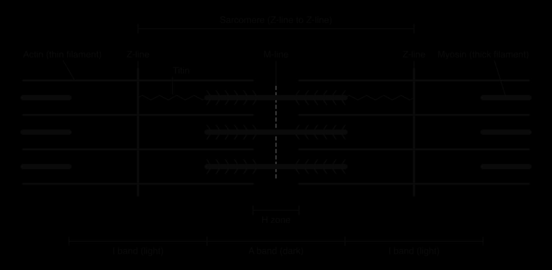
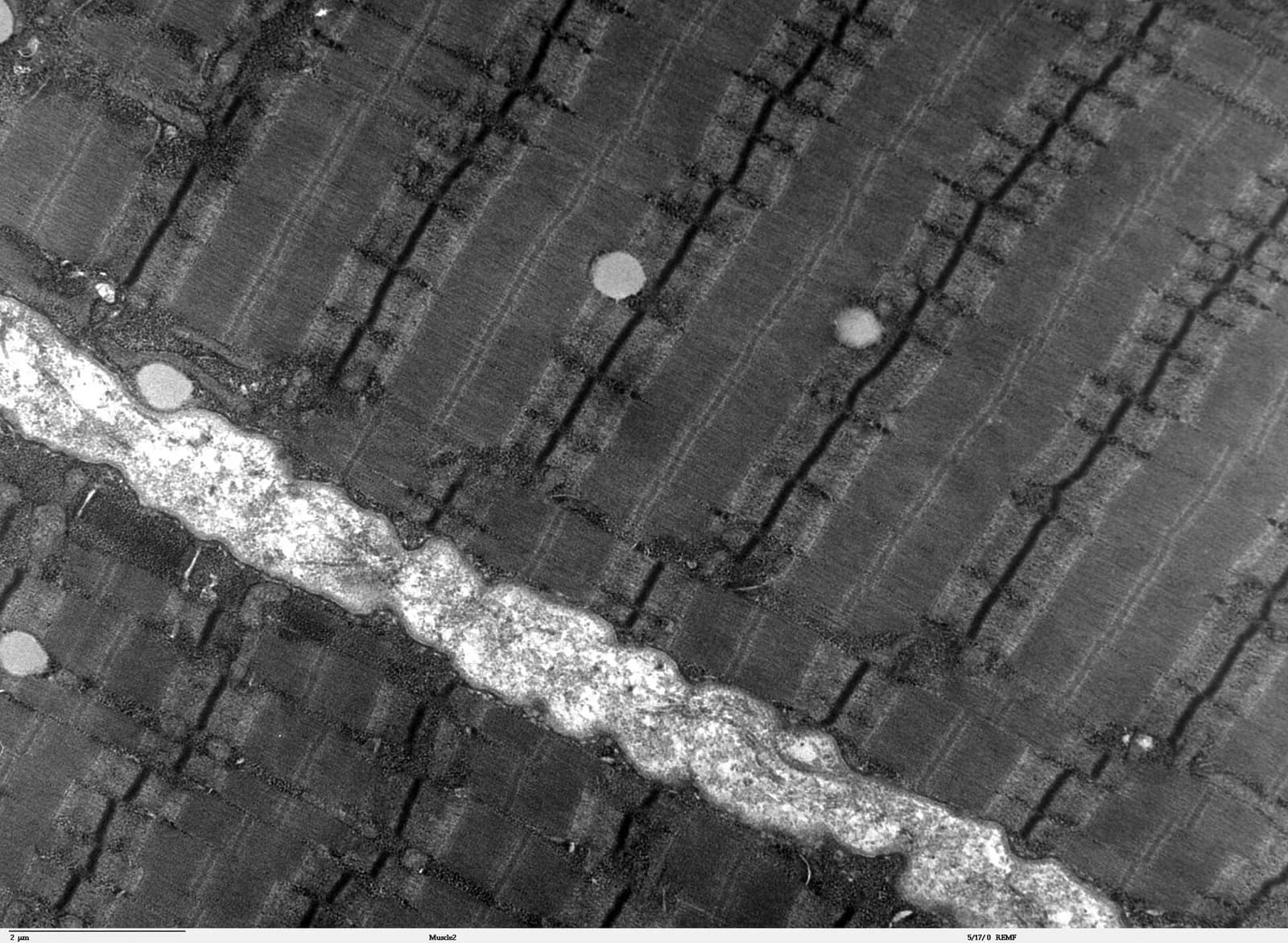
A relaxed sarcomere in human muscle is about 2–3 µm long. That is too small to see the filaments in a light microscope, but the bands are visible, and the transmission electron micrograph of human skeletal muscle shows the filaments and Z-lines directly.
The sliding filament model of muscle contraction
The sliding filament model explains how a sarcomere shortens. During contraction, the thin actin filaments slide inwards over the thick myosin filaments, towards the M-line. Neither type of filament changes length. Because the actin filaments are attached to the Z-lines, the Z-lines are pulled closer together and the sarcomere becomes shorter.
How the bands change during contraction
Comparing a relaxed sarcomere with a contracted one shows which parts change. The overlap between actin and myosin increases, so the regions that contain only one type of filament become shorter. The A band, which is the length of the myosin filaments, stays the same.

Part of the sarcomere | What it contains | Change during contraction |
|---|---|---|
Sarcomere (Z-line to Z-line) | One whole contractile unit | Shortens |
A band (dark) | The full length of the myosin filaments, with overlapping actin | Stays the same length |
I band (light) | Actin filaments only | Shortens |
H zone | Myosin filaments only | Shortens and may disappear |
Actin and myosin filaments | Protein filaments | Stay the same length; they slide past each other |
Evidence for the sliding filament model
The model was proposed in 1954 in two papers published together. Hugh Huxley and Jean Hanson used phase-contrast light microscopy to measure the bands of isolated myofibrils at different lengths. Andrew Huxley and Rolf Niedergerke used interference microscopy to measure the bands in living muscle fibres as they contracted. Both teams found that the A band kept a constant length while the I band and H zone became shorter. These measurements fitted a model in which two sets of filaments of fixed length slide past each other, and earlier electron micrographs had already shown that myofibrils contain two overlapping sets of filaments.
The cross-bridge cycle
Myosin heads provide the force that slides the filaments. Each head can bind to a specific site on an actin molecule, forming a cross-bridge. The head then changes shape, swinging through an angle and pulling the actin filament along. This movement is the power stroke. The head then lets go, returns to its original angle and binds again further along the actin filament, so the filaments move in a series of small steps.
Energy for the cycle comes from the hydrolysis of ATP. Each myosin head has a site that binds ATP and an enzyme activity that hydrolyses it to ADP and inorganic phosphate (Pi). The diagram of the cross-bridge cycle shows the four steps that each head repeats many times a second.
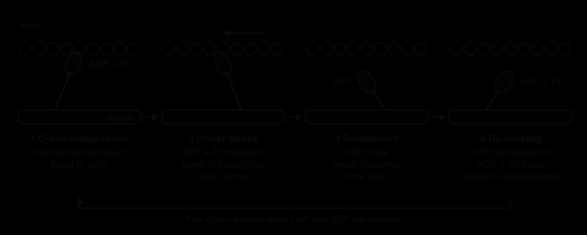
Cross-bridge formation. A myosin head carrying ADP and Pi is in the "cocked" (high-energy) position. When binding sites on actin are exposed, the head binds to actin, forming a cross-bridge.
Power stroke. The head releases Pi and ADP and tilts towards the M-line, pulling the actin filament a few nanometres (about 5–10 nm) towards the centre of the sarcomere.
Detachment. A new ATP molecule binds to the myosin head. This causes the head to detach from actin, breaking the cross-bridge.
Re-cocking. The head hydrolyses the ATP to ADP and Pi. The energy released returns the head to its cocked position, ready to bind to a new site further along the actin filament.
The heads on a myosin filament do not all move together. At any moment some are attached and pulling while others are detaching or re-cocking, so the actin filament is never released completely. The heads on each half of a myosin filament face opposite ways, which is why actin from both ends of the sarcomere is pulled towards the M-line.
Rigor mortis, the stiffening of muscles a few hours after death, shows the role of ATP clearly. After death, cell respiration stops, the ATP in muscle fibres is used up and no new ATP is made. Myosin heads that are bound to actin can no longer detach, so the cross-bridges stay locked and the muscles become rigid until the proteins start to break down.
Control of contraction by calcium ions
In a relaxed muscle fibre, the myosin heads cannot bind to actin because the binding sites are covered. Two regulatory proteins on the thin filament are responsible. Tropomyosin is a long, thin protein that lies in the groove of the actin helix and blocks the binding sites. Troponin is a small protein complex attached to tropomyosin at regular intervals.
An action potential from a motor neuron spreads along the sarcolemma and into the fibre.
The sarcoplasmic reticulum releases calcium ions (Ca²⁺) into the cytoplasm around the myofibrils.
Ca²⁺ binds to troponin, which changes shape and moves tropomyosin away from the binding sites on actin.
Myosin heads bind to the exposed sites and the cross-bridge cycle begins, as long as ATP is available.
When stimulation stops, Ca²⁺ is pumped back into the sarcoplasmic reticulum by active transport (using ATP), tropomyosin covers the binding sites again and the fibre relaxes.
Control by Ca²⁺ means contraction is switched on and off within milliseconds by nerve impulses, while ATP supplies the energy. A fibre therefore needs ATP for two separate jobs during activity: the cross-bridge cycle and the pumping of Ca²⁺ back into the sarcoplasmic reticulum.
 YouTube
Muscle relaxation: titin and antagonistic muscles
A muscle can only exert force by contracting. When the cross-bridges stop cycling, a muscle fibre relaxes and stops pulling, but it has no way of lengthening itself. Two things return a relaxed muscle to its resting length: the elastic protein titin inside the sarcomere, and the pull of an opposing muscle.
Titin, a molecular spring
Titin is the largest protein known. One titin molecule in human muscle contains about 34,350 amino acids, and it stretches from the Z-line to the M-line, half the length of a sarcomere. Part of the molecule lies along the myosin filament and anchors it, keeping the thick filament centred between the Z-lines. The part between the Z-line and the end of the myosin filament (in the I band) is folded, like a coiled spring.
Recoil after stretching. When a sarcomere is stretched, the folded region of titin unfolds and stores potential energy, like a stretched spring. When the stretching force is removed, titin recoils and helps return the sarcomere to its resting length.
Protection against overstretching. Titin resists stretching more and more strongly as it extends, which prevents the filaments being pulled so far apart that they no longer overlap.
Positioning myosin. Titin keeps each thick filament in the centre of the sarcomere, so actin and myosin overlap equally at both ends.
Antagonistic muscles
Because a muscle can pull but cannot push, skeletal muscles are arranged in antagonistic pairs. The two muscles of a pair are attached across the same joint and produce opposite movements. When one contracts, the other relaxes and is stretched, and contraction of the second muscle returns the body part to its first position.
In the upper arm, the biceps bends (flexes) the elbow and the triceps straightens (extends) it. As the biceps contracts, the triceps is stretched; titin in the triceps sarcomeres stores some of the energy of that stretch and releases it as the triceps recoils. Coordinated control by the nervous system ensures that the two muscles of a pair do not contract fully at the same time, except when the joint is being held stiff.
Motor units in skeletal muscle
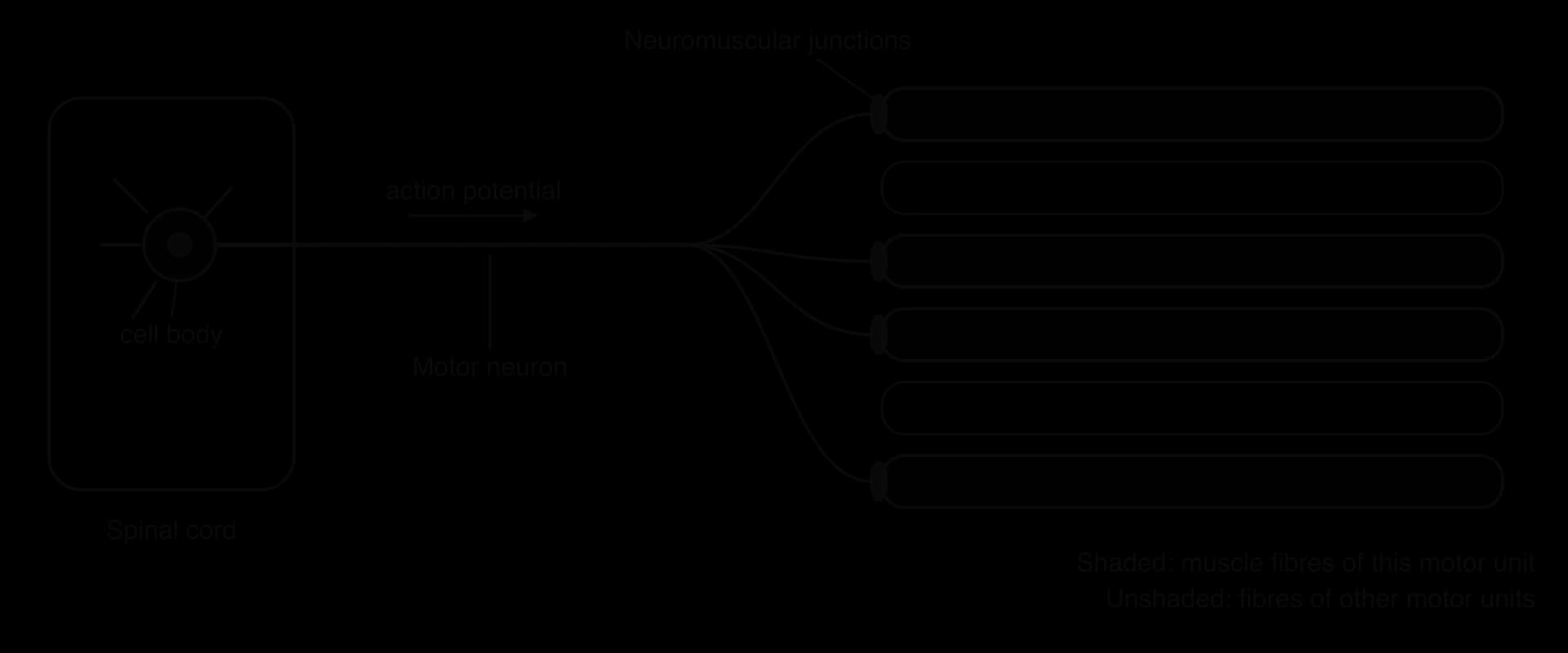
Skeletal muscle fibres contract only when stimulated by a motor neuron. The cell body of each motor neuron lies in the spinal cord or brainstem, and its long axon runs to a muscle, where it branches. Each branch ends on a different muscle fibre, so one motor neuron controls a group of fibres.
The neuromuscular junction
The synapse between a motor neuron and a muscle fibre is a neuromuscular junction. When an action potential arrives at the end of the axon, the neuron releases the neurotransmitter acetylcholine, which diffuses across the narrow gap to the sarcolemma. Acetylcholine binds to receptors on the muscle fibre, which open ion channels and depolarise the sarcolemma. This starts an action potential in the muscle fibre, which leads to the release of Ca²⁺ from the sarcoplasmic reticulum and to contraction. The enzyme acetylcholinesterase then breaks the acetylcholine down, so each impulse causes only one brief contraction.
Myasthenia gravis shows how much motor units depend on this junction. In this autoimmune disease, the body makes antibodies against the acetylcholine receptors on muscle fibres. Fewer receptors respond to each nerve impulse, so muscle fibres contract weakly or not at all, and patients experience weakness that worsens with use, often first in the eyelids and the muscles that move the eyes.
How motor units control the force of contraction
All the fibres in a motor unit contract together whenever its motor neuron fires. The nervous system varies the force of a whole muscle in two ways: by changing how many motor units are active at once (recruitment) and by changing how often each motor neuron fires. A gentle movement uses a few motor units; a maximal lift uses almost all of them.
The size of motor units matches the function of the muscle. In muscles that make fine, precise movements, such as those that move the eyeball, each motor neuron supplies only about ten fibres or fewer. In large muscles used for powerful movements, such as the gastrocnemius in the calf, one motor neuron can supply 1,000–2,000 fibres. Small motor units allow the force of a muscle to be adjusted in very fine steps, while large ones produce great force with few neurons, a relationship between structure and function at the level of tissues and organs.
Skeletons as anchorage for muscles and as levers
A muscle can only move the body if both of its ends are attached to something firm. Skeletons provide these attachment points (anchorage) and form rigid levers that turn the pull of a muscle into movement of a limb, a wing or a jaw. Two main types of skeleton are found in animals that walk, run or fly.
Exoskeletons and endoskeletons
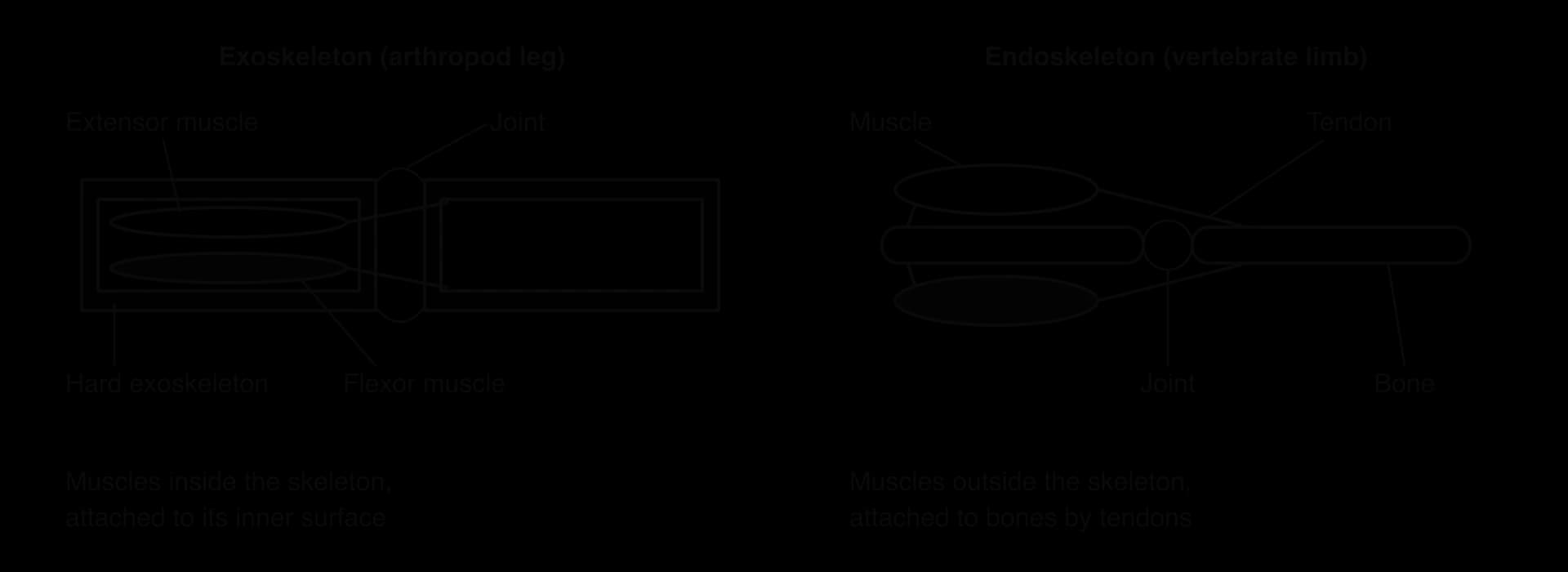
Arthropods (insects, crustaceans, spiders and their relatives) have an exoskeleton: a hard outer covering made of chitin and protein, often strengthened with calcium carbonate in crustaceans. The exoskeleton is a series of hollow tubes and plates joined by thin, flexible membranes at the joints. Muscles lie inside the tubes and are attached to the inner surface of the exoskeleton.
Vertebrates (fish, amphibians, reptiles, birds and mammals) have an endoskeleton: a framework of bone and cartilage inside the body. Muscles lie outside the bones and are attached to them by tendons. The comparison of an arthropod leg with a vertebrate limb shows the same principle, an antagonistic pair of muscles pulling on rigid segments across a joint, built in two different ways.
Feature | Exoskeleton (arthropods) | Endoskeleton (vertebrates) |
|---|---|---|
Position | Outside the body, enclosing the soft tissues | Inside the body, surrounded by muscle |
Material | Chitin and protein, sometimes with calcium carbonate | Bone and cartilage (living tissues) |
Muscle attachment | To the inner surface of the skeleton | To the outer surface of bones, by tendons |
Growth | Cannot grow; shed and replaced by moulting | Grows with the body |
Other functions | Protection, support, reduces water loss | Support, protection of organs, blood cell production |
Bones and joints as levers
A lever is a rigid bar that turns about a fixed point, the fulcrum. A force applied at one point on the lever (the effort) moves a load at another point. In the body, a bone is the rigid bar, a joint is the fulcrum, the contracting muscle provides the effort where its tendon is attached, and the load is the weight of the body part plus anything being held or pushed.
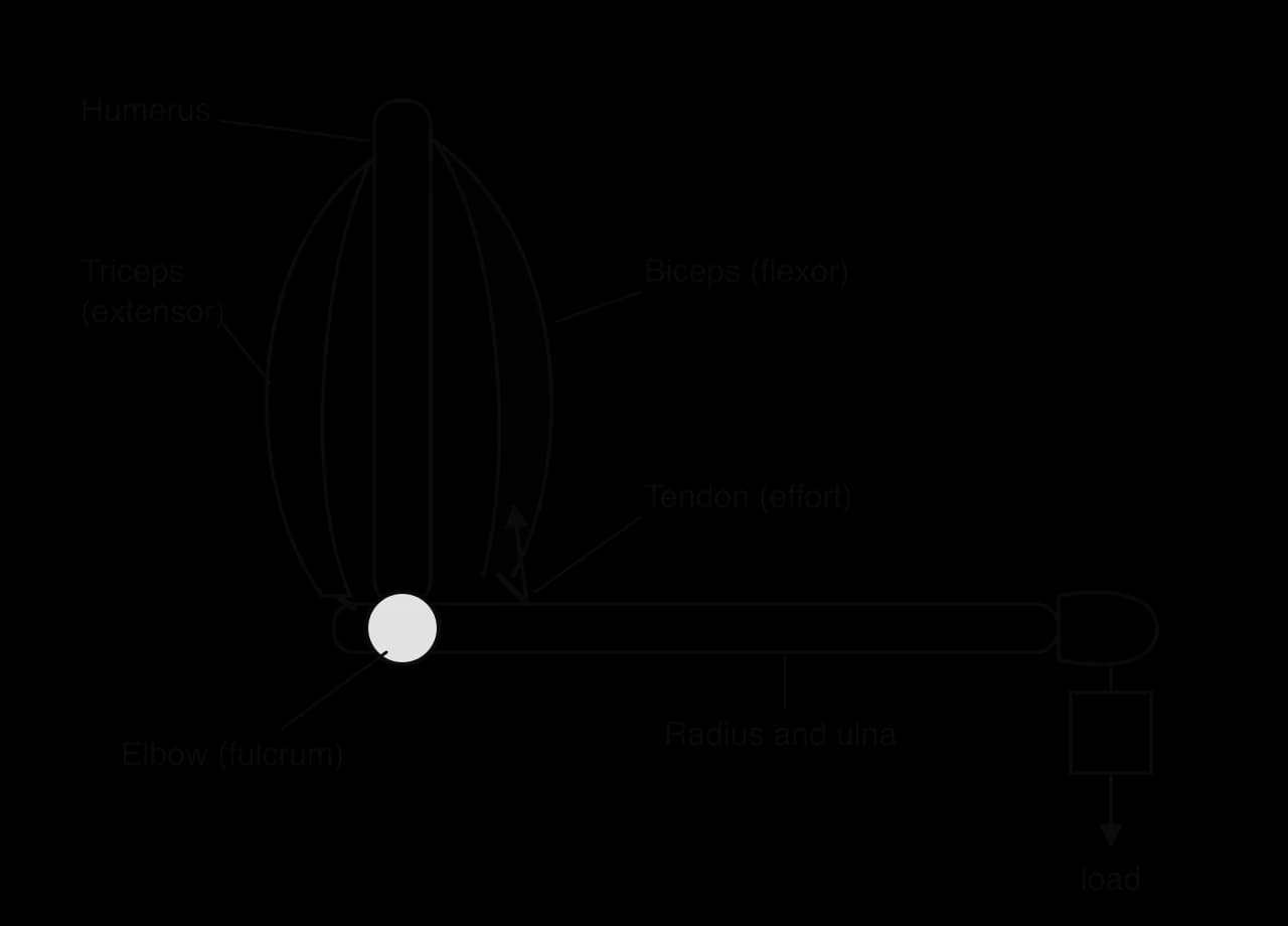
The forearm is a lever in which the effort lies between the fulcrum and the load (a third-class lever). The biceps is attached close to the elbow, so it must exert a force several times larger than the load in the hand. The advantage is speed and range: a contraction of a few centimetres moves the hand through a large arc. Most limb levers in vertebrates are arranged this way, which suits rapid movements such as running and throwing.
Movement at a synovial joint: the human hip
A joint is a place where two or more bones meet. In a synovial joint the ends of the bones are separated by a cavity filled with fluid, which allows free movement. Most joints of the limbs, including the hip, knee, shoulder and elbow, are synovial joints.
The hip joint connects the leg to the trunk. The rounded head of the femur (thigh bone) fits into a deep cup-shaped socket in the pelvis called the acetabulum. Each part of the joint has its own function, as the annotated diagram of the hip joint and the table of its parts show.
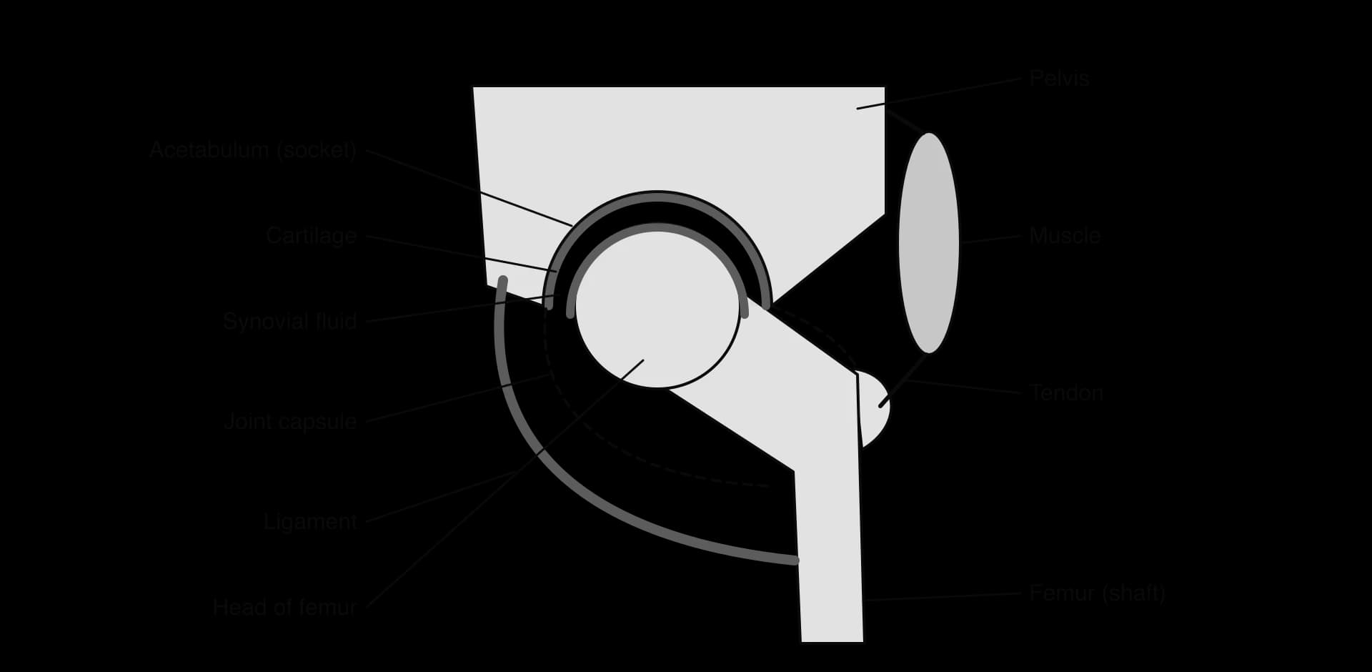
Part | Structure | Function at the hip |
|---|---|---|
Bones (femur and pelvis) | Rigid; the femoral head is a ball, the acetabulum a deep socket | Act as levers and give attachment for muscles; the deep socket makes the joint stable |
Cartilage | Smooth, tough tissue covering the ends of the bones | Reduces friction; absorbs shock when weight is put on the leg |
Synovial fluid | Viscous fluid filling the joint cavity, secreted by the synovial membrane | Lubricates the joint; supplies oxygen and nutrients to the cartilage |
Joint capsule | Tough fibrous sac lined by the synovial membrane | Encloses the joint and holds in the synovial fluid |
Ligaments | Strong bands of fibrous tissue joining bone to bone | Hold the femur in the acetabulum; limit movements that would dislocate the joint |
Muscles | Skeletal muscles in antagonistic groups around the joint | Contract to move the femur (flexion, extension, abduction, adduction, rotation) |
Tendons | Tough, inelastic cords of fibrous tissue joining muscle to bone | Transmit the pull of a contracting muscle to the bone |
When the cartilage of the hip wears away, as in osteoarthritis, bone rubs on bone and movement becomes painful. The low-friction hip replacement developed by the surgeon John Charnley in 1962 replaces the head of the femur with a polished metal ball on a stem and lines the acetabulum with a high-density polyethylene cup, both fixed to the bone with acrylic bone cement. The two artificial surfaces do the job of the cartilage and synovial fluid, and the joint keeps the ball-and-socket shape that allows the full range of movement.
 YouTube
Range of motion of a joint
The range of motion of a joint is the full extent of movement possible at it, measured as an angle in each direction. It depends on the shape of the articulating surfaces, the tightness of the ligaments and joint capsule, and the bulk of surrounding muscle and other tissue.
Movements at a joint are described with standard terms. Flexion bends a joint, decreasing the angle between two bones (raising the thigh forwards at the hip; bending the knee). Extension straightens a joint, increasing the angle. Abduction moves a limb away from the midline of the body (raising the leg sideways), and adduction moves it back towards the midline. Rotation turns a bone about its own long axis.
Hinge joints and ball-and-socket joints
A hinge joint, such as the knee or the elbow, works like the hinge of a door. The shapes of the bone ends and strong side ligaments allow movement in one plane only: flexion and extension. A ball-and-socket joint, such as the hip or the shoulder, allows movement in all three planes: flexion and extension, abduction and adduction, and rotation. Combining these movements lets the leg move in a cone (circumduction).
Movement | Hip (ball-and-socket) | Knee (hinge) |
|---|---|---|
Flexion | Yes: about 120–130° in healthy adults (knee bent) | Yes: about 135°, the largest movement of the knee |
Extension | Yes, but limited: about 10–20° beyond the straight position | Only back to straight (0°) |
Abduction and adduction | Yes | No (prevented by side ligaments) |
Rotation | Yes, inwards and outwards | Very little, and only when the knee is bent |
Planes of movement | Three | One |
Typical values for healthy adults measured with a goniometer; published reference ranges differ by a few degrees between sources.
The hip has a smaller range of motion than the shoulder because its socket is much deeper and its ligaments are stronger. The hip carries the weight of the body, so stability is more important than range there; the shoulder trades stability for range, which is why it dislocates far more often. The two joints show how the form of a structure matches its function.
Measuring joint angles
A goniometer is a protractor with two long arms that pivot about its centre. It measures the angle of a joint directly on the body.
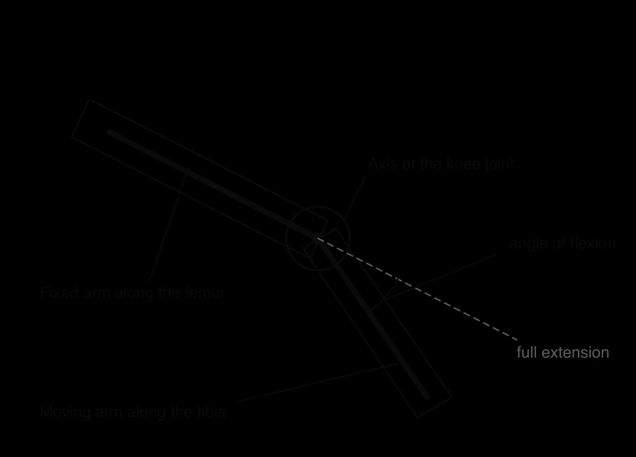
Place the centre of the goniometer over the axis of the joint (for the knee, the side of the knee level with the joint line).
Align the fixed arm with the long axis of one bone (the femur) and the moving arm with the other bone (the tibia), using bony landmarks.
Read the angle with the joint fully extended, then at full flexion. The difference is the range of motion for flexion.
Repeat each measurement three times and calculate a mean, because small changes in where the arms are placed change the reading.
Joint angles can also be measured by computer analysis of images. A photograph or video is taken from the side, perpendicular to the plane of movement, and markers stuck on bony landmarks (hip, knee, ankle) are joined by lines in software such as ImageJ or a video-analysis app, which calculates the angle between them. Image analysis is useful for movements that are too fast to measure by hand, such as the knee during a sprint or a jump.
Antagonistic muscles inside the body: the intercostal muscles
Antagonistic muscles are not only found in the limbs. The intercostal muscles between the ribs move the ribcage during ventilation of the lungs. There are two layers. The external intercostal muscles lie on the outside, and their fibres run downwards and forwards from one rib to the next. The internal intercostal muscles lie underneath, and their fibres run downwards and backwards, at right angles to the external fibres.
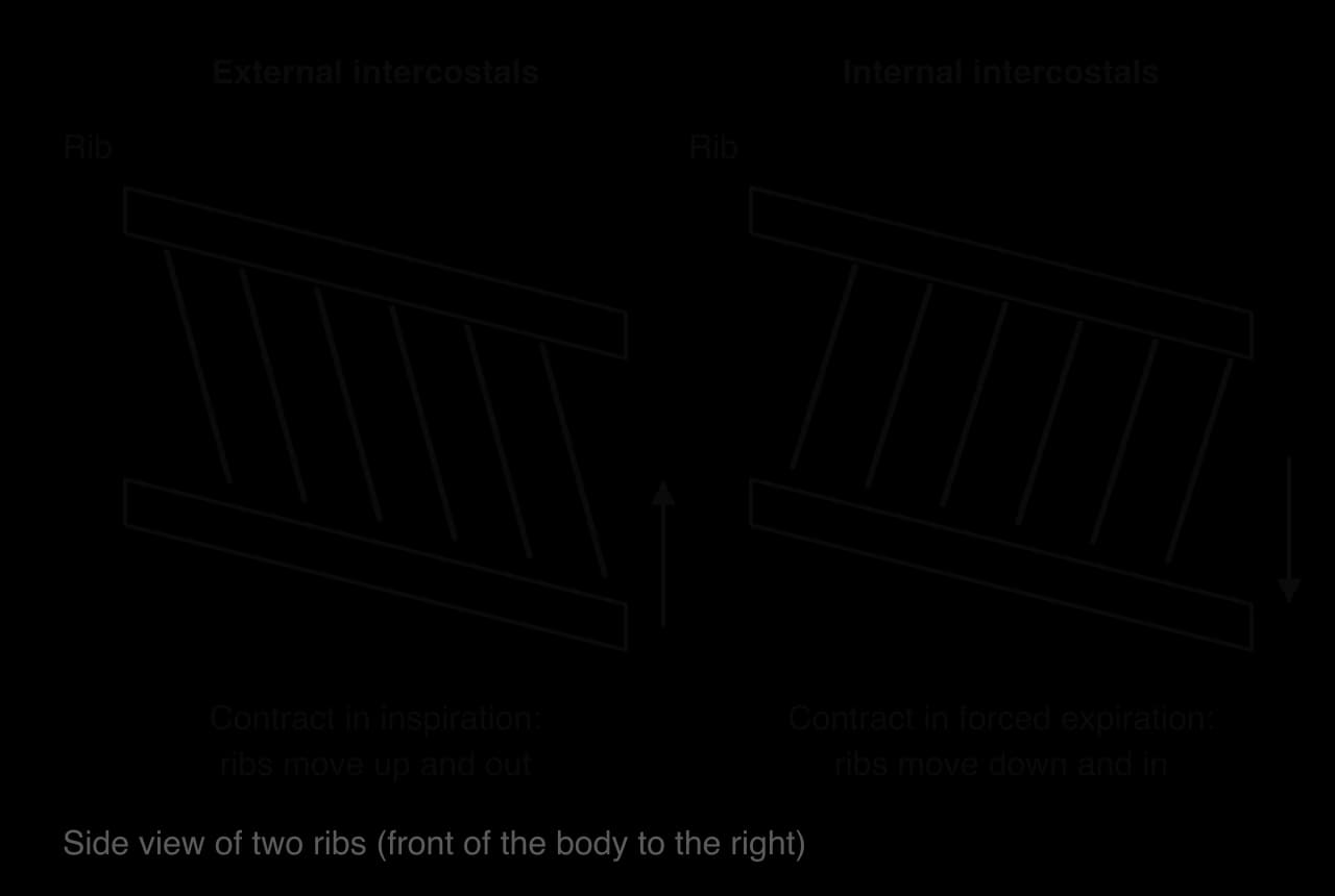
Because of their different fibre orientations, the two layers move the ribs in opposite directions. When the external intercostals contract during inspiration, they pull the ribs upwards and outwards, which increases the volume of the thorax. When the internal intercostals contract during forced expiration, they pull the ribs downwards and inwards, which decreases the volume of the thorax.
Each layer stretches the other when it contracts. Contraction of the external intercostals stretches the relaxed internal intercostals, and the energy of that stretch is stored as potential energy in their titin molecules. When the external layer relaxes, the recoil of titin helps return the internal muscle fibres to their resting length, and the reverse happens when the internal layer contracts. The intercostals are therefore an antagonistic pair that moves a structure inside the body, rather than moving the body from place to place.
Reasons for locomotion
Locomotion uses a great deal of energy, so it must bring benefits that increase an animal's chances of surviving and reproducing. Four main reasons explain why animals move from place to place, and many species move for several of these reasons at different times.
Reason | What it achieves | Example |
|---|---|---|
Foraging for food | Finds food that is patchy or that moves | Cuvier's beaked whale (Ziphius cavirostris) dives to depths of over 1,000 m to hunt squid |
Escaping from danger | Avoids predators or other threats | The great scallop (Pecten maximus) swims away from starfish by rapidly clapping its two shell valves together |
Searching for a mate | Finds a partner for sexual reproduction | Male emperor moths (Saturnia pavonia) fly long distances, following the scent of pheromones released by females |
Migration | Uses resources in different places at different times of year | The Arctic tern (Sterna paradisaea) flies between Arctic breeding grounds and the Antarctic |
Migration is the regular, usually seasonal, movement of animals between two areas, such as a breeding area and a feeding area. Arctic terns that breed in Greenland and Iceland spend the northern winter in the Southern Ocean near Antarctica, so they live in two summers each year. Terns from these colonies tracked with geolocators weighing about 1.4 g flew about 70,900 km a year on average, and the Arctic tern makes the longest regular migration known for any animal.
Locomotion also affects evolution. Motile individuals and motile larvae carry alleles to new populations, and the ability to move to new habitats allows species to colonise places where they meet new selection pressures.
Adaptations for swimming in marine mammals
Whales, dolphins, porpoises and seals are mammals whose ancestors lived on land. They breathe air with lungs, are endothermic and feed their young on milk, but their bodies have been modified by natural selection for swimming. Water is about 800 times denser than air, so it resists movement much more, and an air-breathing animal must return to the surface between dives.
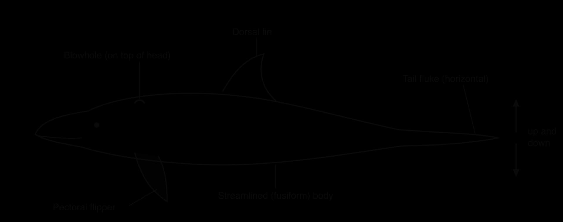
Streamlining
Streamlining is a body shape that allows a fluid to flow smoothly around it, reducing drag. Cetaceans (whales and dolphins) have a fusiform body: widest about a third of the way back and tapering towards both ends. They have no external ears, hind limbs or hair, the male genitals and the mammary glands lie inside slits in the body wall, and a thick layer of blubber smooths the body outline as well as insulating it.
Limbs modified as flippers
The forelimbs of whales and dolphins are flippers. They contain the same bones as a human arm (humerus, radius, ulna, wrist bones and finger bones), but the bones of the arm are short and the fingers are long, and the whole limb is enclosed in a stiff, flat paddle of tissue. Flippers are used for steering and stability, not for propulsion. The hind limbs have been lost, apart from small remnants of the pelvis inside the body.
The tail fluke
Cetaceans swim by moving the tail up and down. The tail ends in a horizontal fluke made of dense connective tissue, with no bones in the fluke itself. Powerful muscles above and below the vertebral column bend the rear of the body up and down, and the fluke pushes against the water on both the upstroke and the downstroke.
Airways for periodic breathing
The nostril of a whale or dolphin has moved to the top of the head, where it forms the blowhole. The animal can breathe while only the top of its head breaks the surface, so it can take a breath quickly while swimming. Muscles keep the blowhole closed during a dive. The airway from the blowhole to the lungs is completely separate from the mouth and the oesophagus, so the animal can open its mouth to catch prey under water without water entering its lungs. Each breath exchanges most of the air in the lungs, so the animal can reload with oxygen in a few breaths between long dives.
Seals are adapted in a similar way but from a different starting point. True seals swim by sweeping their hind flippers from side to side, while sea lions row with their large fore flippers. All of them share streamlined bodies, limbs modified as flippers and nostrils that close tightly under water. These shared features in groups that returned to the sea separately are an example of convergent evolution.
Case study: Cuvier's beaked whale, the deepest-diving mammal
Cuvier's beaked whale (Ziphius cavirostris) is a medium-sized whale, about 5–7 m long, found in deep water in most oceans. It feeds mainly on squid and deep-sea fish, which it finds in the dark by echolocation, so almost all of its foraging is done hundreds or thousands of metres below the surface. Its body shows every adaptation for swimming: it is strongly streamlined, it has small flippers that can be tucked into shallow depressions in the body wall called flipper pockets, and it swims with up-and-down beats of its fluke.
Satellite-linked tags attached with suction cups or small darts record the depth of a whale every few seconds. Two tagging studies have measured its dives directly.
Measurement | Off Southern California | Off Cape Hatteras, North Carolina |
|---|---|---|
Tags analysed | 8 whales | 23 tags; 3,680 dives |
Typical deep dive | Mean 1,401 m deep, lasting 67.4 min | Median duration 59 min |
Deepest dive recorded | 2,992 m | Not reported in this analysis |
Longest dive recorded | 137.5 min | 222 min (and one of 173 min) |
Sources: Schorr et al., PLoS ONE, 2014; Quick et al., Journal of Experimental Biology, 2020.
The two dives of 173 and 222 minutes were made by one whale 17 and 24 days after it had been exposed to naval sonar, so the researchers analysed them separately from the main data. Even typical dives last about an hour without a breath, at depths where no light reaches and the pressure is more than a hundred times that at the surface.
Deep foraging dives are possible because the airway and blowhole allow rapid reloading with oxygen at the surface, and because large amounts of oxygen are stored in the blood and in the muscles, bound to the pigment myoglobin. During a dive the lungs collapse under pressure, and the whale relies on these stores while it swims slowly, using its streamlined shape and steady fluke beats to reduce the energy cost of locomotion. Its adaptations for swimming therefore serve one of the reasons for locomotion, foraging, by giving it access to prey in a part of the ocean that few other air-breathing predators can reach.
Exam practice
Muscle and motility is assessed in Paper 1A through questions on sarcomere bands, the roles of ATP, calcium and titin, and the parts of a synovial joint. Paper 1B questions often give electron micrographs of relaxed and contracted sarcomeres, or joint-angle data from goniometers or image analysis, and ask you to measure, calculate, compare or suggest improvements to the method. In Paper 2, short-answer questions ask you to annotate a sarcomere or hip joint diagram, and extended responses often ask you to explain the sliding filament model, the role of antagonistic muscles or the adaptations of marine mammals.
Drawing questions on the sarcomere are marked for accuracy of proportion as well as labels. Actin filaments must start at the Z-lines and end short of the M-line in a relaxed sarcomere, myosin filaments must be centred, and the band brackets must start and end at filament ends.
Self-check
- To do:
I can distinguish locomotion from movement within the body, using a motile and a sessile species as examples.
- To do:
I can describe the structure of skeletal muscle from the whole muscle to the sarcomere.
- To do:
I can draw and annotate a sarcomere, and state which bands change length during contraction.
- To do:
I can explain the sliding filament model and the cross-bridge cycle, including the roles of ATP, Ca²⁺, troponin and tropomyosin.
- To do:
I can explain how titin and antagonistic muscles return a muscle to its resting length.
- To do:
I can describe a motor unit and the role of the neuromuscular junction.
- To do:
I can compare exoskeletons and endoskeletons and identify the fulcrum, effort and load in a limb lever.
- To do:
I can annotate the hip joint with the roles of bones, cartilage, synovial fluid, ligaments, muscles and tendons.
- To do:
I can compare the range of motion of hinge and ball-and-socket joints and describe how to measure it with a goniometer.
- To do:
I can explain how the internal and external intercostal muscles act antagonistically.
- To do:
I can give named examples of locomotion for foraging, escape, finding a mate and migration.
- To do:
I can explain the adaptations of marine mammals for swimming.