Explain how blood vessels, the heart, xylem and phloem are adapted to transport materials, and evaluate data on coronary heart disease.
Each is in the Biology glossary, with every other term in the subject.
A small branch of an artery leading into a capillary bed, whose smooth muscle can narrow or widen the lumen to control blood flow to a tissue.
A blood vessel that carries blood away from the heart, at high pressure, with a thick wall of smooth muscle and elastic fibres.
The build-up of fatty plaques containing cholesterol in the walls of arteries, which narrows the lumen, reduces elasticity and restricts blood flow.
A valve between an atrium and a ventricle (tricuspid on the right, bicuspid or mitral on the left) that closes during ventricular systole to prevent backflow into the atrium.
One of the two thin-walled upper chambers of the mammalian heart, which receive blood from veins and pump it into the ventricles.
The force per unit area exerted by blood on the walls of blood vessels, measured in mmHg and recorded as systolic over diastolic pressure, such as 120/80 mmHg.
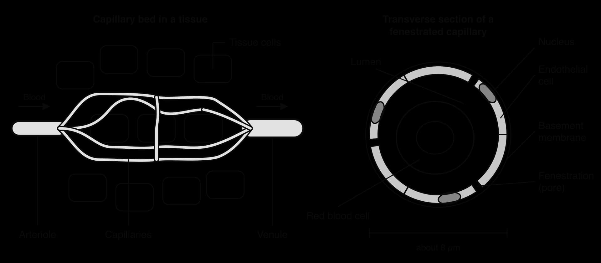
The narrowest type of blood vessel, with a wall made of a single layer of thin endothelial cells, in which materials are exchanged between blood and the surrounding tissue or environment.
The sequence of contraction (systole) and relaxation (diastole) of the atria and ventricles that makes up one heartbeat, together with the opening and closing of the valves.
A relationship in which a change in one variable directly produces a change in another; correlation alone does not demonstrate it.
The explanation that water is pulled up the xylem as a continuous column under tension, created by evaporation from leaf cell walls and transmitted down the column by cohesion between water molecules.
One of the arteries that branch from the aorta and supply the heart muscle itself with oxygenated blood.
Disease caused by narrowing or blockage of the coronary arteries, which reduces the blood supply to heart muscle; also called ischaemic heart disease (CHD).
A relationship between two variables in which a change in one is associated with a change in the other; it may be positive or negative.
A number from −1 to +1 (r) that measures the strength and direction of a linear relationship between two variables; values near 0 show little or no correlation.
In a plant stem or root, the region of mainly parenchyma cells between the epidermis and the vascular tissue, used for support and storage.
The phase of the cardiac cycle in which the heart muscle relaxes and the chambers fill with blood.
A circulatory system in which blood passes through the heart twice in each complete circuit, through separate pulmonary (lungs) and systemic (body) circulations, as in mammals.
The return of stretched elastic fibres in an artery wall to their original length, which pushes blood onward and maintains blood pressure between heartbeats.
The study of the incidence, distribution and causes of diseases in populations, used to identify risk factors and evaluate ways of preventing disease.
A capillary whose endothelial cells have pores (fenestrations) that allow especially rapid exchange of water and solutes, as in kidney glomeruli, intestinal villi and endocrine glands.
The number of times the heart beats per minute (bpm), equal to the pulse rate.
An inadequate blood supply to a tissue, which deprives its cells of oxygen and nutrients.
A hard, waterproof polymer deposited in the cell walls of xylem vessels and other plant tissues, which strengthens them and stops them collapsing under tension.
The space inside a tubular structure such as a blood vessel, xylem vessel or gut, through which material flows.
Excess tissue fluid that has entered the lymphatic vessels, which carry it through lymph nodes and return it to the blood in the subclavian veins.
A thin-walled vessel of the lymphatic system with gaps that let tissue fluid in and valves that keep lymph flowing towards the subclavian veins.
The death of part of the heart muscle because its blood supply has been blocked, usually by a thrombus in a coronary artery; commonly called a heart attack.
The vascular tissue that transports organic compounds, mainly sucrose, in solution from where they are made or stored to where they are used, through living sieve tubes.
A narrow channel of cytoplasm through the cell walls of adjacent plant cells, allowing substances to pass directly between them, for example between companion cells and sieve tube elements.
The movement of water and small solutes out of a capillary through gaps in its wall, driven by the high hydrostatic pressure of the blood, while cells and large proteins are held back.
The rhythmic expansion and recoil of an artery wall caused by the pressure wave that each heartbeat sends through the arteries.
Any characteristic or behaviour that increases the probability of developing a particular disease, such as smoking or high blood pressure for coronary heart disease.
A positive pressure in the xylem of roots, generated when mineral ions are actively transported into the xylem and water follows by osmosis, which pushes water up the stem.
A pocket-shaped valve at the base of the aorta or pulmonary artery that closes when the ventricles relax, preventing backflow of blood into the ventricles.
The perforated end wall between two sieve tube elements in phloem, whose pores allow sap to flow from one element to the next.
A circulatory system in which blood passes through the heart once in each complete circuit of the body, as in bony fish: heart → gills → body → heart.
In translocation, a part of a plant that removes sugars from the phloem for growth, respiration or storage, such as roots, fruits and growing shoots.
A group of specialised cardiac muscle cells in the wall of the right atrium that acts as the pacemaker, starting each heartbeat with an electrical impulse.
Muscle tissue made of unstriated, spindle-shaped cells that contract slowly and involuntarily, found in the walls of arteries, the gut and other organs.
In translocation, a part of a plant that releases sugars into the phloem, such as a photosynthesising leaf or a storage organ being used.
The phase of the cardiac cycle in which the muscle of a heart chamber contracts, pumping blood out of it.
The formation of a blood clot (thrombus) inside an intact blood vessel, which may block it.
The fluid that surrounds the cells of a tissue, formed from blood plasma forced out of capillaries; it has a similar composition to plasma but contains very few proteins and no red blood cells.
The transport of organic compounds, mainly sucrose and amino acids, in solution through the sieve tubes of the phloem from sources to sinks.
A blood vessel that carries blood towards the heart at low pressure, with a thin, flexible wall, a wide lumen and valves to prevent backflow.
One of the two thick-walled lower chambers of the mammalian heart, which pump blood into the arteries at high pressure.
The vascular tissue that transports water and dissolved mineral ions from the roots to the rest of the plant, through dead, hollow, lignified vessels.
Transport systems move materials by mass flow over distances too long for diffusion. In mammals, blood is pumped by the heart through arteries, capillaries and veins; in plants, water rises through xylem and sugars travel through phloem.
Both systems depend on pressure differences, and the structure of each type of vessel matches the pressure it carries and the exchange it allows. Failures of the system, such as blocked coronary arteries, are among the most common causes of death in the world.
45 exam-style questions on this subtopic, written in the style of the IB papers. The first has its markscheme open. Counted on
Researchers measured the rate of transpiration from the leaves of a sunflower (Helianthus annuus) plant during one sunny day. They also measured the pressure in the xylem of the stem and the change in stem diameter compared with its diameter at 06:00.
Time | Transpiration rate / mmol m-2 s-1 | Pressure in xylem / MPa | Change in stem diameter / µm |
|---|---|---|---|
06:00 | 0.4 | −0.3 | 0 |
09:00 | 2.1 | −0.7 | −20 |
12:00 | 4.8 | −1.4 | −65 |
15:00 | 4.2 | −1.3 | −58 |
18:00 | 1.6 | −0.6 | −18 |
21:00 | 0.2 | −0.3 | −3 |
Describe the relationship between the transpiration rate and the pressure in the xylem.
The official deck for this part of the syllabus.
Why can a unicellular organism or a very small animal be supplied by diffusion alone?
Explain why diffusion alone cannot supply the cells of a large, active organism.
With a free account, Augment remembers where you stopped and what you have practiced.
Every cell exchanges materials with its surroundings: it takes in oxygen, glucose, amino acids and ions, and it gets rid of carbon dioxide, urea and other wastes. In a unicellular organism or a very small animal, diffusion across the body surface is fast enough to supply every cell, because no cell is far from the outside. As organisms get larger, the surface area to volume ratio falls and the distance from the surface to the innermost cells rises. Diffusion time increases with the square of the distance, so simple diffusion alone cannot supply the cells of a large, active organism.
Large animals and plants solve this with a mass transport system: a fluid that flows through tubes, driven by a pressure difference, carrying materials quickly over long distances. Diffusion is then needed only over the last short distance, between the fluid and the cells. In mammals the fluid is blood, the tubes are blood vessels and the pressure comes from the heart. In plants, water and minerals move in xylem and sugars move in phloem. The two systems are very different in structure, but both show the same principle of form and function: the structure of each tube matches the pressure it must withstand and the exchange it must allow.
Blood leaves the heart in large arteries, which branch repeatedly into smaller arteries and then into arterioles. Arterioles lead into networks of capillaries, where exchange with the tissues takes place. Capillaries join to form venules, and venules join to form veins, which return blood to the heart. Blood therefore always flows in the same order: artery → arteriole → capillary → venule → vein.
An artery is defined by the direction of flow, not by the oxygen content of its blood. Most arteries carry oxygenated blood, but the pulmonary arteries carry deoxygenated blood from the heart to the lungs. In the same way, a vein is any vessel that returns blood to the heart, including the pulmonary veins, which carry oxygenated blood from the lungs.
A capillary is the smallest blood vessel and the only one whose wall is thin enough for exchange. Capillaries form dense networks called capillary beds in almost every tissue, so that no cell is more than a short distance from one. Exchange takes place either with the internal environment, the fluid that bathes cells in tissues, or with the external environment at exchange surfaces such as the alveoli of the lungs and the villi of the small intestine.
Large surface area. Arterioles branch into very large numbers of capillaries. The total surface area of all the capillaries in an adult human is several hundred square metres, so a large volume of blood is in contact with the tissues at any moment.
Narrow diameter. A capillary has an internal diameter of about 5–10 µm, close to the width of a single red blood cell. Red blood cells pass through in single file, pressed against the wall, which keeps the diffusion distance between haemoglobin and the tissue very short. The narrow width also means that more capillaries fit into a tissue, which adds to the surface area.
Thin wall. The wall is a single layer of flattened endothelial cells on a thin basement membrane, so the diffusion distance across it is usually less than 1 µm.
Permeable wall. Small gaps between the endothelial cells let water and small solutes leak out of the blood. Large plasma proteins and blood cells normally stay inside.
Slow blood flow. Because the total cross-sectional area of the capillary beds is much larger than that of the arteries feeding them, blood moves slowly through capillaries, giving more time for exchange.
In most tissues the capillary wall is continuous, and materials cross it by diffusion through the endothelial cells or through narrow gaps between them. Where exchange needs to be especially rapid, the endothelial cells have pores called fenestrations, about 60–80 nm across, which allow much faster movement of water and solutes. A fenestrated capillary is found in the glomeruli of the kidney, where large volumes of fluid are filtered out of the blood; in the villi of the small intestine, where absorbed nutrients enter the blood; and in endocrine glands, where hormones pass into the blood. The pores are still too small for blood cells, and the basement membrane holds back most plasma proteins.
The walls of arteries and veins are built from the same three layers, but in different proportions. Each layer surrounds the lumen, the space through which blood flows.
Tunica intima, the inner layer: a single layer of smooth endothelial cells on a basement membrane, which gives a low-friction lining. In arteries it is often folded when the vessel is fixed for microscopy, because the elastic tissue recoils after death.
Tunica media, the middle layer: smooth muscle cells and elastic fibres (made of the protein elastin). In arteries this is the thickest layer; in veins it is thin.
Tunica externa, the outer layer: mainly collagen fibres, which are strong and resist overstretching. It anchors the vessel to surrounding tissue.
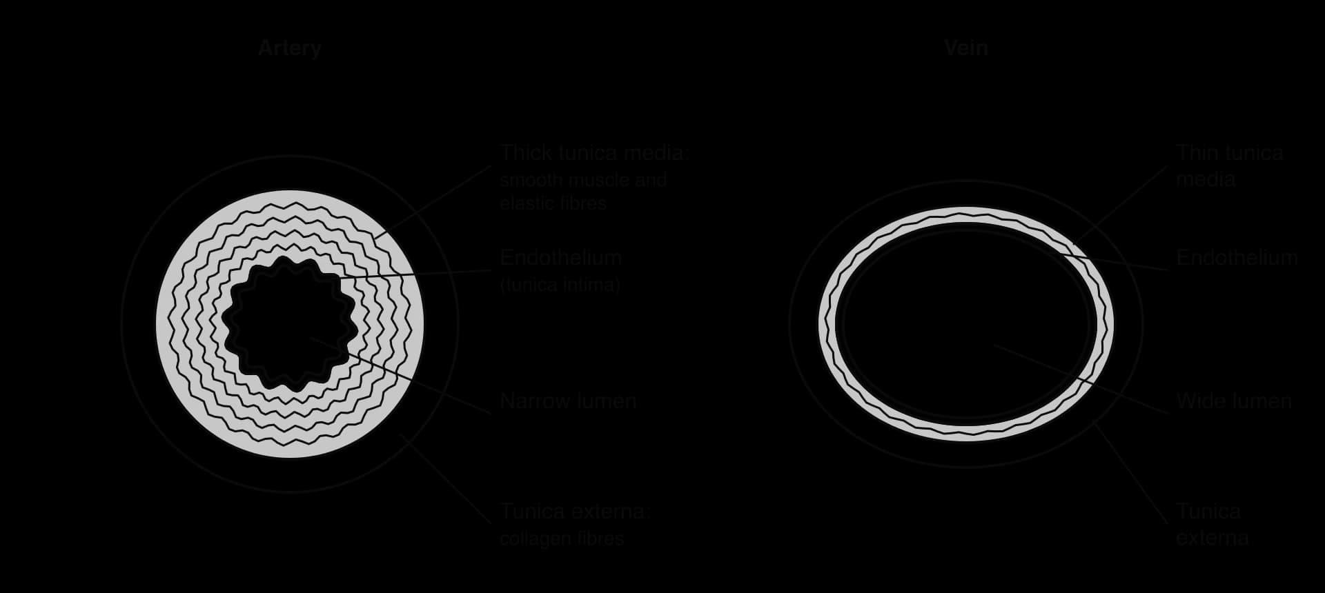
The table compares the three types of vessel. The differences all follow from the pressure each vessel carries and the job it does.
Feature | Artery | Capillary | Vein |
|---|---|---|---|
Direction of flow | Away from the heart | Between arterioles and venules | Towards the heart |
Blood pressure | High and pulsing | Falling, no pulse | Low, no pulse |
Wall thickness compared with lumen | Thick wall, narrow lumen | Wall one cell thick | Thin wall, wide lumen |
Tunica media | Thick: much smooth muscle and elastic tissue | Absent | Thin: little muscle or elastic tissue |
Shape in section | Round, holds its shape | Very small, round | Often flattened or irregular |
Valves | None (except at the base of the aorta and pulmonary artery) | None | Pocket valves in many veins, especially in the limbs |
Main function | Carry blood quickly at high pressure | Exchange with tissues | Return blood at low pressure |
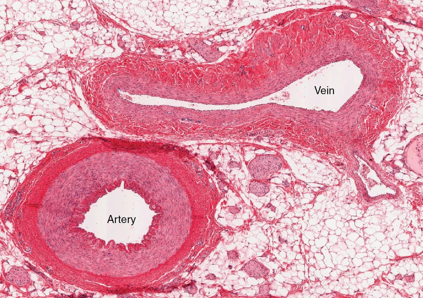
In stained sections, an artery and its matching vein often lie side by side. Several features tell them apart. The artery is round, with a wall that is thick relative to the lumen; the vein is usually flattened or irregular, because its thin wall collapses when there is no blood inside. The tunica media of the artery stains as a thick, even band, and the endothelium of the artery often appears wavy because the elastic tissue has recoiled. The most reliable measurement is the ratio of wall thickness to lumen diameter, which is much higher in an artery.
Each time the heart contracts, it forces a volume of blood into the arteries at high pressure. Blood pressure is the force per unit area that blood exerts on the walls of the vessels. It is measured in millimetres of mercury (mmHg) and written as two numbers, such as 120/80 mmHg: the higher value is the peak pressure while the heart contracts and the lower value is the minimum pressure while the heart relaxes. The pressure in the large arteries never falls to zero between beats because of the structure of the arterial wall.
When a surge of blood enters an artery, the elastic fibres in the tunica media stretch and the artery widens. Some of the energy of each heartbeat is stored in the stretched fibres. Between beats, the fibres shorten again and push on the blood, keeping it moving forwards and keeping the pressure high. This elastic recoil turns the jerky output of the heart into a steadier flow by the time blood reaches the arterioles. It also explains why large arteries close to the heart, such as the aorta, contain the most elastic tissue: they take the largest pressure surges.
The smooth muscle in the tunica media makes the wall thick and strong, so that it can withstand high pressure without bursting. It also allows the diameter of the vessel to change. When the muscle contracts, the lumen narrows (vasoconstriction); when it relaxes, the lumen widens (vasodilation). This control is greatest in arterioles, which have a relatively thick muscular layer for their size. By narrowing or widening, arterioles adjust blood pressure and divert blood to the tissues that need it most, for example to skeletal muscles during exercise and away from the gut.
The collagen of the tunica externa adds tensile strength. Elastic tissue alone could stretch too far under very high pressure; the collagen fibres limit how far the wall can expand and prevent the artery from bulging or rupturing.
Each heartbeat sends a pressure wave along the arteries, and the artery wall expands and recoils as the wave passes. This regular expansion is the pulse. It can be felt wherever an artery runs close to the skin over a firm surface, and its frequency is equal to the heart rate, the number of times the heart beats per minute (beats per minute, bpm).
The two most used sites are the radial artery, on the thumb side of the inside of the wrist, and the carotid artery, at the side of the neck beside the windpipe. The pulse is felt with the index and middle fingertips, not the thumb, because the thumb has a pulse of its own that could be counted by mistake.
Sit quietly for a few minutes so that the heart rate settles to its resting value.
Place two fingertips lightly on the radial artery (or gently on one carotid artery, never both at once).
Count the beats for a fixed time, such as 30 seconds, using a timer.
Multiply to get beats per minute (× 2 for 30 seconds, × 4 for 15 seconds). Counting for a full 60 seconds gives the most accurate result.
Repeat at least three times and calculate the mean.
A normal resting heart rate for an adult is between 60 and 100 bpm. Trained endurance athletes often have resting rates well below 60 bpm, because each beat of their larger, stronger heart pumps more blood. Heart rate rises during exercise, with stress and with fever, as the body needs more oxygen to be delivered.
Several devices measure heart rate automatically. A pulse oximeter clips onto a fingertip and shines red and infrared light through it; the amount of light absorbed by the blood changes with each pulse, which gives the heart rate as well as the oxygen saturation of haemoglobin. Smartwatches and fitness trackers use the same idea with green light reflected from the skin of the wrist. Chest-strap monitors detect the electrical activity of the heart directly, in the same way as an electrocardiogram (ECG).
Method | Advantages | Limitations |
|---|---|---|
Counting the pulse by hand | No equipment except a timer; quick; works anywhere | Human error in counting or timing; hard to count during exercise; gives one reading, not continuous data |
Pulse oximeter | Fast, objective reading; also gives oxygen saturation | Needs still hands; cold fingers, nail varnish and movement can give false readings; some devices are less accurate on darker skin |
Smartwatch or fitness tracker | Continuous recording over hours or days; data stored and graphed | Less accurate during vigorous movement or if loose; accuracy varies between models |
Chest strap (electrical) | Very accurate, even during exercise | More expensive; must be worn against the skin |
By the time blood has passed through the capillaries, most of its pressure has been lost overcoming friction in the narrow vessels. Blood in veins is under low pressure and flows slowly and smoothly, without a pulse. In the legs it must also travel upwards against gravity. Veins have three adaptations that keep blood moving towards the heart.
Valves. Many veins, especially in the arms and legs, contain pocket valves made of folds of the inner wall. Blood flowing towards the heart pushes the flaps flat against the wall and passes freely. If blood starts to flow backwards, it fills the pockets, the flaps meet in the middle and the valve closes. The valves divide a long vein into short sections and prevent backflow.
Thin, flexible walls. The wall of a vein has little muscle or elastic tissue, so the vein is easily compressed. Large veins in the limbs run between skeletal muscles. When these muscles contract, they bulge and squeeze the veins between them, pushing blood along. Because of the valves, blood can only move towards the heart. This mechanism is called the skeletal muscle pump.
Wide lumen. The large lumen offers little resistance to flow, so even the low pressure in veins is enough to move a large volume of blood.
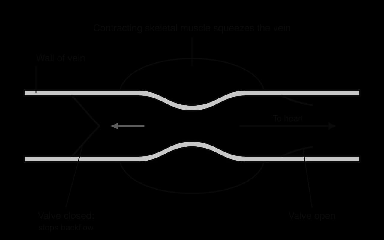
Breathing helps too: when the diaphragm contracts during inhalation, pressure in the thorax falls and blood is drawn into the large veins entering the heart. When valves fail, blood pools in the leg veins, which become stretched and twisted; these are varicose veins. Soldiers who stand still for long periods can faint because, without movement of the leg muscles, too little blood returns to the heart and the brain receives less blood.
 YouTube
Heart muscle works continuously and has a high demand for oxygen and glucose. Its wall is too thick to be supplied by diffusion from the blood inside the chambers, so it has its own blood supply, the left and right coronary arteries, which branch from the aorta just after it leaves the heart and run over the surface of the heart, sending branches into the muscle. Coronary arteries are narrow, so even a small blockage can greatly reduce blood flow to part of the heart.
Atherosclerosis is the build-up of fatty deposits called plaques (or atheromas) in the walls of arteries. It develops over many years and follows a sequence of steps.
The endothelium lining the artery is damaged, for example by high blood pressure, chemicals in tobacco smoke or high blood glucose.
Low-density lipoprotein (LDL), which carries cholesterol in the blood, enters the wall at the damaged site and builds up under the endothelium.
White blood cells (macrophages) move into the wall and take up the LDL, becoming "foam cells". Inflammation follows.
Smooth muscle cells, fibrous tissue and calcium salts are added, and the plaque grows into the lumen. The wall becomes thicker and less elastic.
The plaque narrows the lumen and reduces blood flow. It also raises blood pressure, because the heart must pump harder to force blood through narrower, stiffer arteries, and high blood pressure damages more endothelium, so atherosclerosis and high blood pressure each make the other worse.
A plaque can rupture or its surface can tear. Exposed material from inside the plaque triggers blood clotting: platelets stick to the site and a mesh of fibrin forms a clot. A clot that forms inside an intact blood vessel is a thrombus, and its formation is thrombosis. In a coronary artery that is already narrowed, a thrombus can block the lumen completely. The term occlusion means blockage of a vessel.
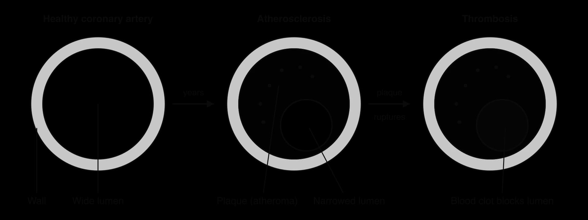
Blockage of a coronary artery has consequences that depend on how complete it is.
Partial blockage reduces the supply of oxygen to part of the heart muscle. This shortage of blood supply is ischaemia. It often causes chest pain called angina, especially during exercise, when the heart works harder and needs more oxygen.
Complete blockage stops the supply to the muscle beyond it. Without oxygen, the cells cannot carry out aerobic cell respiration, ATP runs out and the cardiac muscle cells die. This is a myocardial infarction, commonly called a heart attack. If a large area dies, or if the electrical rhythm of the heart is disrupted, the heart can stop pumping effectively and the person may die.
Disease caused by narrowing or blockage of the coronary arteries is coronary heart disease (CHD), also called ischaemic heart disease. It is the leading cause of death in the world: in 2019 it was responsible for about 16% of all deaths, around 8.9 million people. Narrowed arteries can be treated by inserting a stent, a mesh tube that holds the artery open, or by a bypass operation, in which a section of vein from the leg is grafted to carry blood around the blockage.
Source: WHO Global Health Estimates, 2020 (deaths in 2019).
A risk factor is anything that increases the probability of developing a disease. A risk factor does not make the disease certain, and people with no risk factors can still develop it. The main risk factors for CHD fall into two groups.
Risk factors that cannot be changed | Risk factors linked to lifestyle |
|---|---|
Increasing age: plaques build up over decades | Smoking: damages the endothelium and makes blood clot more easily |
Sex: men develop CHD earlier on average than women | High blood pressure: damages the endothelium |
Genetic factors and family history, such as inherited high blood cholesterol | High LDL cholesterol in the blood, linked to diets high in saturated and trans fats |
Obesity, type 2 diabetes and lack of exercise |
Most of what is known about the causes of CHD comes from epidemiology, the study of the distribution and causes of disease in human populations. Epidemiologists cannot usually do experiments on people, such as making one group smoke. Instead they collect data on large numbers of people and look for patterns, for example whether people who smoke develop CHD more often than people who do not.
A correlation is a relationship between two variables in which a change in one is associated with a change in the other. In a positive correlation both increase together; in a negative correlation one decreases as the other increases. Plotting the data on a scatter graph shows the pattern. The correlation coefficient (r) puts a number on the strength and direction of the relationship. It ranges from +1 (a perfect positive straight-line relationship) through 0 (no linear relationship) to −1 (a perfect negative relationship). Values close to +1 or −1 show a strong correlation; values close to 0 show a weak one.
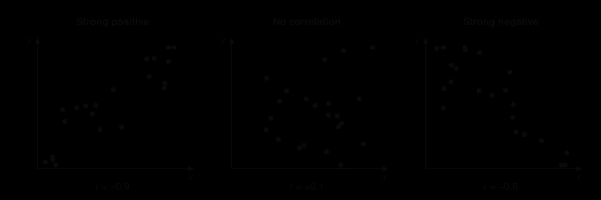
A correlation coefficient helps to test a hypothesis. If a hypothesis predicts that a factor raises the risk of CHD, a correlation close to zero between that factor and CHD would count as evidence against it. A strong correlation is consistent with the hypothesis, but it does not prove it. This is why long-term cohort studies and trials were needed before smoking and high cholesterol were accepted as causes of CHD.
Causation means that a change in one variable directly produces a change in the other. A correlation between two variables can arise without causation for several reasons. A third variable, called a confounding variable, may affect both; for example, people who smoke may also drink more alcohol or take less exercise. The direction of cause may be reversed; for example, people with early heart disease may become less active, rather than inactivity causing the disease. A correlation may also arise by chance, especially in a small sample.
A well-known example is the Seven Countries Study, which began in 1958 and compared men in the USA, Finland, the Netherlands, Italy, Greece, Japan and the former Yugoslavia. Countries with a higher intake of saturated fat had higher death rates from CHD. This correlation between populations was strong, but the countries also differed in smoking, exercise, wealth, sugar intake and many other factors, so it could not show that saturated fat itself caused the difference. Evidence for causation is much stronger when several kinds of study agree: long-term studies that follow individuals over time, randomised controlled trials in which one group changes a single factor, a dose–response relationship (more exposure, more disease) and a known biological mechanism, such as LDL cholesterol entering the artery wall.
In the 1940s, heart disease had become the leading cause of death in the United States, but little was known about why some people developed it and others did not. In 1948 the US Public Health Service began a study in the town of Framingham, Massachusetts. It enrolled 5,209 men and women aged 30–62 who had no signs of heart disease. Every two years each participant had a medical check-up, blood tests and an interview about lifestyle, recording variables such as blood pressure, blood cholesterol, body mass and smoking. The researchers then waited to see who developed coronary heart disease.
This design is a prospective cohort study: a group of people (a cohort) is measured first and then followed forward in time. It is much more powerful than comparing countries, because data are collected on individuals before they become ill, so the suspected cause is known to come before the disease. Comparing participants with and without a factor, while taking account of age and sex, also allows some confounding variables to be controlled.
Year | Finding from the Framingham Heart Study |
|---|---|
1948 | Study begins with 5,209 adults aged 30–62 without heart disease |
1957 | High blood pressure and high blood cholesterol shown to increase the likelihood of heart disease |
1960 | Cigarette smoking found to increase the risk of heart disease |
1961 | Cholesterol level, blood pressure and electrocardiogram abnormalities found to increase the risk; the term "risk factor" introduced |
1967 | Physical activity found to reduce the risk, and obesity to increase it |
1970 | High blood pressure found to increase the risk of stroke |
Source: Framingham Heart Study research milestones; Kannel, Dawber et al., Annals of Internal Medicine, 1961.
The 1961 paper by William Kannel, Thomas Dawber and colleagues described these measurable characteristics as "factors of risk", and the phrase "risk factor" passed into everyday medicine. The idea changed how heart disease was treated. Before Framingham, atherosclerosis was widely seen as an unavoidable part of ageing; afterwards, doctors began to measure blood pressure and cholesterol routinely and to treat them before any symptoms appeared. Later generations of participants were enrolled, including the children of the original cohort from 1971, so the study continues today and has also linked CHD to diabetes and other factors.
Framingham also shows the limits of a cohort study. The original participants were almost all white Americans from one town, so results needed to be checked in other populations. A cohort study still shows correlation, not proof of cause, even though it shows which came first. The case for causation became convincing only when randomised controlled trials showed that lowering blood pressure and LDL cholesterol with drugs reduced the number of heart attacks, and when the biological mechanism of plaque formation was understood.
A tall tree moves hundreds of litres of water a day from its roots to leaves tens of metres above the ground, without any pump. Water travels in xylem, a tissue of long, hollow tubes that runs from the roots, through the stem and into every leaf. The energy for this movement comes from the evaporation of water at the leaf surface, which is driven by heat from the Sun.
Leaves lose water vapour through their stomata, a process called transpiration, which is an unavoidable consequence of opening the stomata for gas exchange (B3.1). The water lost comes from the walls of the mesophyll cells inside the leaf. The walls are made of cellulose fibres with narrow spaces between them, and these spaces are kept wet. Water evaporates from the wet cell walls into the air spaces of the leaf, and the water vapour diffuses out through the open stomata.
As water evaporates from a cell wall, the water that remains is drawn into the narrow spaces between the cellulose fibres. Water adheres strongly to cellulose, and this adhesion in very narrow spaces produces capillary action, which pulls more water into the wall to replace what was lost. That water comes from neighbouring cells and walls, and ultimately from the xylem vessels in the leaf veins. Water is therefore pulled out of the xylem and through the cell walls towards the sites of evaporation.
Pulling water out of the top of the xylem puts the water in the vessels under tension: its pressure falls below atmospheric pressure, so the xylem sap is under negative pressure. This tension is transmitted all the way down the xylem to the roots, because water molecules stick to one another. Cohesion, the attraction between water molecules caused by hydrogen bonds, keeps the water in each vessel as a continuous column that does not break when it is pulled. The whole column is drawn upwards, rather like liquid sucked up a drinking straw. Adhesion of water to the walls of the xylem vessels also helps to hold the column in place against gravity.
At the roots, water moves from the soil into the root hairs by osmosis, because the soil water has a lower solute concentration than the cytoplasm of the root cells. It then crosses the root cortex to the xylem in the centre. The explanation of the whole process is the cohesion–tension theory.
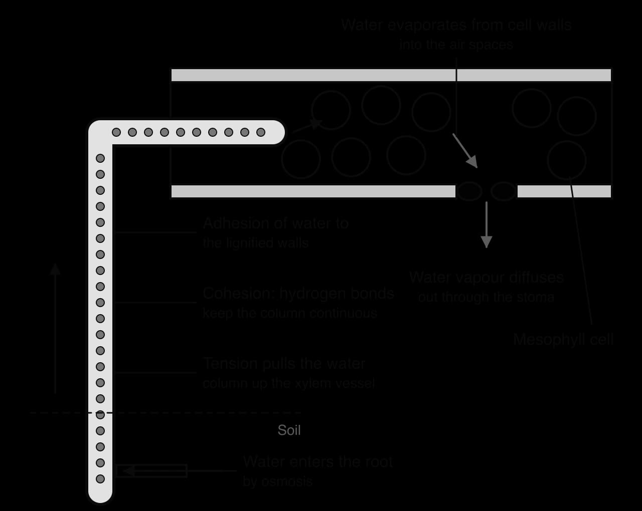
The process is passive: the plant spends no ATP moving water up the xylem. Because the force pulls rather than pushes, the xylem vessels are under tension, and their walls would collapse inwards if they were not strengthened. Several observations support the theory. The trunks of trees shrink very slightly in diameter at midday, when transpiration and tension are greatest. If a xylem vessel is cut, air is pulled in rather than water leaking out. Coloured dye placed at the cut base of a shoot is drawn up into the leaf veins faster when the leaves are in warm, dry, windy conditions. The tallest living tree, a coast redwood (Sequoia sempervirens), is about 116 m tall, close to the height at which the cohesion–tension mechanism is thought to reach its limit (A1.1 Water).
Xylem vessels develop from columns of living cells laid end to end. As they mature, the cells deposit lignin in their walls and then die. Their contents break down and the end walls between them are partly or completely removed, leaving a long, continuous hollow tube. Every feature of the mature vessel suits it for the rapid flow of water under tension.
Feature of xylem vessels | How it helps transport |
|---|---|
No cell contents (no cytoplasm, nucleus or plasma membrane) | Leaves an open lumen with nothing to slow the flow; water does not have to cross membranes |
End walls absent or incomplete | Forms a continuous tube from root to leaf, so water flows without obstruction |
Walls thickened with lignin, often as rings or spirals | Resists the inward pull of tension, so vessels do not collapse; rings and spirals let young vessels stretch as the stem grows |
Pits: thin, unlignified areas in the wall | Let water move sideways between vessels and out into the surrounding tissues; water can bypass a vessel blocked by an air bubble |
Narrow lumen | Increases the effect of adhesion to the walls and makes the column less likely to break |

Xylem also contains tracheids, narrower dead cells with tapering ends, and in flowering plants these sit alongside the vessels. Together with the lignified fibres of the xylem, they help to support the plant: wood is mostly xylem.
 YouTube
Flowering plants are divided into monocotyledons (such as grasses) and dicotyledons (such as sunflowers, beans and most trees). In a young dicotyledonous stem seen in transverse section, the tissues are arranged in a regular pattern, with the vascular bundles in a ring.
Epidermis: a single outer layer of cells, covered with a waxy cuticle that reduces water loss and protects against infection.
Cortex: several layers of mainly thin-walled parenchyma cells between the epidermis and the vascular bundles. Cells near the outside often have thickened corners (collenchyma), which give support. The cortex can store starch and, in green stems, carry out photosynthesis.
Vascular bundles: arranged in a ring near the outside of the stem. Each bundle contains xylem towards the centre and phloem towards the outside, with a thin layer of dividing cells, the cambium, between them. Placing the strong, lignified bundles near the edge of the stem resists bending, in the same way that a hollow tube is stiffer than a solid rod of the same mass.
Pith: a central region of large parenchyma cells used for storage.
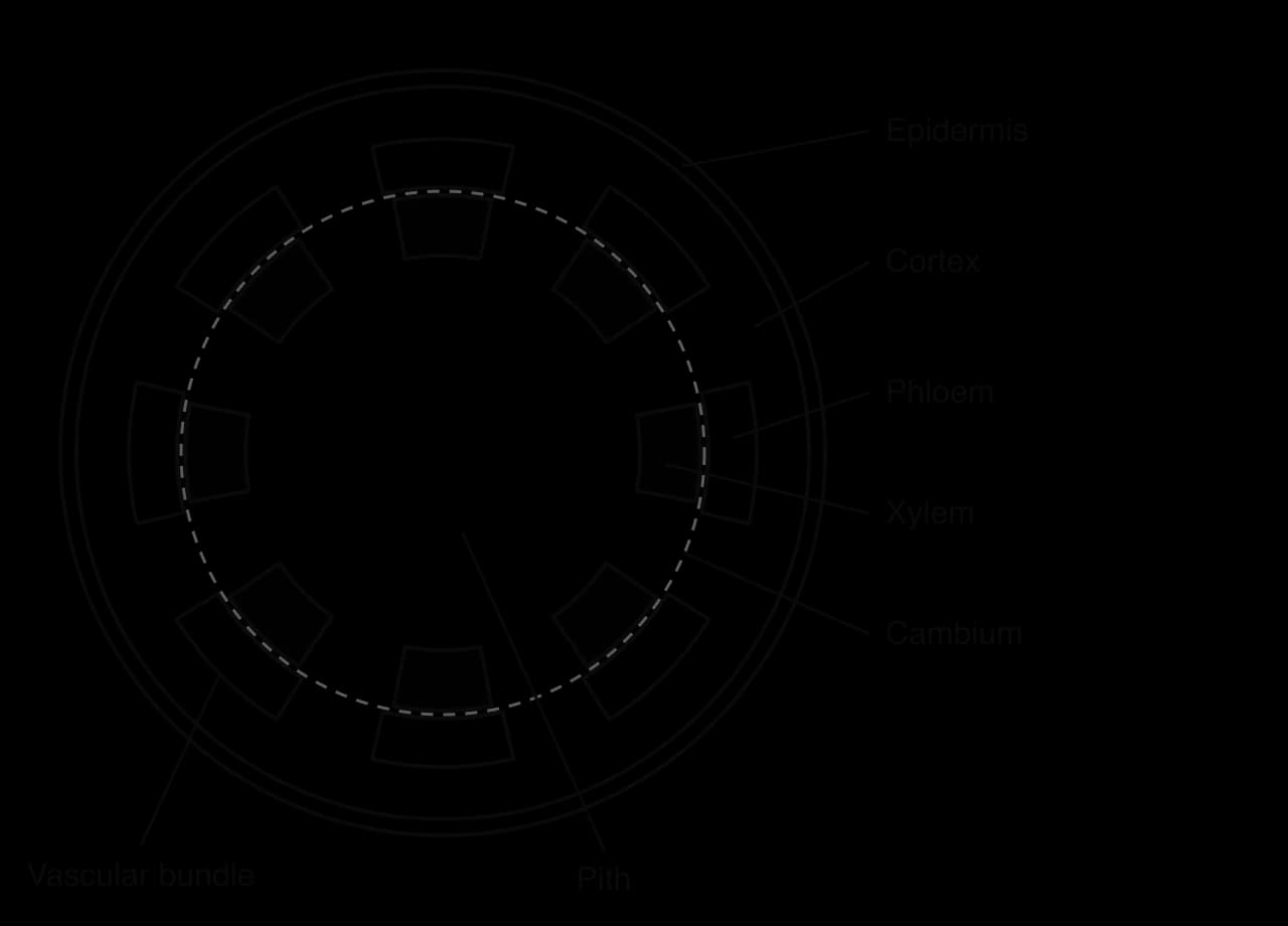
A plan diagram, or tissue map, shows the outlines of the tissues and their relative sizes and positions. It does not show individual cells. Plan diagrams are drawn from micrographs or from slides seen through a light microscope at low power, and they follow the general rules for biological drawings.
Use a sharp pencil and single, clear, continuous lines, with no shading.
Draw only the boundaries between tissues, keeping the proportions the same as in the specimen (for example, the width of the cortex compared with the diameter of the stem).
Draw ruled, straight label lines that end exactly on the tissue and do not cross; write labels outside the drawing, horizontally.
Give the drawing a title, and add a magnification or scale bar if one is asked for.
Annotate when asked: an annotation is a short note on the function of a labelled structure, such as "xylem: transports water and mineral ions".
In a transverse section of a young dicotyledonous root, the arrangement is quite different from the stem. The vascular tissue is gathered in the centre, in a column called the stele, rather than in a ring of bundles.
Epidermis: the outer layer, without a waxy cuticle, so that water can enter. Many epidermal cells grow long, thin extensions called root hairs, which greatly increase the surface area for the absorption of water and mineral ions.
Cortex: a wide region of parenchyma cells, which often stores starch. Water moves across the cortex towards the centre, both through the cell walls and from cell to cell.
Endodermis: a single ring of cells around the stele. Their walls contain a waterproof band called the Casparian strip, which forces water and ions to pass through the cytoplasm of the endodermal cells, so the plant controls which ions enter the xylem.
Stele: the central vascular tissue. The xylem forms a star or cross shape, often with four arms in dicotyledons, and the phloem lies in groups between the arms.
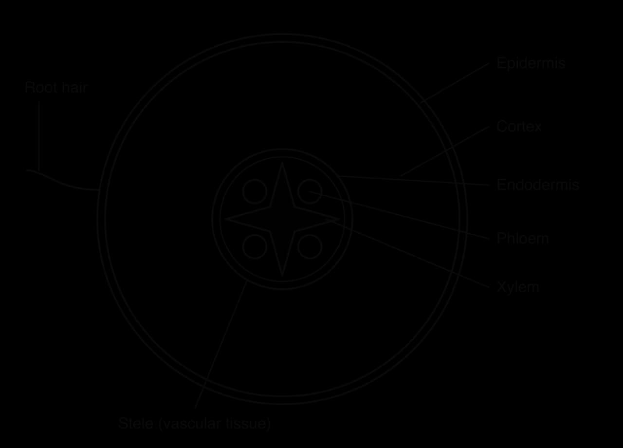
The central position of the vascular tissue suits the root: a root is pulled rather than bent, as the shoot above sways in the wind, and a central core resists pulling forces as a rope does. Water absorbed by the root hairs also has a short, direct route across the cortex to the xylem.
Feature | Dicotyledonous stem | Dicotyledonous root |
|---|---|---|
Position of vascular tissue | In separate bundles arranged in a ring | In one central stele |
Arrangement of xylem and phloem | In the same bundle: xylem inside, phloem outside | Xylem in a star shape; phloem between the arms |
Centre of the organ | Pith | Xylem |
Epidermis | Covered by waxy cuticle | No cuticle; root hairs |
Endodermis | Not distinct | Clear ring around the stele |
Main mechanical force resisted | Bending | Pulling |
Cells in tissues are bathed in tissue fluid, which fills the spaces between cells and forms the internal environment through which every exchange passes. Tissue fluid is formed from blood plasma that leaks out of capillaries, and most of it later drains back in.
Two opposing forces act on water across the capillary wall. Hydrostatic pressure, the blood pressure inside the capillary, pushes fluid out. The plasma proteins that stay in the blood, mainly albumin, lower the water potential of the blood, so water tends to move back in by osmosis. The balance between the two changes along the capillary.
At the arteriole end, the blood pressure is relatively high (about 35 mmHg in a typical capillary), higher than the opposing osmotic effect of the plasma proteins (about 25 mmHg). Fluid is forced out through the gaps in the capillary wall, carrying water and small solutes such as glucose, amino acids, fatty acids, oxygen and ions. This is pressure filtration. Blood cells and large proteins are too big to pass and stay in the capillary.
At the venule end, the pressure has fallen (to about 15 mmHg), because fluid has been lost and friction has reduced the pressure. The osmotic effect of the proteins, which are now slightly more concentrated, is greater than the blood pressure, so water returns to the capillary by osmosis, bringing carbon dioxide and other wastes with it.
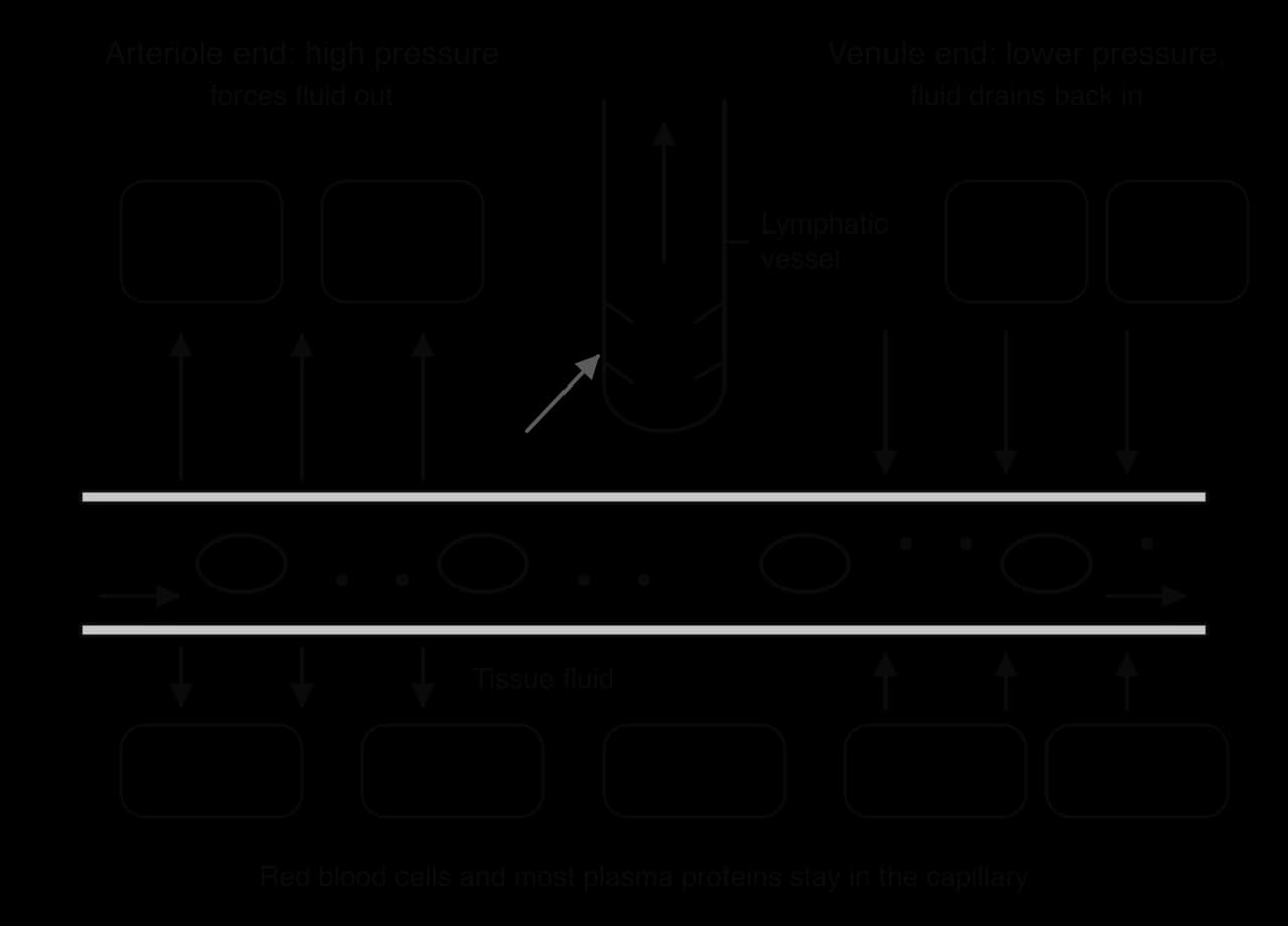
Once in the tissue fluid, substances move between the fluid and the cells across their plasma membranes. Oxygen diffuses into cells, because cells use it in aerobic respiration and keep its concentration low inside. Glucose enters by facilitated diffusion through carrier proteins, and some ions and amino acids enter by active transport. Carbon dioxide, urea and other wastes made by the cells diffuse out into the tissue fluid and then into the blood. Because the blood keeps arriving with fresh supplies and removing wastes, the concentration gradients across the capillary wall and the cell membrane are maintained.
Component | Blood plasma | Tissue fluid |
|---|---|---|
Water, glucose, amino acids, ions | Present | Present, at similar concentrations |
Oxygen | Higher concentration (arriving at the tissues) | Lower: used by cells |
Carbon dioxide and urea | Lower | Higher: released by cells |
Plasma proteins (albumin, antibodies, clotting proteins) | High concentration | Very low: too large to be filtered |
Red blood cells and platelets | Present in blood | Absent |
White blood cells | Present in blood | A few, which squeeze between endothelial cells |
More fluid leaves the capillaries at the arteriole end than returns at the venule end. The excess is collected by a separate system of vessels, the lymphatic system, and returned to the blood. Without it, fluid would build up in the tissues, causing swelling (oedema).
Lymphatic capillaries are blind-ended tubes among the tissue cells. Their walls are very thin, and the endothelial cells overlap loosely, leaving gaps that act as one-way flaps: fluid, and even large proteins and bacteria, can enter but cannot easily leave. Once inside a lymphatic vessel, the fluid is called lymph.
Valves in the larger lymph ducts allow lymph to flow only away from the tissues. As in veins, lymph is moved by the squeezing of nearby skeletal muscles and by pressure changes in the thorax during breathing.
Return to the blood: lymph ducts join to form larger ducts, mainly the thoracic duct, which empties into the left subclavian vein beneath the collarbone; a smaller duct empties into the right subclavian vein. Along the way, lymph passes through lymph nodes, where white blood cells remove pathogens (C3.2).
The importance of drainage is shown by lymphatic filariasis, a tropical disease caused by parasitic roundworms, mainly Wuchereria bancrofti, which are spread by mosquitoes. The adult worms live in lymph vessels and block them. Tissue fluid cannot drain from the affected limbs, which swell greatly, a condition called elephantiasis. The World Health Organization launched a global programme to eliminate the disease in 2000, based on mass treatment of whole communities with drugs that kill the parasites.
Bony fish, such as cod and trout, have a single circulation: blood passes through the heart once for each complete circuit of the body. The fish heart has two main chambers, one atrium and one ventricle. The ventricle pumps deoxygenated blood to the gills, where it picks up oxygen in the gill capillaries. The oxygenated blood then flows directly on to the capillaries of the body organs and returns to the heart in veins. The sequence is heart → gills → body → heart.
Mammals, birds and other tetrapods have a double circulation, in which blood passes through the heart twice in each complete circuit. The heart is divided into a right side and a left side, separated by a wall. The right side pumps deoxygenated blood to the lungs and back to the left side of the heart: this is the pulmonary circulation. The left side then pumps oxygenated blood to all the other organs and back to the right side: this is the systemic circulation. The sequence is right side of heart → lungs → left side of heart → body → right side of heart.
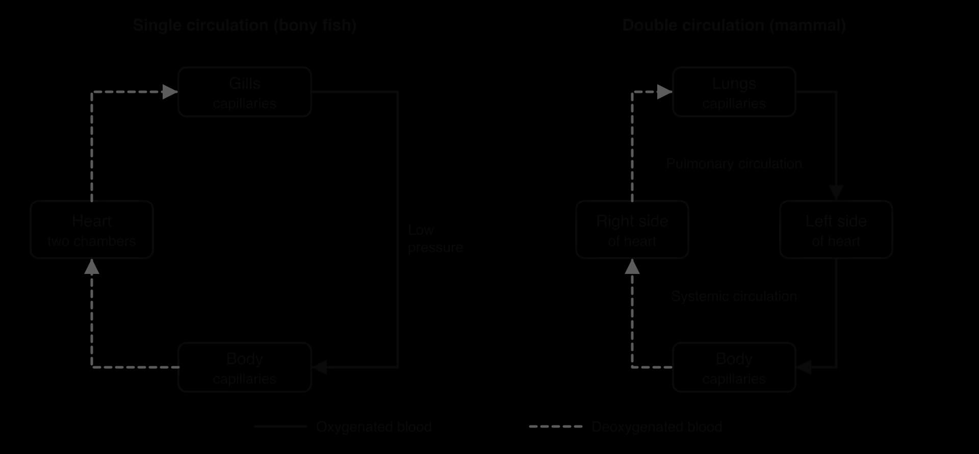
Blood loses much of its pressure when it passes through a capillary bed. In a fish, the blood leaving the gill capillaries has to flow on through the body capillaries with no further pumping, so blood reaches the body organs at low pressure and flows slowly. In a mammal, blood returns to the heart after the lungs and is pumped again, so it reaches the body at high pressure. This delivers oxygen and glucose quickly enough to support a high metabolic rate, including the heat production of an endothermic animal.
Separating the two circulations also allows each to run at a suitable pressure. The pulmonary circulation runs at a much lower pressure than the systemic circulation, which protects the delicate capillaries of the lungs and stops fluid being forced into the alveoli. In addition, oxygenated and deoxygenated blood are kept apart in the heart, so blood leaving for the body carries as much oxygen as possible.
Feature | Single circulation (bony fish) | Double circulation (mammal) |
|---|---|---|
Passes through the heart per circuit | Once | Twice |
Chambers of the heart | One atrium, one ventricle | Two atria, two ventricles |
Gas exchange organ | Gills | Lungs |
Capillary beds between leaving and returning to the heart | Two (gills, then body) | One in each circuit |
Pressure of blood reaching the body | Low | High |
Blood in the heart | Deoxygenated only | Oxygenated (left) and deoxygenated (right), kept separate |
The heart is a muscular pump with four chambers. Its wall is made of cardiac muscle, a tissue found only in the heart (B2.3). Cardiac muscle cells are branched and joined end to end by intercalated discs, which allow electrical impulses to pass quickly from cell to cell, so the muscle of each chamber contracts as one unit. The cells contain many mitochondria, because the heart contracts continuously and needs a steady supply of ATP from aerobic respiration. Cardiac muscle is myogenic: it contracts without needing a nerve impulse, and it does not tire.
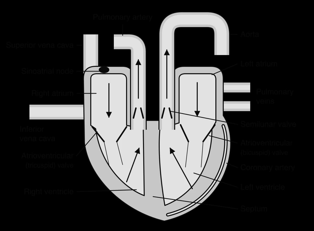
The diagram shows the heart in the frontal plane, as if the person were facing the viewer, so the right side of the heart appears on the left of the page. Each structure has a specific job in moving blood in one direction at high pressure.
Structure | Form | Function |
|---|---|---|
Atria (right and left) | Two upper chambers with thin muscular walls | Receive blood from veins and pump it a short distance into the ventricles |
Ventricles (right and left) | Two lower chambers with thick muscular walls; the left wall is about three times thicker than the right | Pump blood out into the arteries at high pressure; the left ventricle generates enough pressure to send blood around the whole body, the right only to the nearby lungs |
Atrioventricular (AV) valves | Tricuspid on the right, bicuspid (mitral) on the left; flaps held by tendons (chordae tendineae) | Close when the ventricles contract, preventing backflow into the atria; the tendons stop the flaps turning inside out |
Semilunar valves | Three pocket-shaped cusps at the base of the aorta and of the pulmonary artery | Close when the ventricles relax, preventing backflow from the arteries into the ventricles |
Septum | Thick wall of muscle between the left and right sides | Keeps oxygenated and deoxygenated blood separate |
Sinoatrial (SA) node | A small group of specialised cardiac muscle cells in the wall of the right atrium | Acts as the pacemaker: starts each heartbeat by sending out an electrical impulse |
Coronary arteries and veins | Vessels on the surface of the heart | Supply the heart muscle with oxygen and glucose, and remove carbon dioxide |
Blood flows through the heart in only one direction, kept that way by the valves. Starting with deoxygenated blood returning from the body:
Deoxygenated blood from the body enters the right atrium through the superior and inferior venae cavae.
It passes through the tricuspid (right AV) valve into the right ventricle.
The right ventricle pumps it through a semilunar valve into the pulmonary artery, which carries it to the lungs.
Oxygenated blood returns from the lungs in the pulmonary veins to the left atrium.
It passes through the bicuspid (left AV) valve into the left ventricle.
The left ventricle pumps it through a semilunar valve into the aorta, which distributes it to the body.
The cardiac cycle is the sequence of events in one heartbeat. Contraction of a chamber is systole and relaxation is diastole. At a resting heart rate of 75 beats per minute, one cycle lasts 60 ÷ 75 = 0.8 s. The two sides of the heart go through the cycle at the same time; the description follows the left side.
Each cycle is started by the SA node. Its cells depolarise spontaneously at a regular rate, sending an electrical impulse across the walls of both atria, which contract together. The impulse cannot pass directly to the ventricles, because a layer of non-conducting tissue separates them from the atria. It reaches the ventricles only through the atrioventricular (AV) node, which delays it by about 0.1 s, giving the atria time to empty before the ventricles contract. The impulse then travels down the septum to the apex, the lowest point of the heart, and up through the ventricle walls, so the ventricles contract from the bottom upwards, squeezing blood up towards the arteries. Nerves and hormones such as adrenaline change the rate at which the SA node fires, but they do not start the beat.
Atrial systole (about 0.1 s). The left atrium contracts. Its pressure rises slightly above that of the ventricle, and blood is pushed through the open AV valve to fill the ventricle completely. The semilunar valve is closed, because pressure in the aorta is much higher than in the ventricle.
Ventricular systole (about 0.3 s). The ventricle contracts and its pressure rises steeply. As soon as ventricular pressure exceeds atrial pressure, the AV valve is forced shut, making the first heart sound ("lub"). For a moment both valves are closed and pressure builds with no change in volume. When ventricular pressure exceeds the pressure in the aorta, the semilunar valve is pushed open and blood is ejected into the aorta. Aortic pressure rises to its maximum, the systolic pressure.
Diastole (about 0.4 s). The ventricle relaxes and its pressure falls. When it drops below aortic pressure, blood starts to flow back and fills the cusps of the semilunar valve, which closes, making the second heart sound ("dub"). This causes a small notch in the aortic pressure trace. When ventricular pressure falls below atrial pressure, the AV valve opens and blood that has collected in the atrium flows passively into the ventricle. Meanwhile, the elastic recoil of the aorta keeps blood moving round the body.
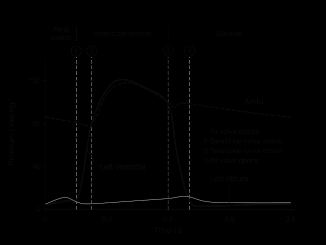
The pressure graph can be read with one rule: a valve opens when the pressure behind it is higher than the pressure in front of it, and closes when the pressure difference reverses. The AV valve closes at point 1, where ventricular pressure rises above atrial pressure, and opens at point 4, where it falls below. The semilunar valve opens at point 2, where ventricular pressure rises above aortic pressure, and closes at point 3, where it falls below.
Blood pressure is usually measured in the brachial artery of the upper arm with a sphygmomanometer: an inflatable cuff stops the blood flow, and the pressure is recorded as the cuff is slowly released, either by listening with a stethoscope or automatically with a digital monitor. The systolic pressure is the highest pressure in the arteries during ventricular systole; the diastolic pressure is the lowest pressure, during diastole. A typical healthy reading for a young adult is about 120/80 mmHg. Hypertension (high blood pressure) is diagnosed when readings are repeatedly 140 mmHg or more systolic and/or 90 mmHg or more diastolic. Hypertension increases the risk of atherosclerosis, CHD and stroke.
When transpiration is very slow, for example at night, in very humid air, or in spring before a deciduous tree has opened its leaves, there is little evaporation to create tension in the xylem. Some plants can still move water upwards by root pressure. Cells around the xylem in the root use ATP to transport mineral ions actively into the xylem vessels. The accumulation of ions lowers the water potential of the xylem sap, so water follows from the surrounding root cells by osmosis. The endodermis stops the ions leaking back out. Water entering the closed xylem system produces a positive pressure potential that pushes the water column upwards.
Root pressure produces two visible effects. In spring, sap flows from cuts in the trunks of birch trees (Betula) before the leaves have opened; the flow is driven by root pressure and stops once the leaves are out and transpiration takes over. On cool, humid mornings, small plants such as grasses and strawberries push drops of liquid out of pores at the tips and edges of their leaves, a process called guttation; this water is forced out by root pressure, not by dew. Root pressure can lift water only a few metres, so it cannot explain transport in tall trees during the day.
Transpiration pull | Root pressure | |
|---|---|---|
Pressure in the xylem | Negative (tension) | Positive |
Source of energy | Evaporation, driven by heat from the Sun (passive for the plant) | ATP used to actively transport ions in the root |
Direction of force | Pulls water up from the leaves | Pushes water up from the roots |
When most important | In the day, with open stomata and dry air | At night, in humid air, and in spring before leaves open |
Sugars made by photosynthesis in leaves must be carried to every part of the plant that cannot make enough of its own. The movement of organic compounds, mainly sucrose, in phloem is called translocation. Unlike xylem, phloem moves its contents in either direction, up or down, according to where they are made and where they are needed.
A source is a part of the plant that releases sugars into the phloem: mature photosynthesising leaves, and storage organs when their reserves are being used. A sink is a part that takes sugars out of the phloem, for use in growth and respiration or for storage: roots, growing shoot tips, developing fruits and seeds, and storage organs that are filling. The same organ can change role. A potato tuber is a sink in summer, when it fills with starch, and a source in spring, when its starch is broken down to feed the growing shoots.
Phloem is made of living cells. Sap flows through sieve tubes, formed from sieve tube elements joined end to end. Each sieve tube element is paired with a companion cell that keeps it alive and loads it with sugar.
Sieve plates: the end walls between sieve tube elements are perforated by large pores, so that sap flows from one element to the next. The pores can be sealed quickly with callose if the phloem is damaged.
Reduced cytoplasm and organelles: a mature sieve tube element has no nucleus, no vacuole and very few organelles, leaving only a thin layer of cytoplasm against the wall. This leaves the lumen open, so sap meets little resistance.
Companion cells: each has a nucleus, which controls the activity of both cells, and many mitochondria, which supply ATP for the active loading of sucrose. They often have infoldings of the plasma membrane that increase the surface area for transport.
Plasmodesmata: narrow channels of cytoplasm through the cell walls connect each companion cell to its sieve tube element, so sugars, proteins and ATP can pass between them.
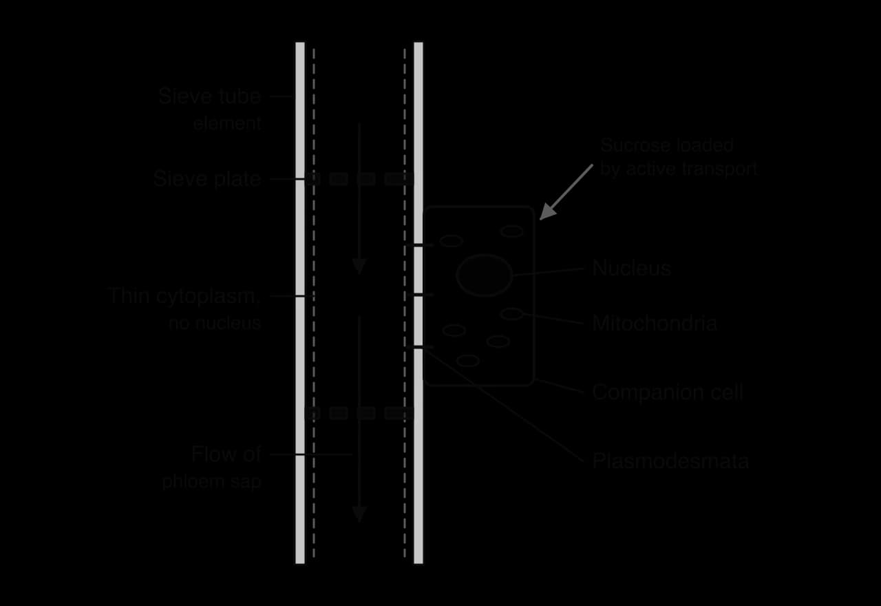
Loading at the source. Companion cells use ATP to pump hydrogen ions (H⁺) out across their plasma membranes. The H⁺ ions diffuse back in through cotransporter proteins, carrying sucrose with them, so sucrose is concentrated inside the companion cells against its gradient. It then moves through plasmodesmata into the sieve tube elements. This is active transport, which is why companion cells have so many mitochondria.
Water enters by osmosis. The high sucrose concentration lowers the water potential in the sieve tubes at the source. Water moves in from the nearby xylem by osmosis, raising the pressure inside the sieve tubes.
Flow under pressure. At the sink, sucrose is removed from the sieve tubes, so the water potential there is higher and water leaves. The pressure at the source end is therefore higher than at the sink end, and sap flows along this pressure gradient, from high to low pressure, carrying sucrose and other solutes with it.
Unloading at the sink. Sucrose leaves the sieve tubes by diffusion or active transport and is used in respiration or converted to starch for storage. Converting it to starch keeps the sucrose concentration low in the sink, so unloading continues.
Strong evidence that phloem sap is under pressure came from aphids, insects that feed by pushing a needle-like mouthpart called a stylet into a single sieve tube. In 1953, Kennedy and Mittler anaesthetised feeding aphids and cut off their bodies, leaving the stylet in the plant. Sap kept flowing out of the cut stylet for hours, pushed out by the pressure in the sieve tube. The sap collected this way was a concentrated solution, mainly of sucrose, showing what phloem carries.
Feature | Xylem | Phloem |
|---|---|---|
Cells | Dead, hollow vessels | Living sieve tube elements with companion cells |
End walls | Absent or incomplete | Sieve plates with pores |
Walls | Thickened with lignin | Cellulose, not lignified |
What is carried | Water and mineral ions | Sucrose, amino acids and other organic compounds in water |
Direction | Upwards only, from roots to leaves | Up or down, from sources to sinks |
Pressure in the tube | Usually negative (tension) | Positive |
Energy | Passive (evaporation) | ATP used for active loading |
Transport is assessed in every part of the papers. Paper 1A multiple-choice questions often show a micrograph of an artery and a vein, a plan diagram of a stem or root, or a heart diagram to identify. Paper 1B and Paper 2 Section A use data: heart rates before and after exercise, epidemiological data on CHD with correlation coefficients, blood pressure readings and (at HL) pressure graphs of the cardiac cycle. Paper 2 Section B extended responses include "explain how water is transported from roots to leaves", "compare the structure of arteries and veins" and, at HL, "describe the events of the cardiac cycle" or "explain the movement of sucrose in phloem". The command terms seen most often are identify, distinguish, explain, evaluate, outline and draw.
Questions that ask you to compare arteries and veins or xylem and phloem are best answered as a table or as paired statements ("arteries have thick walls, whereas veins have thin walls"). Each point needs both sides of the comparison to earn the mark.
I can explain how capillaries, including fenestrated capillaries, are adapted for exchange.
I can distinguish arteries and veins in micrographs using wall thickness relative to lumen diameter.
I can explain how elastic fibres and smooth muscle help arteries withstand and maintain high pressure.
I can measure heart rate from the radial or carotid pulse and compare it with digital methods.
I can explain how valves and skeletal muscles return blood to the heart in veins.
I can explain the causes and consequences of coronary artery occlusion and evaluate epidemiological data, including correlation coefficients.
I can explain the cohesion–tension theory and the adaptations of xylem vessels.
I can draw annotated plan diagrams of a dicotyledonous stem and root.
I can explain the formation and reuptake of tissue fluid and the drainage of lymph. (HL)
I can compare single and double circulation and trace blood through the mammalian heart. (HL)
I can describe the cardiac cycle and interpret pressure graphs and blood pressure readings. (HL)
I can explain root pressure and translocation in phloem. (HL)
a. as the transpiration rate increases, the pressure in the xylem becomes more negative / tension increases;
b. pressure is most negative (−1.4 MPa) at 12:00 when transpiration is highest;
c. both return to their starting values by 21:00 / at night;
[2 max]
Explain why the diameter of the stem decreased during the day.
2 marks
a. the water in the xylem is under tension / negative pressure;
b. water adheres to the xylem walls;
c. (so) the walls of the xylem are pulled inwards;
d. greater tension at midday causes the greatest decrease in diameter;
e. water is drawn out of the living tissues of the stem / bark, which shrink;
[2 max]
Explain how the cohesion of water allows it to be transported to the leaves under tension.
3 marks
a. hydrogen bonds form between water molecules / water molecules are cohesive;
b. water forms a continuous column in the xylem;
c. water evaporates from the leaves / is lost by transpiration;
d. (so) water molecules are pulled up / tension is created at the top of the column;
e. cohesion transmits the tension down the column to the roots;
f. the column does not break despite the tension;
[3 max]
Suggest why the walls of xylem vessels are strengthened with lignin.
1 mark
to stop the vessels collapsing under tension / negative pressure;
Suggest why an air bubble in a xylem vessel stops water transport in that vessel.
1 mark
it breaks the continuous column so cohesion / tension can no longer pull water up;
Hydrogen bonds are individually weak, but together they have major effects in living organisms.
Describe how hydrogen bonds form between water molecules.
4 marks
Outline the role of hydrogen bonds in the structure of DNA.
4 marks
Explain how the properties of water allow it to be transported to the top of tall trees.
7 marks
Fenestrated capillaries have pores in their walls that allow faster exchange of materials. In which location are fenestrated capillaries most likely to be found?