Explain how lungs and leaves are adapted for gas exchange, how breathing works and how to measure lung volumes and stomatal density, and (HL) how haemoglobin loads and unloads oxygen.
Each is in the Biology glossary, with every other term in the subject.
A tiny air sac at the end of a bronchiole in the lungs, with a wall one cell thick and a dense capillary network, where gas exchange takes place.
The movement of the oxygen dissociation curve to the right when carbon dioxide concentration rises, because haemoglobin's affinity for oxygen falls, so more oxygen is released.
At constant temperature, the pressure of a fixed mass of gas is inversely proportional to its volume, so increasing the volume lowers the pressure.
One of the narrow, branching airways in the lungs that carry air from the bronchi to the alveoli.
Binding in which each oxygen molecule that binds to haemoglobin changes its conformation and increases the affinity of the remaining haem groups for oxygen (and the reverse on release).
A dome-shaped sheet of muscle below the lungs; it contracts and flattens during inspiration, increasing the volume of the thorax.
A flowering plant whose seeds have two seed leaves (cotyledons), typically with net-veined leaves and vascular bundles arranged in a ring in the stem.
The outer layer of cells covering a leaf, stem or root; in leaves it is transparent, secretes the cuticle and contains stomata.
Breathing out: the volume of the thorax decreases, raising the pressure in the lungs so that air flows out; it is mostly passive at rest.
The extra volume of air that can be breathed out with maximum effort after a normal expiration.
The haemoglobin of the fetus, with two γ chains instead of β chains; it has a higher affinity for oxygen than adult haemoglobin, so it takes up oxygen from maternal blood at the placenta.
The uptake of one gas and release of another across a surface, by diffusion, such as the uptake of oxygen and loss of carbon dioxide in respiration.
A surface across which gases diffuse between an organism and its surroundings; it is permeable, thin, moist and has a large surface area.
The gas exchange organ of a fish, made of filaments with thin lamellae and many capillaries, ventilated by a one-way flow of water.
One of a pair of cells surrounding a stoma that change shape to open or close it, controlling gas exchange and water loss.
Breathing in: contraction of the diaphragm and external intercostal muscles increases the volume of the thorax, lowering pressure so that air flows into the lungs.
The extra volume of air that can be breathed in with maximum effort after a normal inspiration.
Muscles between the ribs: the external intercostals raise the ribs in inspiration and the internal intercostals lower them in forced expiration; they form an antagonistic pair.
A graph of the percentage saturation of haemoglobin with oxygen against the partial pressure of oxygen; it is S-shaped because of cooperative binding.
The layer of tall, closely packed cells below the upper epidermis of a leaf, containing many chloroplasts; the main site of photosynthesis.
The pressure exerted by one gas in a mixture of gases, proportional to its concentration; pO₂ is used as a measure of oxygen concentration, in kilopascals (kPa).
A diagram showing the outlines and positions of the tissues in an organ, without individual cells, drawn with clear, continuous lines and no shading.
The extent to which repeated measurements give consistent results; it is improved by taking repeated readings or replicate samples and calculating a mean.
An instrument that measures the volume of air breathed in and out, recording lung volumes against time.
The layer of loosely packed, rounded cells with large air spaces in the lower part of a leaf; its moist cell surfaces are the site of gas exchange.
A pore in the epidermis of a leaf, between two guard cells that open and close it, through which carbon dioxide, oxygen and water vapour diffuse.
The number of stomata per unit area of leaf surface, usually expressed per mm².
A mixture of phospholipids and proteins secreted onto the moist lining of the alveoli that reduces surface tension, preventing the alveoli from collapsing.
The volume of air breathed in or out in one normal breath at rest, about 0.5 dm³ in an adult.
The loss of water vapour from the leaves and stems of a plant, by evaporation from mesophyll cell walls and diffusion out through the stomata.
A strand of transport tissue containing xylem and phloem; in a leaf it forms a vein, with xylem towards the upper surface.
The movement of air or water into and out of a gas exchange organ, which maintains concentration gradients for oxygen and carbon dioxide at the exchange surface.
The number of breaths taken per minute; multiplied by tidal volume it gives the volume of air breathed per minute.
The maximum volume of air that can be breathed out after a maximum inspiration; the sum of tidal volume, inspiratory reserve volume and expiratory reserve volume.
A layer of wax secreted by the epidermis of a leaf that is almost impermeable to water, reducing water loss by evaporation.
Gas exchange is the movement of oxygen and carbon dioxide between an organism and its surroundings. Every cell that carries out aerobic respiration needs a supply of oxygen and must get rid of carbon dioxide, and every photosynthesising cell needs carbon dioxide. A person at rest takes in roughly 0.25 dm³ of oxygen every minute, and the lungs must deliver it to the blood without pause for a whole lifetime.
Animals and plants solve the same physical problem in different ways. A mammal pumps air in and out of lungs and carries the gases in its blood; a leaf has no pump and relies on diffusion through pores. Comparing them shows how the same principles (surface area, distance and concentration gradient) shape organisms at very different levels of organisation.
44 exam-style questions on this subtopic, written in the style of the IB papers. The first has its markscheme open. Counted on
Organisms are adapted to the physical environments in which they live.
Compare and contrast water and air as habitats for animals, with reference to the black-throated loon (Gavia arctica) and the ringed seal (Pusa hispida).
7 marks
a. similarity: in both media, animals must overcome resistance / drag to move;
b. similarity: both conduct heat away from an animal warmer than its surroundings;
c. similarity: both supply oxygen for respiration (although water holds far less oxygen than air);
d. water is much denser than air, so it provides more buoyancy / support;
e. the loon has dense bones / expels air from its feathers to reduce buoyancy when diving;
f. air gives little support, so the loon must generate lift with its wings to fly;
g. water is more viscous than air, so drag is greater in water;
h. the loon and the seal have streamlined bodies to reduce drag when swimming;
i. water has a higher thermal conductivity, so heat is lost faster in water than in air at the same temperature;
j. the seal has a thick layer of blubber / the loon has dense, waterproof plumage that traps air for insulation;
k. water has a higher specific heat capacity, so water temperatures are more stable than air temperatures;
l. the seal hauls out onto the ice, where it loses heat more slowly than in water;
m. legs set far back on the loon's body give propulsion in water but make walking on land difficult;
[7 max]
Answers must include at least one similarity and one difference for full marks.
The official deck for this part of the syllabus.
Define gas exchange.
State why gas exchange is needed by (a) cells that respire aerobically and (b) cells that photosynthesise.
With a free account, Augment remembers where you stopped and what you have practiced.
Gas exchange takes place by diffusion. Gases move from a region where their concentration is higher to a region where it is lower, so the rate depends on the area available, the distance the gas must travel and the difference in concentration. All organisms must exchange gases, but the difficulty increases with size.
A single-celled organism such as Amoeba can exchange gases across its plasma membrane: its surface area is large compared with its volume, and no part of the cell is far from the surface. As organisms become larger, two things change. The surface area-to-volume ratio falls, so there is less surface for each unit of volume of respiring tissue. The distance from the centre of the body to the surface also increases, and diffusion is fast only over very short distances (a fraction of a millimetre). A large animal cannot get enough oxygen to its inner cells by diffusion through its skin.
Large multicellular organisms therefore have specialised gas exchange surfaces, such as lungs, gills or the spongy tissue of leaves, together with a transport system (blood in animals) that carries gases between the exchange surface and the cells. Large, active animals also have a high rate of respiration, which increases the demand.
The availability of oxygen in the surroundings matters too. At high altitude each breath contains less oxygen. Tibetan highlanders, whose ancestors have lived above 4,000 m for thousands of years, carry a variant of the gene EPAS1 that is associated with adaptation to low oxygen, and which they inherited from Denisovans, an extinct group of archaic humans.
All efficient gas exchange surfaces share four properties, each of which increases the rate of diffusion:
Property | Why it matters | Example in human lungs |
|---|---|---|
Permeable | O₂ and CO₂ must be able to pass through the cells and their membranes | Thin cell membranes; O₂ and CO₂ pass through the phospholipid bilayer |
Thin tissue layer | A short distance means faster diffusion | Alveolus wall and capillary wall are each one cell thick; total barrier under 1 µm in places |
Moist | Gases dissolve in water before they diffuse through cells | A film of liquid lines each alveolus |
Large surface area | More area allows more gas to diffuse per second | Hundreds of millions of alveoli with a total area of the order of 70 m² |
The four properties can be combined into one relationship (Fick's law): the rate of diffusion is proportional to surface area × concentration difference ÷ thickness of the barrier. Doubling the area or halving the thickness doubles the rate.
Diffusion stops when the concentrations on the two sides of a surface become equal. To keep oxygen diffusing into the blood, the oxygen concentration on the air side must be kept high and the concentration on the blood side kept low. Animals do this in three ways:
Dense networks of blood vessels. A capillary runs close to every part of the exchange surface, so there is a short distance for diffusion over the whole area.
Continuous blood flow. Blood that has picked up oxygen is carried away and replaced by blood with a low oxygen concentration and a high carbon dioxide concentration, so the gradients never disappear.
Ventilation. In ventilation, fresh air (or water) is moved over the surface, bringing in oxygen and removing carbon dioxide. Lungs are ventilated with air; gills are ventilated with water.
Fish face an extra difficulty because water holds far less oxygen than the same volume of air and is denser and more viscous to pump. Bony fish ventilate their gills with a one-way flow of water: the mouth and the flaps covering the gills (the opercula) act together as a pump, so water flows in through the mouth, across the gills and out behind the opercula, never stopping. Blood in the gill lamellae flows in the opposite direction to the water, so blood always meets water with a higher oxygen concentration than itself along the whole length of the lamella.
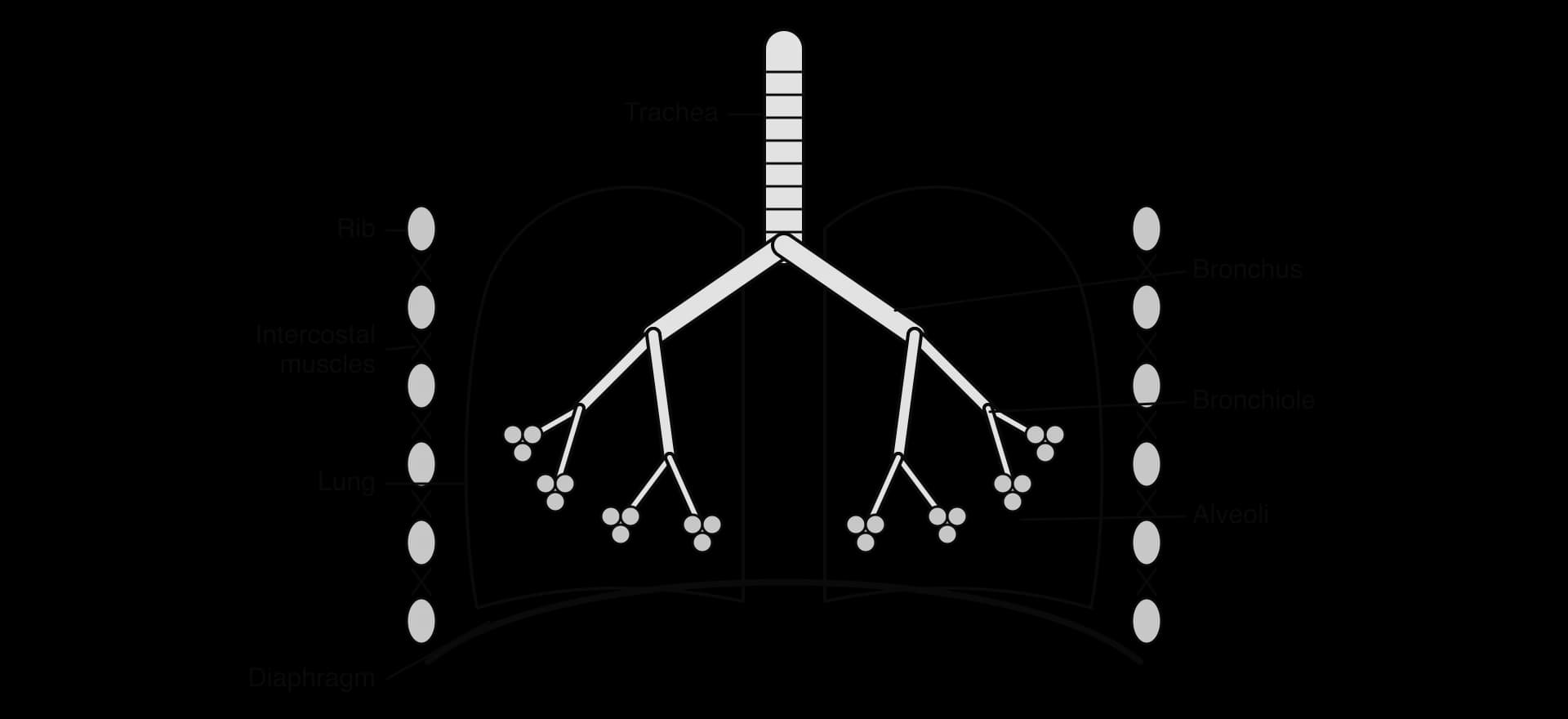
In mammals, air enters through the nose and mouth and passes down the trachea, which divides into two bronchi, one to each lung. Each bronchus divides again and again into narrower tubes called bronchioles. The smallest bronchioles end in clusters of tiny air sacs, the alveoli (singular alveolus), where gas exchange takes place.
The lungs show four key adaptations for gas exchange:
A branched network of bronchioles. Repeated branching delivers air to every part of each lung and divides the lung into a very large number of alveoli. Rings or plates of cartilage keep the trachea and bronchi open.
A high surface area. An adult human lung contains several hundred million alveoli. Their total surface area is of the order of 70 m², many times the area of the skin.
Extensive capillary beds. Each alveolus is covered by a dense network of capillaries, so blood flows close to almost every part of the alveolar wall. The capillaries are narrow, so red blood cells pass in single file and are pressed against the capillary wall, close to the air.
Surfactant. The inside of each alveolus is lined with a thin film of water, needed for gases to dissolve. Water has a high surface tension, which would pull the walls of small alveoli together so that they collapsed. Cells in the alveolar wall secrete surfactant, a mixture of phospholipids and proteins that spreads over the water and reduces surface tension, so the alveoli stay open and are easy to inflate.
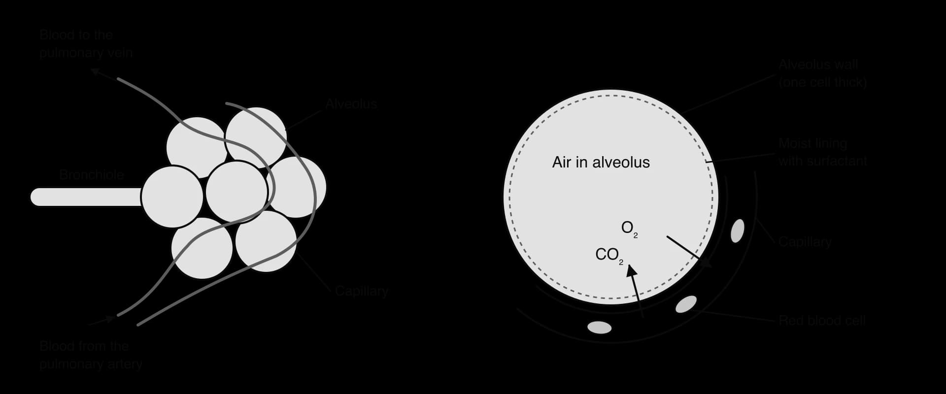
Blood arriving at the lungs in the pulmonary artery is low in oxygen and high in carbon dioxide, because it has come from the respiring tissues. The air in the alveoli is high in oxygen and low in carbon dioxide, because ventilation keeps replacing it. Oxygen therefore diffuses from the air into the blood, and carbon dioxide diffuses from the blood into the air. The blood leaving in the pulmonary vein is almost fully loaded with oxygen.
The degree to which haemoglobin is loaded with oxygen can be measured with a pulse oximeter, which shines red and infrared light through a fingertip. In healthy people the oxygen saturation of arterial blood is normally about 95–100%. Pulse oximeters were widely used to monitor patients with COVID-19, whose damaged alveoli could not load the blood fully.
The lungs have no muscle of their own. They are inflated and deflated by changes in the volume of the thorax (chest cavity), which are produced by muscles. The lungs follow these changes because they are sealed inside the thorax by the pleural membranes.
Air flows because of differences in pressure. Boyle's law states that, at a constant temperature, the pressure of a fixed amount of gas is inversely proportional to its volume. If the volume of a container of gas increases, the pressure inside falls, and if the volume decreases, the pressure rises. Air always flows from a region of higher pressure to a region of lower pressure.
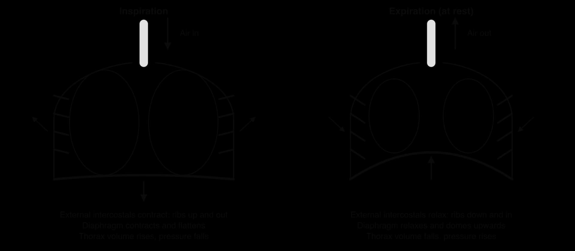
During inspiration (breathing in), two sets of muscles contract:
The diaphragm, a sheet of muscle below the lungs, contracts. It flattens and moves downwards.
The external intercostal muscles, between the ribs, contract and pull the ribs upwards and outwards.
Both movements increase the volume of the thorax, and the lungs expand with it.
By Boyle's law, the pressure inside the lungs falls below atmospheric pressure.
Air flows into the lungs down this pressure gradient until the pressures are equal.
During quiet breathing at rest, expiration (breathing out) is mostly passive. The diaphragm and external intercostal muscles relax. The diaphragm is pushed back up into a dome by the organs of the abdomen, the ribs move down and in under their own weight, and the stretched elastic tissue of the lungs recoils. The volume of the thorax decreases, the pressure in the lungs rises above atmospheric pressure, and air flows out.
In forced expiration, during exercise or when coughing, muscles are used to breathe out as well. The internal intercostal muscles contract and pull the ribs downwards and inwards. The muscles of the abdominal wall contract, pushing the abdominal organs and the diaphragm upwards. The thorax becomes smaller faster and further than in passive expiration, so more air is expelled.
Inspiration | Forced expiration | |
|---|---|---|
Diaphragm | Contracts; flattens and moves down | Relaxes; pushed up by abdominal organs |
External intercostal muscles | Contract; ribs move up and out | Relax |
Internal intercostal muscles | Relax | Contract; ribs move down and in |
Abdominal muscles | Relax | Contract; push diaphragm up |
Volume of thorax | Increases | Decreases |
Pressure in lungs | Falls below atmospheric | Rises above atmospheric |
Air movement | In | Out |
The muscles of ventilation work in antagonistic pairs: muscles can only pull when they contract, so a different muscle is needed to move a structure back. The external and internal intercostal muscles are an antagonistic pair acting on the ribs; the diaphragm and the abdominal muscles act in opposition on the floor of the thorax.
 YouTube
The volume of air moved in and out of the lungs can be measured with a spirometer, a device that records volume as a person breathes through a mouthpiece, or more simply by breathing out into an upturned, water-filled container (displacing the water) or into a balloon. Electronic spirometers draw a trace of volume against time.
Volume | Meaning | Typical adult value |
|---|---|---|
Tidal volume | Volume of air breathed in or out in one normal breath at rest | About 0.5 dm³ |
Inspiratory reserve volume | Extra volume that can be breathed in with maximum effort after a normal inspiration | About 3 dm³ |
Expiratory reserve volume | Extra volume that can be breathed out with maximum effort after a normal expiration | About 1.2 dm³ |
Vital capacity | Maximum volume that can be breathed out after a maximum inspiration; tidal volume + inspiratory reserve + expiratory reserve | About 4–5 dm³ |
Residual volume | Volume that remains in the lungs even after maximum expiration | About 1.2 dm³ |
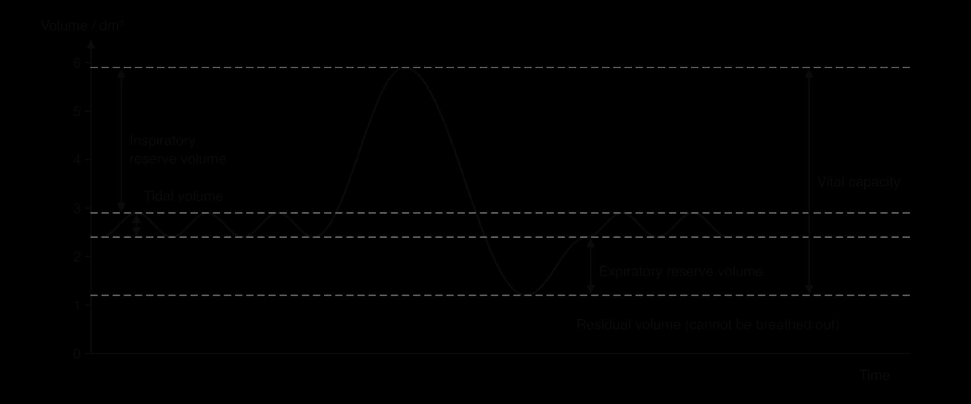
The ventilation rate is the number of breaths per minute, about 12–20 at rest in an adult. Multiplying tidal volume by ventilation rate gives the volume of air moved into the lungs per minute (minute ventilation). During exercise both tidal volume and ventilation rate increase, because respiring muscles need more oxygen and produce more carbon dioxide.
In a plant, the leaf is the main organ of gas exchange. During the day, photosynthesis uses carbon dioxide and releases oxygen faster than respiration does the reverse, so a leaf in light takes in CO₂ and gives out O₂. The leaf has no ventilation system: gases move entirely by diffusion through pores and air spaces. Its structure provides a large, moist surface inside the leaf, while limiting water loss from the outside.
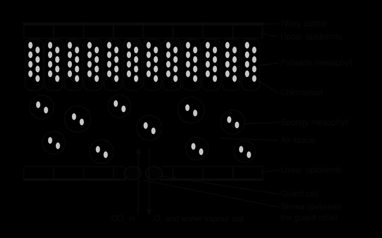
Waxy cuticle. The waxy cuticle, a layer of wax secreted by the epidermis, covers the leaf surface, especially the upper surface. It is almost impermeable to water and gases, so it reduces water loss by evaporation.
Epidermis. The epidermis is a single layer of tightly packed, transparent cells, which protects the leaf and allows light to pass through to the cells below.
Stomata and guard cells. Stomata are pores, mostly in the lower epidermis, through which gases enter and leave. Each stoma lies between two guard cells, which can change shape to open or close it, and so control gas exchange and water loss.
Air spaces. The spongy mesophyll contains large air spaces connected to the stomata, so CO₂ can diffuse quickly to all the mesophyll cells.
Spongy mesophyll. Its loosely packed, rounded cells have moist cell walls with a very large total surface area exposed to the air spaces. This is the actual gas exchange surface of the leaf: CO₂ dissolves in the water on the cell walls and diffuses into the cells.
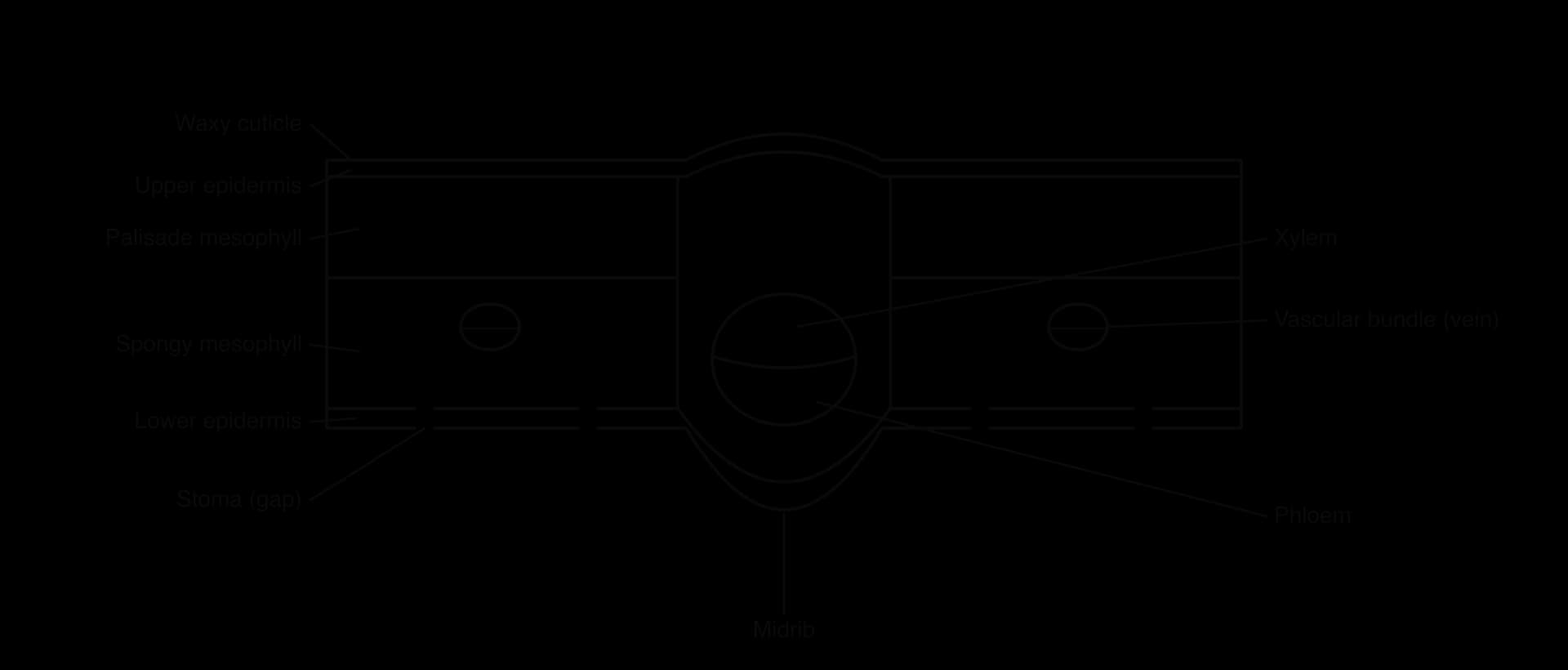
Veins. Each vein (vascular bundle) contains xylem, which brings water to keep the mesophyll cells moist and replace water lost by transpiration, and phloem, which carries away the sugars made in photosynthesis.
The palisade mesophyll, just below the upper epidermis, is the main site of photosynthesis. Its tall, closely packed cells contain many chloroplasts and receive most of the light. It uses CO₂ that diffuses up from the air spaces of the spongy layer.
A plan diagram shows the distribution of tissues in an organ, as outlines only, without drawing individual cells. A plan diagram of a transverse section (TS) through the leaf of a dicotyledonous plant (one with two seed leaves, net-veined leaves and vascular bundles in a ring in the stem) shows the layers from top to bottom and the veins.
For CO₂ to diffuse into the leaf, the stomata must be open and the surfaces of the mesophyll cells must be moist. Water therefore evaporates from the cell walls into the air spaces, and the water vapour diffuses out through the open stomata into the drier air outside. This loss of water vapour from the leaves is transpiration.
Transpiration is an unavoidable consequence of gas exchange: any opening that allows CO₂ in also allows water vapour out. Stomata open to take in CO₂ for photosynthesis, not to lose water. The water lost is replaced by water drawn up through the xylem from the roots (B3.2 Transport), so transpiration also moves water and dissolved minerals through the plant and cools the leaves.
Factor | Effect of an increase | Explanation |
|---|---|---|
Temperature | Rate increases | Water evaporates faster from cell walls and vapour molecules diffuse faster; warm air can also hold more water vapour, so it is relatively drier |
Humidity | Rate decreases | Moist air outside reduces the concentration gradient of water vapour between the air spaces and the air |
Wind speed | Rate increases | Moving air carries away the humid layer of air next to the leaf, keeping the gradient steep |
Light intensity | Rate increases, then levels off | Stomata open in light for photosynthesis; once they are fully open, more light has little further effect |
Plants control the opening of stomata through the day. In most plants the stomata open in the morning, when light allows photosynthesis, and close at night, when no CO₂ is needed for photosynthesis and keeping them open would only lose water. When water is scarce, a plant hormone (abscisic acid) causes the guard cells to close the stomata even in daylight. The plant then saves water but takes in less CO₂, so photosynthesis slows. The opening of stomata is therefore always a compromise between taking in carbon dioxide and losing water.
Stomatal density is the number of stomata per unit area of leaf surface, usually given per mm². It can be found from a micrograph of the leaf surface, or from a leaf cast. To make a cast, a thin layer of clear nail varnish is painted on the leaf surface, left to dry, and then peeled off with clear sticky tape and stuck on a microscope slide. The cast is an imprint of the epidermis, with the outline of each stoma.
Find the diameter of the field of view at the magnification used (with a stage micrometer or from the microscope's specification).
Calculate the area of the field of view: .
Count the stomata in the field of view. Count stomata on the edge on two sides only, so that none is counted twice.
Repeat the count in several different fields of view, on different parts of the leaf and on different leaves, and calculate the mean.
Divide the mean number of stomata by the area of the field of view.
Counts vary from one field of view to the next, because stomata are not spread evenly over a leaf, and they vary between leaves of the same plant. A single count could therefore be unusually high or low. Repeating the measurement and calculating a mean gives a value that is more representative of the leaf, and the spread of the repeated counts shows how variable the data are. Repeated measurements improve the reliability of a result: a reliable result is one that would be obtained again if the investigation were repeated.
Comparisons can be made between the upper and lower surfaces of a leaf, between species from different habitats, or between leaves grown in different conditions. In most dicotyledons the lower surface has many more stomata than the upper surface, which is exposed to direct sunlight and would lose more water.
Herbarium collections provide a record of how leaves have changed as the concentration of carbon dioxide in the atmosphere has risen. Herbaria hold dried, labelled plant specimens, some collected more than two centuries ago. In a study published in 1987, stomata were counted on leaves of eight species of temperate forest trees collected over about 200 years, from the time before the Industrial Revolution to the 1980s.
Over that period, atmospheric CO₂ rose by about 60 parts per million, from about 280 to about 340 ppm. Stomatal density in the specimens fell by about 40%. To test whether CO₂ itself could cause the change, plants of some of the same species were grown in growth chambers at CO₂ concentrations from 225 to 340 ppm. Plants grown at higher CO₂ developed leaves with fewer stomata, reproducing the trend seen in the herbarium leaves.
Measure | Value |
|---|---|
Species studied | Eight species of temperate forest trees |
Period covered by herbarium specimens | About 200 years |
Rise in atmospheric CO₂ over the period | About 60 ppm (about 280 to 340 ppm) |
Change in stomatal density | A fall of about 40% |
Growth-chamber test | CO₂ from 225 to 340 ppm; fewer stomata at higher CO₂ in some of the same species |
Source: Woodward, Nature 327:617, 1987.
The result is explained by the compromise between gas exchange and water loss. When CO₂ is more concentrated in the air, it diffuses into the leaf faster through each stoma, so fewer stomata are needed for the same rate of photosynthesis. Fewer stomata also mean less transpiration, so a leaf with lower stomatal density saves water. The finding showed that plants respond to the rise in CO₂ caused by human activity, and stomatal density in fossil leaves is now used to estimate the CO₂ concentration of past atmospheres. The two lines of evidence, a correlation in historical specimens and an experiment in controlled conditions, together make a much stronger case than either alone.
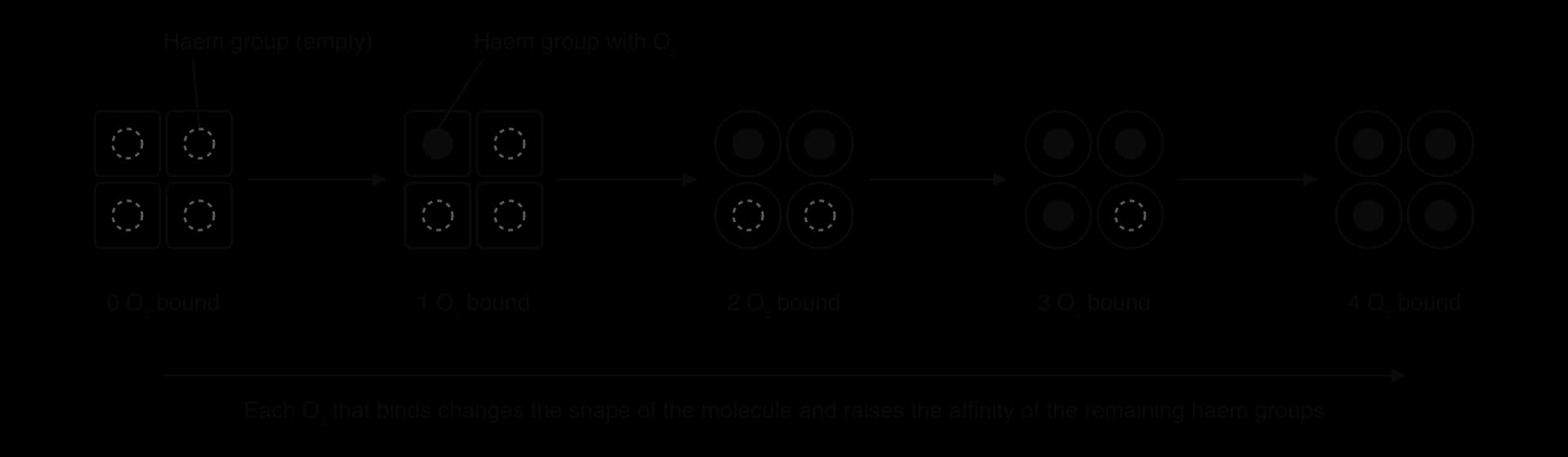
Haemoglobin is the oxygen-carrying protein in red blood cells. Each molecule has four polypeptide chains (two α and two β chains in adult haemoglobin), and each chain holds a haem group with an iron ion (Fe²⁺) at its centre. Each haem group can bind one molecule of O₂, so one haemoglobin molecule carries up to four O₂ molecules.
The four haem groups do not bind oxygen independently. When one O₂ binds to a haem group, the polypeptide around it changes shape, and this conformational change is passed to the other chains. The change increases the affinity of the remaining haem groups for oxygen, so the second O₂ binds more easily than the first, and the third and fourth more easily still. This is cooperative binding. It also works in reverse: when one O₂ is released, the others are released more easily.
Carbon dioxide also binds to haemoglobin, but not at the haem groups. It binds to amino groups on the polypeptide chains, at sites away from the oxygen-binding sites, which are called allosteric sites. Binding of CO₂ changes the conformation of haemoglobin so that its affinity for oxygen falls, and oxygen is released. Hydrogen ions, produced when CO₂ dissolves in the blood plasma and forms carbonic acid, have the same effect.
This allosteric effect matches supply to demand. In actively respiring tissues, where CO₂ concentration is high, haemoglobin binds CO₂ and releases more of its oxygen. In the lungs, where CO₂ concentration is low, CO₂ leaves haemoglobin and its affinity for oxygen rises again, so it loads oxygen.
Carbon monoxide shows what happens when a molecule competes for the haem group itself. It binds to haem about 200–250 times more strongly than oxygen, so even a low concentration in the air occupies many haem groups and reduces the amount of oxygen the blood can carry. It also increases the affinity of the remaining haem groups, so less oxygen is released in the tissues.
A fetus gets its oxygen from its mother's blood at the placenta. The two bloodstreams do not mix: oxygen must diffuse from the mother's haemoglobin, across the placenta, to the fetus's haemoglobin. This can happen only if the fetal haemoglobin binds oxygen more strongly than the mother's haemoglobin at the same oxygen concentration.
Fetal haemoglobin (HbF) has two γ (gamma) chains in place of the two β chains of adult haemoglobin. This changes its structure so that it binds a molecule in the red blood cells that lowers oxygen affinity (2,3-bisphosphoglycerate) less strongly. Fetal haemoglobin therefore has a higher affinity for oxygen than adult haemoglobin. At the placenta, where the oxygen concentration is fairly low, maternal haemoglobin releases oxygen and fetal haemoglobin takes it up. After birth, production switches to adult haemoglobin over the first months of life.
Fetal haemoglobin has become a target in medicine. In sickle cell disease, faulty β chains make adult haemoglobin form long fibres when it releases oxygen. Casgevy, a gene-editing treatment approved in the UK and the USA at the end of 2023, switches fetal haemoglobin production back on in a patient's own blood stem cells, so that their red blood cells contain working HbF (D1.3 Mutations and gene editing).
The affinity of haemoglobin for oxygen is shown by an oxygen dissociation curve. It plots the percentage saturation of haemoglobin with oxygen (how many of its haem groups carry O₂) against the partial pressure of oxygen (pO₂), which measures the concentration of oxygen in the surrounding blood plasma.
The curve is sigmoid (S-shaped) because of cooperative binding:
At low pO₂ the curve is shallow: binding the first O₂ is difficult, because haemoglobin with no oxygen has a low affinity.
In the middle range the curve is steep: once one O₂ has bound, the affinity of the other haem groups rises, so a small increase in pO₂ causes a large increase in saturation.
At high pO₂ the curve levels off, because nearly all the haem groups are occupied and the haemoglobin is almost saturated.
This shape suits the function of haemoglobin. In the alveoli, pO₂ is about 13 kPa, on the flat top of the curve, so haemoglobin becomes almost fully saturated, even if pO₂ falls a little. In respiring tissues pO₂ is about 5 kPa at rest and much lower in active muscle, on the steep part of the curve. A small fall in pO₂ there makes haemoglobin release a large amount of oxygen, exactly where it is needed.
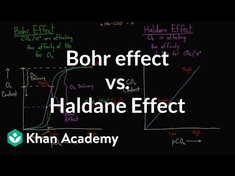
When the concentration of carbon dioxide rises, the whole dissociation curve moves to the right. This is the Bohr shift. At any given pO₂, haemoglobin is less saturated, because CO₂ (binding allosterically) and H⁺ ions lower its affinity for oxygen. Oxygen therefore dissociates from haemoglobin more readily.
The Bohr shift benefits actively respiring tissues. Muscle that is working hard produces a lot of CO₂, so the blood passing through it has a high CO₂ concentration. Haemoglobin there gives up more of its oxygen than it would at the same pO₂ in resting tissue: on the graph of the Bohr shift, at 4 kPa saturation is much lower on the high-CO₂ curve. In the lungs, CO₂ diffuses out of the blood, the curve moves back to the left, and haemoglobin loads oxygen fully.
 YouTube
Gas exchange is assessed in Paper 1A through questions on the properties of exchange surfaces, ventilation and leaf structure. Paper 1B uses spirometer traces, stomatal counts and data on transpiration rates. Paper 2 asks for explanations of ventilation, plan diagrams of leaves and, at HL, sketches and interpretation of dissociation curves. Common command terms are outline, explain, draw, determine, calculate and compare and contrast.
I can explain why gas exchange is harder for larger organisms.
I can state the four properties of gas exchange surfaces and explain each.
I can explain how blood flow and ventilation maintain concentration gradients in lungs and gills.
I can describe the adaptations of mammalian lungs, including surfactant.
I can explain inspiration and expiration using muscle action, volume and pressure.
I can determine lung volumes and ventilation rate from a spirometer trace.
I can describe the adaptations of a leaf for gas exchange and draw a plan diagram of a dicotyledonous leaf.
I can explain transpiration as a consequence of gas exchange and the effects of temperature, humidity, wind and light.
I can calculate stomatal density and explain why repeated counts are needed.
I can explain cooperative binding, allosteric binding of CO₂ and the advantage of fetal haemoglobin. (HL)
I can sketch and interpret oxygen dissociation curves, including the Bohr shift. (HL)
Describe the properties of gas-exchange surfaces.
4 marks
a. permeable to oxygen and carbon dioxide;
b. thin (tissue layer) / short diffusion distance;
c. moist, so gases can dissolve before diffusing across;
d. large surface area (relative to the volume of the organism);
e. dense network of blood vessels / good blood supply;
f. ventilated with air or water, which maintains concentration gradients;
[4 max]
Outline how the hydrophobic properties of lipids are used by organisms.
4 marks
a. lipids are non-polar / hydrophobic, so they are insoluble in water;
b. waxy cuticle on leaves reduces water loss;
c. oils / waxes on feathers or fur make them waterproof;
d. hydrophobic tails of phospholipids form a barrier in membranes;
e. fat / blubber forms an insulating layer;
f. fats can be stored without affecting the water potential of cells / without drawing in water by osmosis;
[4 max]
One additional mark is available for the construction of the answer.
Animals use energy released by cell respiration to move.
Explain how ATP is produced by chemiosmosis in mitochondria.
7 marks
Describe how antagonistic muscles move the ribcage during ventilation of the lungs.
4 marks
Suggest reasons why some animals undertake long-distance migrations despite the large amounts of energy used.
4 marks
Organisms show adaptations to the ways in which they live.
Outline the adaptations of mammalian lungs for gas exchange.
4 marks
Compare and contrast exoskeletons and endoskeletons.
7 marks
Outline adaptations for movement in one named motile organism and one named sessile organism.
4 marks