
Explain how DNA is copied semi-conservatively, how PCR and gel electrophoresis amplify and separate DNA, and how DNA profiles are analysed; at HL, leading and lagging strands, the replication enzymes and proofreading.
Each is in the Biology glossary, with every other term in the subject.
A mixture of DNA fragments of known lengths run in one lane of a gel, used as a scale to estimate the sizes of unknown fragments.
An enzyme that joins DNA fragments, such as Okazaki fragments, by forming a phosphodiester bond between the 3′ end of one and the 5′ end of the next.
An enzyme that links free nucleotides into a new DNA strand complementary to a template strand, forming covalent phosphodiester bonds between them.
A prokaryotic enzyme that removes RNA primers from new DNA strands and replaces them with DNA nucleotides.
The main replicating enzyme in prokaryotes: it extends primers by adding nucleotides to the 3′ end of the new strand (5′ to 3′) and proofreads it.
An enzyme that builds short RNA primers complementary to the template, giving DNA polymerase III a 3′ end to extend.
Analysis of an individual’s DNA, usually the lengths of several short tandem repeat loci, to give a pattern of fragments that is almost unique to that individual.
The production of exact copies of a DNA molecule, with base sequences identical to the original, using each existing strand as a template.
DNA replication is the process in which a cell makes an exact copy of its DNA before it divides. Every new cell needs a complete and accurate set of genetic instructions, so replication underlies reproduction, growth and the repair of tissues in every living organism.
The copying process depends on the structure of DNA itself: two antiparallel strands held together by complementary base pairs. The same principles are used in the laboratory, where the polymerase chain reaction copies a chosen piece of DNA millions of times and gel electrophoresis sorts the copies by size. Together these techniques make DNA profiling, disease diagnosis and many other applications possible.
41 exam-style questions on this subtopic, written in the style of the IB papers. The first has its markscheme open. Counted on
Nucleic acids store genetic information and allow it to be copied and expressed.
Draw a labelled diagram of a section of a DNA molecule consisting of two nucleotides on each strand.
4 marks
a. phosphate shown as circle, deoxyribose as pentagon and base as rectangle, with at least two of these labelled;
b. base and phosphate both attached to the sugar of each nucleotide;
c. phosphate of one nucleotide linked to the sugar of the next in each strand / sugar–phosphate backbone labelled;
d. bases of the two strands paired, with A paired with T and C paired with G;
e. hydrogen bonds labelled between paired bases;
f. covalent bonds labelled in the backbone;
g. strands shown antiparallel / sugars on one strand drawn inverted relative to the other;
[4 max]
Do not award d. if U is shown or if A is paired with G or C.
Explain how the base sequence of a gene is used to produce a polypeptide.
7 marks
a. transcription produces mRNA using a DNA template;
b. RNA polymerase separates the DNA strands / breaks hydrogen bonds between bases;
c. RNA nucleotides pair with complementary bases on the template strand / A with U, T with A, G with C and C with G;
One of 1 short multiple-choice questions on D1.1. Pick an answer to see why it's right or wrong.
DNA replication is semi-conservative. What does that mean?
The official deck for this part of the syllabus.
Define DNA replication.
When does DNA replication take place in a eukaryotic cell?
With a free account, Augment remembers where you stopped and what you have practiced.
A technique that separates DNA fragments (or proteins) by size by moving them through a gel in an electric field; smaller DNA fragments move further towards the positive electrode.
An enzyme that unwinds the DNA double helix and separates the two strands by breaking the hydrogen bonds between complementary bases.
The new DNA strand built discontinuously, away from the replication fork, as a series of Okazaki fragments that are later joined.
The new DNA strand built continuously in the same direction as the replication fork moves, from a single RNA primer.
A 1958 experiment that grew E. coli on heavy nitrogen (¹⁵N) and then light nitrogen (¹⁴N) and separated their DNA by density, showing that DNA replication is semi-conservative.
A short section of DNA built discontinuously on the lagging strand during replication, each starting from its own RNA primer.
A specific base sequence at which DNA replication begins; prokaryotes usually have one per chromosome, eukaryotes many.
A technique that amplifies a chosen DNA sequence by repeated cycles of denaturation, primer annealing and extension, doubling the number of copies in each cycle.
A short single strand of nucleic acid that pairs with a complementary sequence on a template and provides a 3′ end from which DNA polymerase can start building a new strand.
Checking of a new DNA strand by DNA polymerase III, which removes a nucleotide with a mismatched base from the 3′ end and replaces it with a correctly paired one.
The Y-shaped region where the DNA double helix is being unwound and separated by helicase, so that both strands can act as templates.
Replication in which each new DNA molecule consists of one strand from the original molecule and one newly built strand.
A region of DNA in which a short base sequence (typically 2–6 bases) is repeated many times in a row; the number of repeats varies between individuals.
A heat-stable DNA polymerase from the thermophilic bacterium Thermus aquaticus, used in PCR because it is not denatured at about 95 °C.
An organism that lives and grows best at high temperatures, typically above 45 °C, such as Thermus aquaticus in hot springs.
A cell carries its genetic information as a sequence of bases in DNA. When a cell divides, each of the two new cells must receive a full copy of that sequence. DNA replication therefore takes place before every cell division, and in eukaryotes it happens during interphase, before mitosis begins.
Replication is needed for three purposes:
Reproduction. A single-celled organism such as Escherichia coli reproduces by dividing in two, so it must copy its whole chromosome first. In sexually reproducing organisms, DNA is replicated before meiosis so that gametes can be formed.
Growth. A multicellular organism grows by producing more cells by mitosis. A human develops from a single zygote into an adult with tens of trillions of cells, and the DNA is replicated before each of the divisions on the way.
Tissue replacement and repair. Cells that wear out or are damaged are replaced by the division of other cells. The lining of the small intestine, the outer layer of the skin and the blood cells made in bone marrow are replaced continually throughout life.
In each case the copies must be identical to the original. A change to the base sequence during copying is a mutation, and it would be passed on to every cell descended from the one in which it happened. Replication is therefore a basis of continuity between generations of cells and of organisms, one of the central themes of biology.
A DNA molecule is a double helix of two strands of nucleotides. The two strands are held together by hydrogen bonds between the bases, and the pairing is always the same: adenine (A) pairs with thymine (T), and cytosine (C) pairs with guanine (G). This rule is called complementary base pairing. Because of it, the base sequence of one strand fixes the base sequence of the other. If one strand reads ATGCCA, the strand opposite it must read TACGGT.
During replication the two strands separate, and each one acts as a template for a new strand. Free nucleotides pair with the exposed bases on each template according to the base-pairing rules, and they are then linked together to form a new strand. The result is two DNA molecules, each identical in base sequence to the original.
The word "conservative" means kept unchanged, and "semi" means half. In semi-conservative replication half of each original molecule (one strand) is kept intact in each daughter molecule. The diagram of a parent molecule and its two daughters shows this pattern.
The two features work together to make copying very reliable. Complementary base pairing means there is only one correct base to pair with each base on the template, so the template strand specifies the sequence of the new strand exactly. Semi-conservative replication means that every new molecule keeps an original strand. If an error were made in a new strand, the original strand opposite it would still carry the correct sequence, which allows the error to be detected and corrected.
Hydrogen bonding contributes to accuracy too. A correctly paired A–T forms two hydrogen bonds and a C–G pair forms three, and the shapes of correctly paired bases fit together with a constant width across the helix. An incorrect base fits poorly and is much less likely to be held in place long enough to be linked into the new strand.
Replication is carried out by enzymes. Two of them have the central roles: helicase, which separates the strands, and DNA polymerase, which builds the new strands.
A template can only be read once its bases are exposed. Helicase is an enzyme that moves along the DNA, unwinding the double helix and separating the two strands by breaking the hydrogen bonds between complementary bases. The point where the double helix opens into two single strands is called the replication fork. As helicase moves along, the fork moves with it, exposing more template.
Helicase uses energy from ATP to break the hydrogen bonds. It does not break the covalent bonds within each strand: the sugar–phosphate backbone of each strand stays intact, so the base sequence of the template is unchanged.
DNA polymerase is the enzyme that links free nucleotides together to form a new strand, using the existing strand as a template. Free nucleotides in the nucleoplasm (or in the cytoplasm of a prokaryote) move by random motion. When one with the correct complementary base meets an exposed base on the template, it forms hydrogen bonds with it. DNA polymerase then joins this nucleotide to the end of the growing new strand by a covalent phosphodiester bond, in a condensation reaction. A separate DNA polymerase works on each of the two template strands.
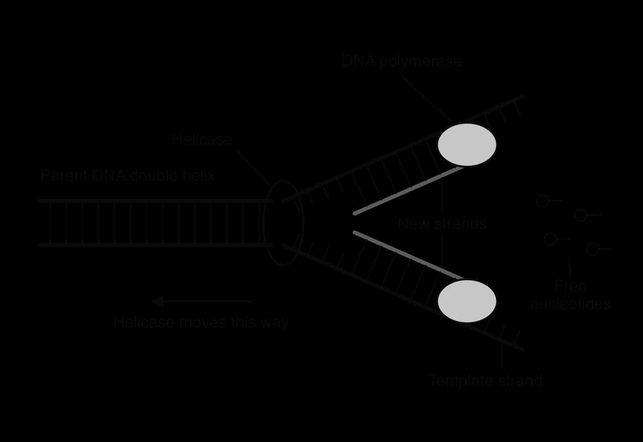
The steps of replication in order are:
Helicase unwinds the double helix and breaks the hydrogen bonds between the bases, separating the two strands.
Each single strand acts as a template, with its bases exposed.
Free nucleotides pair with the exposed bases by complementary base pairing (A with T, C with G), forming hydrogen bonds.
DNA polymerase links each new nucleotide to the growing strand with a covalent phosphodiester bond.
Each new DNA molecule winds into a double helix, consisting of one original strand and one new strand.
 YouTube
The double helix model proposed by Watson and Crick in 1953 suggested that each strand could act as a template. That idea was a hypothesis, and three models of copying were possible:
Conservative replication: the original double helix stays intact and a completely new double helix is built alongside it.
Semi-conservative replication: the strands separate, and each daughter molecule has one original strand and one new strand.
Dispersive replication: each strand of each daughter molecule is a patchwork of original and newly made DNA.
Each model makes a different prediction about how the original DNA is shared out among the molecules of later generations. The diagram of the three models shows the DNA molecules that each one predicts after one and two rounds of replication.
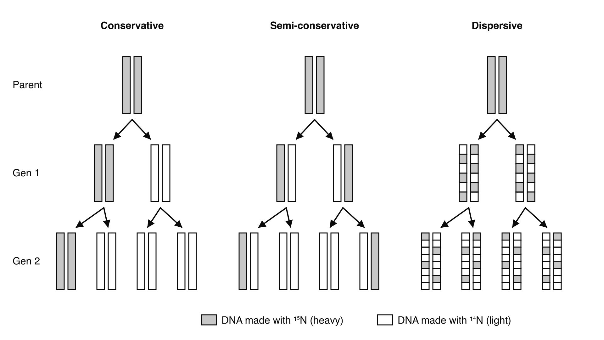
The Meselson–Stahl experiment, carried out by Matthew Meselson and Franklin Stahl in 1958, tested the models with E. coli. Nitrogen is part of every DNA base, and it has two stable isotopes: the common ¹⁴N and the heavier ¹⁵N. DNA that contains ¹⁵N is slightly denser than DNA that contains ¹⁴N, and the difference can be detected.
The bacteria were grown for 14 generations in a medium whose only source of nitrogen was ¹⁵NH₄Cl, so that almost all their DNA contained ¹⁵N ("heavy" DNA).
The bacteria were then transferred to a medium containing only ¹⁴N, so that all new DNA strands made from then on contained the lighter isotope.
Samples were taken at intervals that matched the generation time of the bacteria. DNA was extracted from each sample.
The DNA was mixed with a solution of caesium chloride and spun at very high speed in an ultracentrifuge. The caesium chloride formed a gradient of density down the tube, and each DNA molecule settled at the level that matched its own density. Heavy DNA formed a band lower in the tube than light DNA.

Before transfer (generation 0), all the DNA formed a single heavy band.
After one generation in ¹⁴N, all the DNA formed a single band of intermediate (hybrid) density, between the heavy and light positions.
After two generations, there were two bands of equal intensity: one hybrid and one light.
The single hybrid band after one generation rules out conservative replication, which predicts one heavy band and one light band. Each molecule must contain both ¹⁵N and ¹⁴N. Both semi-conservative and dispersive replication predict a hybrid band at this stage, so the first generation alone cannot separate them.
The results of the second generation distinguish between them. Semi-conservative replication predicts that the two heavy strands stay intact, each paired with a new light strand, while the new light strands pair with more new light strands: half the molecules hybrid, half light. Dispersive replication predicts that every molecule becomes a little lighter each generation, giving a single band moving steadily upwards, never a separate light band. The observed hybrid and light bands matched the semi-conservative prediction only.
Model | Predicted bands after generation 1 | Predicted bands after generation 2 | Matches the results? |
|---|---|---|---|
Conservative | One heavy, one light | One heavy, one light (3 times as much light) | No |
Semi-conservative | One hybrid | One hybrid, one light (equal amounts) | Yes |
Dispersive | One hybrid | One band between hybrid and light | No |
The experiment was designed so that the competing hypotheses made different, testable predictions. Two of the three models were falsified by the results, while the semi-conservative model survived the test. The experiment also depended on technology: without the ultracentrifuge and density-gradient method, the tiny difference in density between ¹⁴N and ¹⁵N DNA could not have been measured.
Many techniques in molecular biology need a large amount of one particular piece of DNA, but a sample from a crime scene, a patient or a fossil may contain only a few molecules. The polymerase chain reaction solves this problem. It uses the principles of replication in a test tube to make millions of copies of a chosen sequence in a few hours.
A PCR reaction is set up in a small tube containing:
the template DNA that contains the target sequence;
free nucleotides carrying the four bases A, T, C and G;
two kinds of primer, one for each strand;
Taq polymerase, a heat-stable DNA polymerase;
a buffer solution that keeps the pH suitable for the enzyme (with magnesium ions that the enzyme needs).
The tube is placed in a thermal cycler (a PCR machine), which changes the temperature quickly and precisely through a programmed cycle.
The two primers are made with base sequences complementary to the two ends of the target sequence, one on each strand. They have two jobs. DNA polymerase cannot start a new strand on its own; it can only add nucleotides to an existing strand, so the primer gives it a starting point. The primers also define which part of the DNA is copied: only the region between the two primers is amplified.
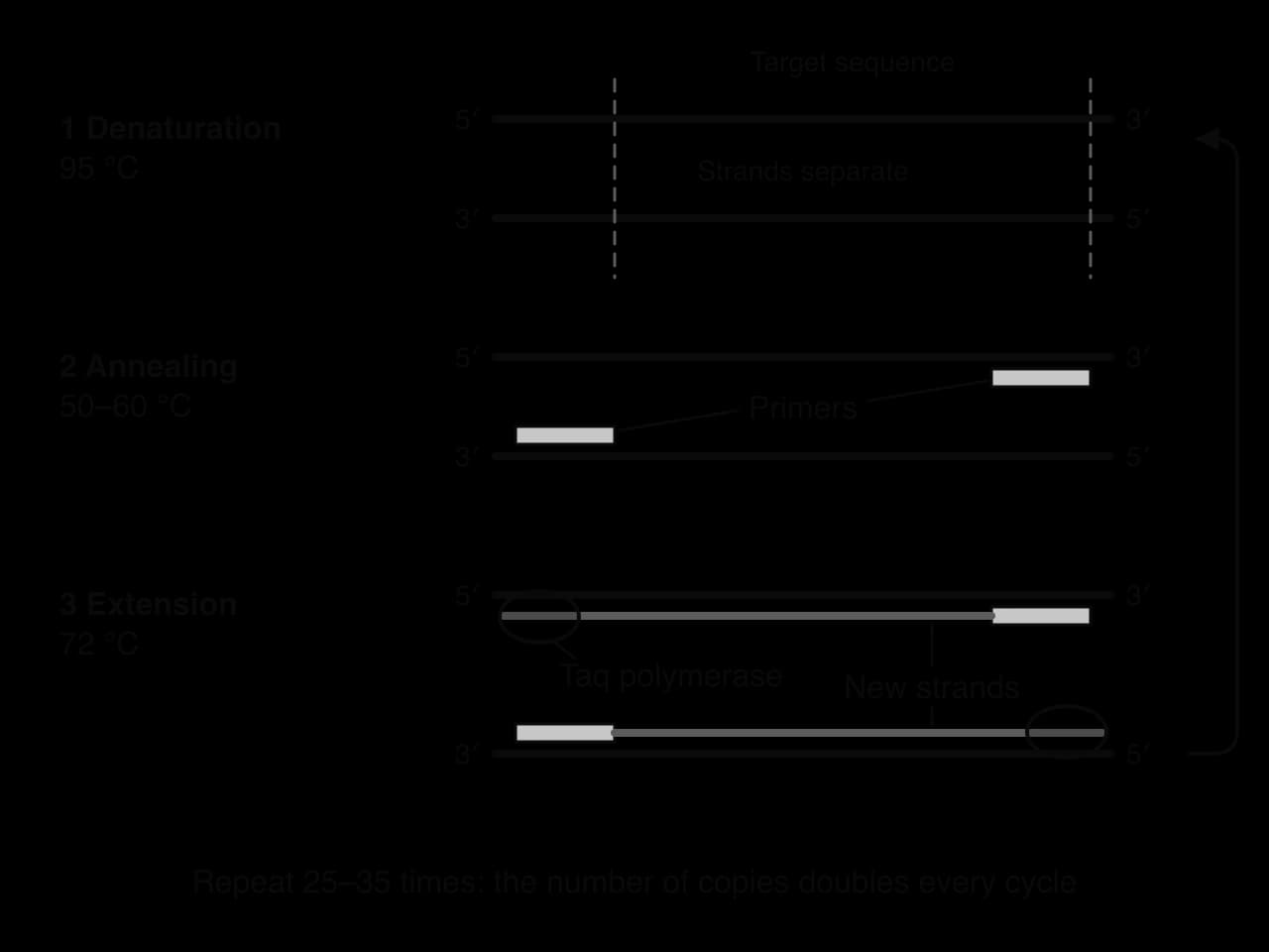
Denaturation (about 95 °C). The high temperature breaks the hydrogen bonds between the bases, so the double-stranded DNA separates into single strands. This does the job that helicase does in a cell.
Annealing (about 50–60 °C). Cooling allows the primers to form hydrogen bonds with their complementary sequences on the single strands. The primers are present in large excess, so they bind much faster than the long template strands can re-pair with each other.
Extension (about 72 °C). Taq polymerase binds at each primer and adds free nucleotides, building a new strand complementary to the template. 72 °C is close to the optimum temperature of Taq polymerase.
One cycle takes a few minutes. At the end of it, every double-stranded target molecule has become two. The cycle is repeated, typically 25 to 35 times, and the products of each cycle become templates for the next.
An ordinary DNA polymerase, such as the one from E. coli or from humans, is denatured at 95 °C and would be destroyed in the first denaturation step. Early PCR therefore needed fresh enzyme to be added after every cycle. The problem was solved by using Taq polymerase, the DNA polymerase of Thermus aquaticus, a bacterium that lives in hot springs. T. aquaticus was first described in 1969 from a hot spring in Yellowstone National Park, USA, where it grows best at about 70 °C.
Because T. aquaticus is a thermophile, its proteins have structures that keep their shape at temperatures that would denature most enzymes. Taq polymerase survives repeated heating to 95 °C, so one addition at the start lasts through the whole run, and the reaction can be fully automated. Kary Mullis devised PCR in 1983 and shared the 1993 Nobel Prize in Chemistry for it. PCR is now a standard technique in medicine, forensic science and research.
Because the number of target molecules doubles in every cycle, the increase is exponential. Starting from one double-stranded molecule, there are copies after cycles.
The graph of copies against cycle number rises slowly at first and then very steeply. After 10 cycles one molecule has become 1024; after 30 cycles it has become over a billion. In practice the increase slows after many cycles, because primers and nucleotides start to run out.
 YouTube
PCR produces many copies of DNA, but they are invisible in solution. Gel electrophoresis separates DNA fragments according to their size and makes them visible as bands, so that the fragments in different samples can be compared.
Every nucleotide in DNA has a phosphate group, and each phosphate carries a negative charge. DNA is therefore negatively charged and moves towards the positive electrode when placed in an electric field. The amount of charge is proportional to the length of the fragment, so all DNA fragments have about the same charge for their mass, and charge alone cannot separate them.
The gel does the separating. It is a slab of agarose, a polysaccharide from seaweed, which forms a mesh of fibres with small pores. Short fragments slip through the pores easily and move quickly; long fragments are slowed down by the mesh. After a set time, the short fragments have travelled further from the starting point than the long ones.
A gel is cast in a tray with a comb at one end, which leaves a row of small wells when it is removed.
The gel is placed in a tank and covered with a buffer solution that conducts electricity. The wells are at the end with the negative electrode.
The DNA samples, mixed with a dense loading dye, are pipetted into the wells. A DNA ladder is loaded into one well.
A voltage is applied. The DNA moves out of the wells towards the positive electrode. The current is switched off before the fastest fragments reach the end of the gel.
The gel is stained with a dye that binds to DNA and glows under ultraviolet or blue light, showing each group of fragments of the same length as a band.
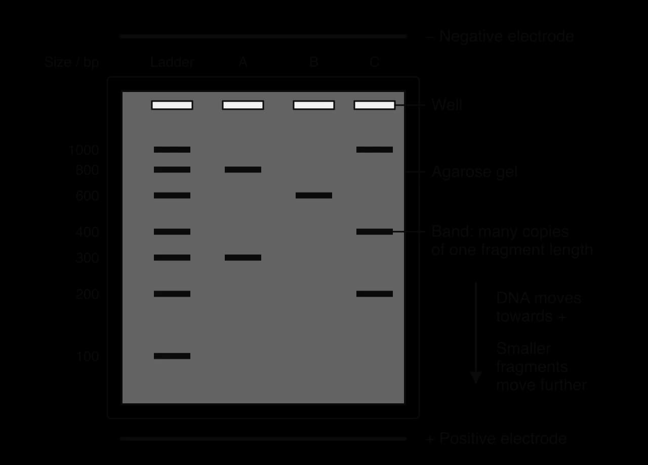
A DNA ladder is a mixture of fragments of known lengths. Its bands act like a ruler: the size of an unknown fragment is estimated by comparing how far its band has moved with the positions of the ladder bands. Sizes are given in base pairs (bp). Each band contains millions of copies of a fragment of one length, which is why PCR is usually done first: a few molecules would not make a visible band.
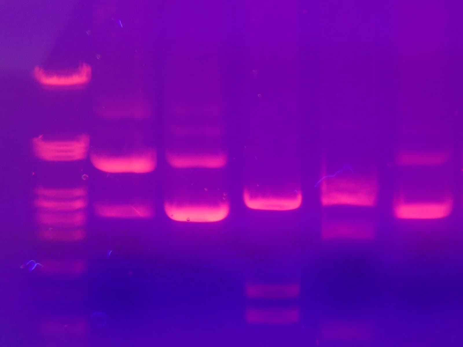
PCR and gel electrophoresis are used together in a very wide range of fields: forensic science, medicine, agriculture, conservation and the study of evolution. The most familiar application is DNA profiling.
Most of the human genome is identical in everyone, but some regions vary a great deal from person to person. The most useful of these are short tandem repeats (STRs): places where a short sequence of bases, such as GATA, is repeated several times one after another. The number of repeats at a given STR locus (position on a chromosome) varies between people. One person might have 8 repeats on the chromosome inherited from the mother and 11 on the one from the father, while another might have 9 and 9.
To make a DNA profile, DNA is extracted from a sample (blood, saliva, hair roots, skin cells). PCR is used to amplify several STR loci, with primers that bind on either side of each repeat region. The lengths of the copied fragments depend on the number of repeats, so gel electrophoresis separates them into a pattern of bands. Modern laboratories label the primers with fluorescent dyes and separate the fragments in fine capillary tubes, but the principle is the same: fragments are amplified by PCR and separated by size.
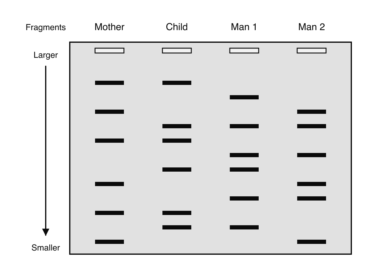
Each person has two copies of each STR locus, one inherited from each parent, so each locus usually gives one or two bands. A child receives half of their bands from the mother and half from the father. Identical twins are the only people with identical profiles, because they develop from the same zygote.
In a paternity test, the profile of a child is compared with the profiles of the mother and of a possible father. Every band in the child must come from one parent or the other. The bands that match the mother are identified first; the remaining bands must have come from the biological father.
In forensic science, a DNA profile from a sample left at a crime scene is compared with the profiles of suspects or with a national database. If every band matches, the suspect is very likely to be the source of the sample. If even one locus does not match, the suspect is excluded. DNA profiling can therefore prove innocence as well as identify a likely source.
Alec Jeffreys discovered DNA fingerprinting at the University of Leicester in 1984. Its first criminal use, in Leicestershire, cleared an innocent suspect who had confessed to one of two murders and then, in 1988, led to the conviction of Colin Pitchfork, the first person convicted using DNA evidence.
A related method can establish relatedness between people who lived centuries apart. In 2012 a skeleton was found under a car park in Leicester on the site of a medieval friary. Its mitochondrial DNA, which is inherited only through the mother, matched that of two living people descended from the sister of King Richard III through an unbroken female line, and the remains were identified as those of the king.
A match at one STR locus is weak evidence, because many people share the same number of repeats at any single locus. Each locus is inherited independently of the others, so the probability that two unrelated people match at every locus is found by multiplying the probabilities for each locus. Using more loci makes a chance match much less likely.
This is an example of a general principle of science: reliability increases with the number of measurements. Forensic laboratories now test many loci; the United States national DNA database has used 20 core STR loci since 2017. With this many loci, the chance that an unrelated person has the same full profile is less than one in a billion.
Diagnosing infectious disease. During the COVID-19 pandemic, from early 2020, RT-PCR was used worldwide to detect the RNA of the SARS-CoV-2 virus in swabs. The viral RNA is first copied into DNA by the enzyme reverse transcriptase, and the DNA is then amplified by PCR, so even a few viral particles can be detected.
Diagnosing genetic disease. PCR can amplify a gene from a small sample of cells and test it for a particular allele, for example to find out whether a person is a carrier of an inherited disorder or to test an embryo.
Detecting genetically modified crops. Primers that match an inserted gene amplify it only if it is present, so PCR shows whether a food or a field crop contains genetically modified material.
Studying evolution and biodiversity. DNA from museum specimens, fossils and environmental samples can be amplified and compared, which helps to classify species and to work out evolutionary relationships.
The carbon atoms of deoxyribose are numbered 1′ to 5′ (read "one prime" to "five prime"). In a strand of DNA, the phosphate of each nucleotide is attached to carbon 5′ of its sugar, and it links to carbon 3′ of the sugar in the next nucleotide. All the nucleotides in a strand face the same way, so a strand has two different ends. This property is called directionality.
The 5′ end has a phosphate group attached to carbon 5′ of the last sugar.
The 3′ end has a hydroxyl (–OH) group on carbon 3′ of the last sugar.
In a double helix the two strands are antiparallel: they run in opposite directions, so the 5′ end of one strand lies opposite the 3′ end of the other.
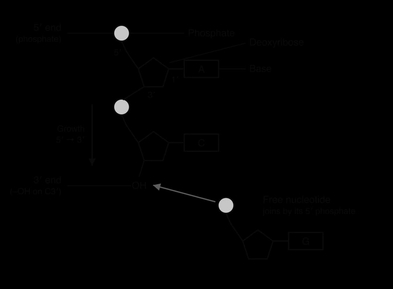
DNA polymerases can only add a nucleotide to the free 3′ –OH group at the end of an existing strand. The free nucleotide is joined by its 5′ phosphate to that 3′ carbon, forming a phosphodiester bond. The strand being built therefore grows only at its 3′ end, which means every new strand is synthesised in the 5′ to 3′ direction.
Because the template and the new strand are antiparallel, DNA polymerase moves along the template strand in its 3′ to 5′ direction while the new strand grows 5′ to 3′. DNA polymerases also need an existing 3′ end to add to, so they cannot start a new strand from nothing. Each new strand therefore begins with a short primer.
Replication begins at specific sequences called origins of replication. The circular chromosome of E. coli has a single origin; the much longer chromosomes of eukaryotes each have many origins, so that the whole genome can be copied within a few hours. At each origin, helicase opens the double helix and two replication forks form, moving away from each other in opposite directions.
At a replication fork the two template strands run in opposite directions, but DNA polymerase can build a new strand only in the 5′ to 3′ direction. The two new strands are therefore built in different ways.
The leading strand is built in the same direction as the fork moves. DNA polymerase can follow the fork as it opens and add nucleotides continuously, so this strand is built in one long piece and needs only one RNA primer, at the origin.
The lagging strand is built in the opposite direction to fork movement, away from the fork. DNA polymerase can only work on template that has already been exposed, so it builds a short section and then has to start again closer to the fork as more template is exposed. This strand is built discontinuously, in short pieces called Okazaki fragments, each starting with its own RNA primer.

Replication in prokaryotes uses a team of enzymes, each with a particular job. Helicase opens the double helix at each fork. The other four enzymes and their functions are:
DNA primase builds short RNA primers, complementary to the template, at the start of each new strand. On the leading strand this happens once; on the lagging strand it happens again for every Okazaki fragment. Primase can start a new strand from nothing, which DNA polymerases cannot.
DNA polymerase III is the main replicating enzyme. It adds DNA nucleotides to the 3′ end of each primer, extending the new strand 5′ to 3′ along the template. It is fast, adding about 1000 nucleotides per second in E. coli, and it also proofreads the new strand.
DNA polymerase I removes the RNA primers and replaces them with DNA nucleotides. It works from the 3′ end of the preceding fragment, removing the ribonucleotides of the primer ahead of it one by one and filling the gap with DNA.
DNA ligase seals the nick left between neighbouring fragments once the primer has been replaced. It forms the final phosphodiester bond between the 3′ end of one fragment and the 5′ end of the next, so the lagging strand becomes one continuous strand.
Enzyme | Function | Leading strand | Lagging strand |
|---|---|---|---|
Helicase | Unwinds DNA; breaks hydrogen bonds between bases | Acts at the fork for both strands | Acts at the fork for both strands |
DNA primase | Makes RNA primers | Once, at the origin | Once for each Okazaki fragment |
DNA polymerase III | Adds nucleotides to the 3′ end of the primer; proofreads | Continuously, towards the fork | In fragments, away from the fork |
DNA polymerase I | Replaces RNA primers with DNA | Once | Once for each fragment |
DNA ligase | Joins fragments by forming phosphodiester bonds | Rarely needed | Joins every pair of fragments |
Eukaryotes use the same principles, with different enzymes. Their linear chromosomes also raise a problem at the very end of the lagging strand, where the last primer cannot be replaced; the repeated telomere sequences at chromosome ends protect genes from the small loss of DNA in each round.
Complementary base pairing makes replication accurate, but not perfect. Occasionally a nucleotide with the wrong base is added to the new strand, for example a G opposite a T. Left uncorrected, this mismatch would become a mutation in one of the daughter molecules after the next round of replication.
Proofreading takes place in four steps:
DNA polymerase III adds a nucleotide to the 3′ end of the new strand.
If the base is mismatched, it does not pair properly with the template, and the distorted shape of the new end is detected by the enzyme.
DNA polymerase III moves back by one nucleotide and removes the incorrect nucleotide from the 3′ end, by hydrolysis of its phosphodiester bond (its 3′ to 5′ exonuclease activity).
The enzyme then adds the correct nucleotide and continues building the strand.
The effect is large. DNA polymerase selecting nucleotides by base pairing alone makes roughly one error in every 10⁵ nucleotides added. Proofreading removes about 99 in every 100 of these errors, and other repair enzymes that act after replication (mismatch repair) correct most of the rest. Taken together, the error rate of replication is about one in 10⁹ to 10¹⁰ nucleotides.
Stage of error correction | Approximate errors per nucleotide copied |
|---|---|
Selection of nucleotides by DNA polymerase (base pairing alone) | 1 in 10⁵ |
After proofreading by DNA polymerase III | 1 in 10⁷ |
After mismatch repair following replication | 1 in 10⁹ to 10¹⁰ |
Source: Alberts et al., Molecular Biology of the Cell, 7th edition, 2022.
Even with this accuracy, a human cell copying about 6 billion base pairs of DNA makes a few errors in every division. These rare uncorrected errors are one of the sources of mutation, and therefore of genetic variation (see D1.3 Mutations and gene editing).
DNA replication is assessed in Paper 1A through questions on the roles of enzymes, the results of the Meselson–Stahl experiment and the steps of PCR. Paper 1B and Paper 2 Section A use gel images and DNA profiles, in which you deduce fragment sizes, paternity or a match to a suspect. Paper 2 Section B often asks for an extended description of replication or of PCR, and at HL an explanation of the differences between the leading and lagging strands. Common command terms are outline, describe, explain, deduce and, for profiles, identify.
The DNA profiles in the question come from a sample of blood found at a crime scene and from three suspects.

I can outline why DNA replication is needed for reproduction, growth and tissue replacement.
I can explain semi-conservative replication and how complementary base pairing makes copying accurate.
I can describe the roles of helicase and DNA polymerase.
I can explain how the Meselson–Stahl results support semi-conservative replication and rule out the other models.
I can outline the three stages of PCR with their temperatures, and explain the roles of primers and Taq polymerase.
I can calculate the number of copies produced by PCR after a given number of cycles.
I can explain how gel electrophoresis separates fragments by size and estimate fragment sizes using a DNA ladder.
I can analyse DNA profiles for paternity and forensic cases, and explain why many loci are used.
I can explain why new DNA strands are built only in the 5′ to 3′ direction. (HL)
I can distinguish leading and lagging strands and state the functions of primase, DNA polymerase III, DNA polymerase I and DNA ligase. (HL)
I can outline proofreading by DNA polymerase III. (HL)
[7 max]
Outline the roles of helicase and DNA polymerase in DNA replication.
4 marks
a. helicase unwinds the double helix;
b. helicase breaks hydrogen bonds between (complementary) bases / separates the two strands;
c. each strand then acts as a template;
d. DNA polymerase adds free nucleotides to form a new strand;
e. nucleotides are added according to complementary base pairing / A with T and C with G;
f. DNA polymerase links nucleotides into a strand by covalent bonds (between sugar and phosphate);
g. the result is two DNA molecules each with one old and one new strand / semi-conservative replication;
[4 max]
One additional mark is available for the construction of the answer.
Genetic information varies between individuals and between species.
Outline what single-nucleotide polymorphisms (SNPs) are and how they arise.
4 marks
Describe how the polymerase chain reaction (PCR) and gel electrophoresis can be used to amplify and separate DNA.
4 marks
Explain the current and potential future uses of whole genome sequencing, and why these uses are becoming more widespread.
7 marks
Which process in a multicellular organism depends on DNA replication?