Explain how internal and external risk factors combine to cause injury, distinguish acute from cumulative trauma, classify injuries to each tissue and, at HL, link overuse injuries to technique and how correcting it lowers risk.
Each is in the SEHS glossary, with every other term in the subject.
A superficial wound in which the outer layers of skin are scraped off by friction against a rough surface, such as a graze from a fall on a road or artificial pitch.
An injury caused by a single, sudden or excessive application of force, or by a force from an unexpected direction, such as a ligament torn in a tackle or a bone broken in a fall.
A ligament inside the knee running from the back of the femur to the front of the tibia, which stops the tibia sliding forward on the femur and limits rotation of the knee.
A faulty movement pattern, often developed through habit, fatigue, weakness or previous injury, that places abnormal or excessive load on a tissue and increases the risk of injury.
A pocket of fluid between layers of skin caused by repeated friction, for example between a shoe and the heel; a cumulative skin injury.
A traumatic brain injury caused by a blow to the head, neck or body that transmits an impulsive force to the brain, producing a temporary disturbance of brain function rather than visible structural damage.
An injury caused by a direct blow, in which small blood vessels are damaged and blood collects in the tissue, causing a bruise, swelling and pain.
Outline the internal and external risk factors for sports injury, with examples.
Explain how internal and external risk factors interact to make an individual susceptible to injury, using a risk factor model.
Explain why female athletes have a higher rate of anterior cruciate ligament injury than male athletes, evaluating the possible causes.
Distinguish between acute trauma and cumulative trauma, using the balance between load and tissue capacity.
Describe injuries to connective tissue, muscle, bone, skin and the brain, including the grading of sprains and strains.
Distinguish between a concussion and other injuries, and outline its signs and symptoms.
Interpret injury data, including incidence per 1,000 hours of exposure.
Explain how faulty technique contributes to common overuse injuries (HL).
Evaluate gait retraining, technique coaching and equipment changes as ways of correcting biomechanical maladaptations (HL).
An injury is damage to the body’s tissues that results when the force applied to a tissue exceeds what it can withstand. In sport, injuries rarely have a single cause: a mix of factors inside the athlete and in the environment makes some individuals far more likely to be hurt than others in the same situation.
Understanding the causes of injury serves both lenses of the course. For health, it explains why a new runner develops shin pain, why an older adult who falls is more likely to break a bone, and how regular physical activity can be kept safe for everyone. For performance, it explains why leading players miss major tournaments, as several did with knee ligament injuries before the 2023 FIFA Women’s World Cup.
With a free account, Augment remembers where you stopped and what you have practiced.
An injury caused by the repeated application of force, each load too small to cause damage alone, so that microscopic damage builds up faster than the tissue can repair it.
An injury in which the articulating surfaces of the bones in a joint are forced completely out of contact, usually stretching or tearing the joint capsule and ligaments.
An inward collapse of the knee during landing, cutting or squatting, combining hip adduction, hip internal rotation and knee abduction, which increases load on the ACL and the kneecap.
A feature of the environment in which an individual exercises or competes that increases the risk of injury, such as equipment, surface, weather, rules, opponents or coaching.
A break or crack in a bone, caused by a force greater than the bone can withstand or by repeated loading; classified as open or closed and by the pattern of the break.
An intervention that changes how a person walks or runs, usually using real-time feedback and practice, to reduce the load on an injured or at-risk tissue.
A layer of cartilage near each end of a long bone in children and adolescents where the bone grows in length; it is weaker than surrounding bone and ligaments until it closes.
The specific action or situation at the moment an injury occurs, such as a landing, tackle or sudden change of direction, which provides the load that exceeds a tissue’s capacity.
Damage to the body’s tissues that occurs when the force applied to a tissue exceeds what it can withstand, causing pain and loss of function.
A characteristic of the individual that increases the risk of injury, such as age, sex, previous injury, fitness, fatigue, anatomy or technique; also called an intrinsic risk factor.
A cut or tear in the skin, often deep and irregular, caused by a sharp object or by a blunt impact that splits the skin.
Pain on the outside of the elbow at the attachment of the wrist extensor muscles, caused by repeated overloading of their common tendon, as in a faulty tennis backhand; also called tennis elbow.
The relationship between the load placed on a tissue and its capacity to tolerate it; injury occurs when load exceeds capacity, suddenly or because repeated loading without recovery lowers capacity.
Pain along the inner border of the lower half of the tibia caused by repeated loading in running and jumping, thought to involve stress reactions in the bone; also called shin splints.
A cause of pain and swelling just below the knee in active adolescents, caused by repeated pulling of the patellar tendon on the growing tibial tuberosity during growth spurts.
An injury caused by cumulative trauma, in which repeated loading of a tissue without enough recovery leads to gradual damage; called a chronic injury when it persists.
Pain around or behind the kneecap, made worse by running, squatting or stairs, caused by excessive load on the joint between the patella and the femur; also called runner’s knee.
Equipment worn by an individual to reduce the risk or severity of injury, such as helmets, mouthguards, shin guards, pads and eye protection.
The angle between the line of pull of the quadriceps and the line of the patellar tendon at the knee; it is larger on average in women because of a wider pelvis.
A hormone produced mainly during pregnancy that increases the laxity of ligaments, especially those of the pelvis, so that the pelvis can widen for childbirth.
Any characteristic of an individual or their environment that increases the probability of injury, without on its own causing it.
An injury to a ligament caused by stretching it beyond its normal range, graded from microscopic tearing of fibres (grade I) to a partial tear (grade II) or complete rupture (grade III).
An injury to a muscle or the junction between muscle and tendon, caused by overstretching or a forceful, often eccentric, contraction; graded from mild (grade I) to complete rupture (grade III).
A small crack in a bone caused by repeated loading, when microscopic damage accumulates faster than the bone can repair it; a cumulative injury common in runners and jumpers.
A partial dislocation, in which the bones of a joint move partly out of their normal position and then return, often spontaneously.
A painful condition of a tendon, usually caused by repeated overloading, in which the tendon’s structure is disorganised and its capacity to tolerate load is reduced.
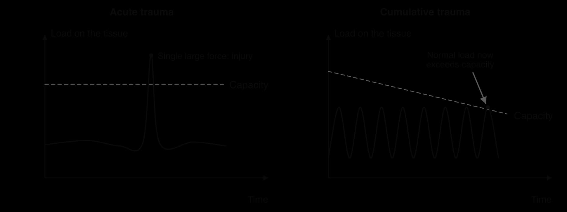
Every tissue in the body (bone, ligament, tendon, muscle, cartilage, skin) can withstand a certain amount of force before it is damaged. The force placed on a tissue is its load; the maximum load it can tolerate without damage is its capacity. Injury happens when load exceeds capacity, either all at once or after repeated loading has worn the capacity down.
Capacity is not fixed. Tissues adapt to the loads placed on them: bone becomes denser, tendons stiffer and stronger, and muscles larger when training is progressive and recovery is adequate. The same tissues lose capacity with inactivity, fatigue, previous injury, poor nutrition or ageing. The load–capacity balance therefore links injury to training principles such as progressive overload and recovery: a sensible programme raises capacity faster than it raises load.
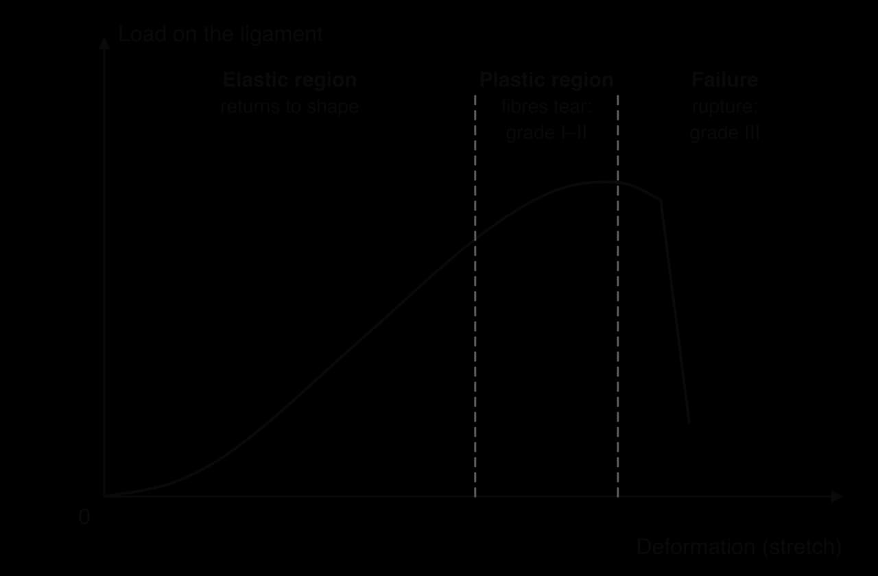
Engineers describe how a material responds to load with a load–deformation (stress–strain) curve, and the same idea applies to a ligament or tendon. At low loads the tissue stretches and springs back to its original length. Beyond a certain point, some of its collagen fibres tear and it no longer returns fully to shape. If the load keeps rising, the tissue fails completely.
A risk factor does not cause an injury on its own; it makes injury more likely. Risk factors are divided into two groups. Internal risk factors are characteristics of the individual. External risk factors are features of the environment in which the individual exercises or competes. Some can be changed (modifiable), such as strength, technique or footwear; others cannot (non-modifiable), such as age, sex or a previous injury.
The distinction matters for prevention. Non-modifiable factors identify who is at greater risk, so that they can be monitored and supported. Modifiable factors are the targets of prevention programmes, protective equipment, rule changes and coaching.
Internal risk factors describe the athlete’s body and its condition. The main ones are age, sex differences, pregnancy, the effects of training, congenital (inborn) factors and previous injury.
Children and adolescents are growing, and growth changes where the body is weakest. At each end of a long bone there is a growth plate, a layer of cartilage where new bone forms. Until growth stops, usually in the late teens, the growth plate is weaker than the surrounding ligaments and tendons. An injury that would sprain a ligament in an adult can therefore fracture the growth plate in a child.
During growth spurts, bones lengthen faster than muscles and tendons, which become relatively tight and pull hard on their attachments. Repeated pulling by the patellar tendon on the front of the tibia causes Osgood–Schlatter disease, pain and swelling just below the knee in active adolescents. In a Finnish study of 389 students, 21% of those who were active in sport at the age of 13 had suffered from it, compared with 5% of those who were not; among athletes treated at a sports clinic, symptoms first appeared at an average age of 13.1 years.
Sources: Kujala, Kvist and Heinonen, American Journal of Sports Medicine, 1985.
At the other end of life, tissue capacity falls. Older adults have lower bone mineral density, especially women after the menopause, so a fall is more likely to cause a fracture. Tendons and ligaments become stiffer and less able to stretch, muscle mass and strength decline, and healing is slower. Balance and reaction time also decrease, which raises the risk of falls. Regular strength and balance training slows all these changes, one of the strongest health arguments for staying active in older age.
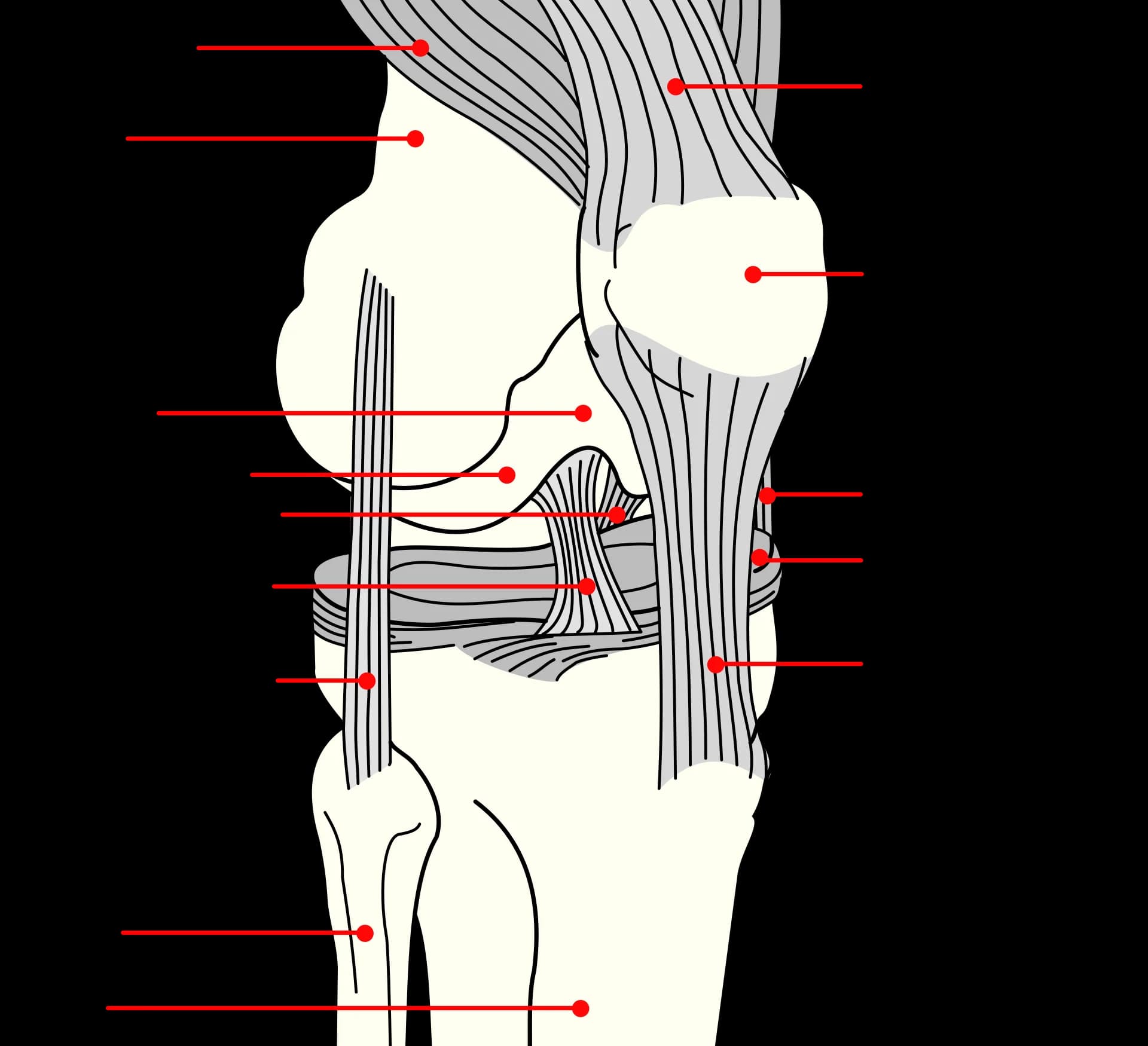
Male and female athletes are injured at different rates and in different ways. The clearest example is injury to the anterior cruciate ligament (ACL), one of the two ligaments that cross inside the knee joint.
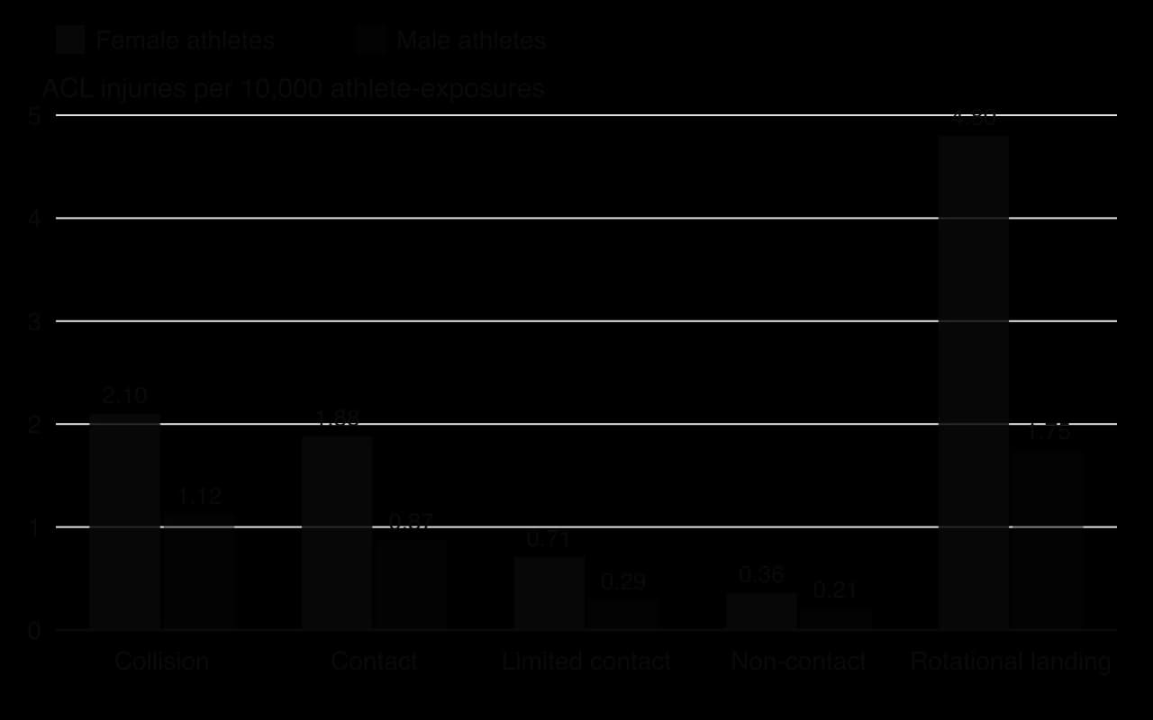
Most ACL injuries happen without contact with another player, during cutting (changing direction), sudden stopping or landing from a jump. A meta-analysis of studies that recorded ACL injuries and exposure by sex found that, in contact sports such as football and basketball, female athletes were injured three times as often as male athletes. Gymnastics and similar sports with high-impact landings had the highest rates for both sexes.
Why the difference exists is still debated, and several explanations probably act together:
Anatomy. Women on average have a wider pelvis relative to leg length, which increases the Q-angle (the angle between the line of pull of the quadriceps and the patellar tendon). A larger Q-angle may increase inward collapse of the knee, although studies linking it directly to ACL injury are inconsistent. The notch in the femur through which the ACL passes, and the ACL itself, also tend to be smaller.
Hormones. The ACL has receptors for oestrogen and relaxin, and some studies suggest that ligament laxity and injury risk vary across the menstrual cycle. The evidence is mixed, and the effect, if real, is probably small compared with other factors.
Neuromuscular control. On average, female athletes land with straighter knees and hips and more inward movement of the knee, and rely more on the quadriceps than the hamstrings to stabilise the joint. These patterns increase strain on the ACL, and unlike anatomy they can be trained.
Environment. Many female athletes have less access to strength and conditioning coaching, play on poorer pitches, wear boots designed for men’s feet and may have congested schedules. These are external factors that are easily mistaken for differences in the body itself.
Pregnancy changes several risk factors at once. The hormone relaxin increases the laxity of the ligaments, especially those of the pelvis, to allow the pelvis to widen for childbirth. As the baby grows, the centre of mass moves forward and upward, changing posture and balance and increasing the curve of the lower back. Body mass rises, increasing the load on joints.
These changes do not mean that pregnant women should avoid exercise. Regular moderate physical activity is recommended during an uncomplicated pregnancy for its health benefits. They do mean that activities with a high risk of falls or abdominal impact, and movements at the extreme of joint range, carry more risk, and that balance becomes less reliable later in pregnancy.
How an athlete trains is one of the most important, and most modifiable, internal risk factors.
Fatigue. A fatigued muscle produces less force and absorbs less energy, and fatigue slows reactions and impairs coordination. Fatigued athletes tend to land with stiffer knees and poorer control of the joint. Fatigue therefore lowers the capacity of tissues and changes technique at the same time.
Training load errors. A sudden increase in training volume or intensity, too little recovery between sessions, or a congested match schedule loads tissues faster than they can adapt. Overtraining syndrome is the extreme form, with persistent fatigue and poor performance.
Poor conditioning. Muscles that are weak, or have low endurance, cannot protect joints. An athlete returning from the off-season with low fitness is at higher risk in pre-season.
Muscle imbalance. When one muscle group is much stronger or tighter than its antagonist, joint movement and loading are altered. Weak hamstrings relative to the quadriceps are linked with hamstring strains and ACL injury; tight hip flexors change pelvic posture.
Congenital factors are features present from birth. A marked difference in leg length changes the loading of the hips, knees and back, and is linked with overuse problems in runners. Flat feet (low arches) or high arches change how the foot absorbs force. Generalised joint hypermobility (unusually loose joints) increases the risk of dislocations and sprains in some sports. These factors cannot be removed, but their effects can often be reduced with strength training, footwear or orthotics.
The strongest single predictor of a future injury is a previous injury. In a study of 12 elite Swedish men’s football teams over two seasons, players injured in the first season were 2.7 times as likely to be injured in the second season as those who had not been injured. Players with a previous hamstring injury, groin injury or knee trauma were two to three times more likely to suffer the same injury again.
Sources: Hägglund, Waldén and Ekstrand, British Journal of Sports Medicine, 2006.
Previous injury raises risk for several reasons. Healed tissue is often weaker and less elastic than the original (scar tissue in a muscle or ligament). Strength, flexibility and proprioceptor feedback from the joint may not have fully returned. The athlete may move differently to protect the area, overloading other tissues. And an athlete who returns too early, before the tissue’s capacity has recovered, is exposed to normal training loads with a reduced capacity.
Internal risk factor | How it raises risk | Modifiable? | Example |
|---|---|---|---|
Age (young) | Growth plates weaker than ligaments; tight muscle–tendon units during growth spurts | No | Osgood–Schlatter disease in a 13-year-old footballer |
Age (older) | Lower bone density, stiffer tendons, weaker muscles, slower healing, poorer balance | Partly (training) | Wrist fracture after a fall in an older runner |
Sex differences | Anatomy, hormones, neuromuscular control; often combined with external differences | Neuromuscular control is | ACL injury in female football and basketball players |
Pregnancy | Ligament laxity (relaxin), forward shift of centre of mass, higher body mass | No (but activity can be adapted) | Pelvic girdle pain; loss of balance |
Effects of training | Fatigue, load errors, poor conditioning, muscle imbalance lower capacity | Yes | Hamstring strain late in a match |
Congenital factors | Leg-length difference, foot shape or hypermobility alter loading | Effects can be reduced | Overuse knee pain in a runner with one longer leg |
Previous injury | Weaker scar tissue, lost strength and proprioception, altered movement | Partly (full rehabilitation) | Second hamstring strain in the same season |
External risk factors are outside the athlete’s body. Many of them can be controlled by the athlete, coaches, governing bodies or facility managers, which makes them important targets for prevention.
Personal protective equipment works by spreading force over a larger area or increasing the time over which it acts, which lowers the peak force on the tissue. Whether it reduces injury depends on its use and fit. A cycle helmet left unbuckled, shin guards that are too small, or a mouthguard that does not fit the teeth gives far less protection. Equipment designed for adults and worn by children, or designed for men and worn by women, may not fit properly. Protective equipment can also change behaviour: some athletes take more risks when they feel protected.
Equipment that does not suit the athlete raises injury risk. A tennis racket with a grip that is too small or strings that are too tight transmits more vibration to the forearm. A bicycle that is set up for the wrong body size loads the knees and back. Running shoes that are badly worn absorb less force. Football boots with long studs on a hard pitch grip the ground too well, so the foot stays fixed while the body rotates, twisting the knee.
The surface determines how much force returns to the body and how much grip the shoe has. Hard surfaces such as concrete increase impact forces, while very soft surfaces such as sand make muscles work harder and may strain tendons. Uneven or wet surfaces cause slips and ankle sprains. The interaction between the shoe and the surface matters most: too little friction causes slipping, but too much prevents the foot from rotating when the body turns, which is a mechanism of knee ligament injury.
In cold conditions, muscles and tendons are less extensible and athletes may not warm up fully, which raises the risk of strains. Rain makes surfaces slippery and reduces visibility. Heat causes dehydration and fatigue, and therefore poorer coordination. Frozen or very dry ground is harder and gives more impact force.
Rules exist partly to prevent injury. Rules against high tackles in rugby, dangerous play in football or body-checking in youth ice hockey reduce the forces on players, but only if officials enforce them. In contact sports, opponents are the source of most acute injuries through tackles, collisions and accidental kicks; differences in size between opponents, for example in youth sport where children of the same age differ greatly in maturity, increase the risk.
Coaching shapes both training load and technique. A coach who increases training too quickly, ignores fatigue or teaches a faulty technique adds risk; one who plans recovery and teaches safe landing reduces it. The coach also influences how players think about risk: in a climate focused on beating others, players may hide pain or play on when injured to protect their place. Links between the motivational climate, an athlete’s perceived ability and risk-taking are an active area of research.
External risk factor | Example of how it raises risk | How it can be modified |
|---|---|---|
Personal protective equipment | No mouthguard in hockey; loose helmet in cycling | Make it compulsory; check fit and condition |
Equipment and footwear | Long studs on hard ground; worn running shoes | Choose for the surface and body size; replace when worn |
Surface | Hard, uneven or wet surface; high shoe–surface friction | Maintain pitches; choose suitable shoes |
Weather | Cold muscles; slippery ground; heat and dehydration | Extended warm-up; reschedule; hydration |
Rules and officiating | Dangerous tackles not penalised | Rule changes; consistent enforcement |
Opponents | Collisions; mismatched size in youth sport | Grouping by maturity or weight; tackle technique |
Coaching | Rapid increase in training load; poor technique taught | Coach education; planned progression |
Injuries are rarely explained by one factor. A widely used model, developed by the Canadian sports medicine researcher Willem Meeuwisse, describes how factors combine in a sequence. Internal risk factors make an athlete predisposed to injury. External risk factors act on the predisposed athlete to make them susceptible. An inciting event, the specific situation at the moment of injury, then provides the load that exceeds capacity.

A later version of the model added a loop. Repeated participation changes the athlete: training may lead to adaptation (greater capacity), maladaptation (for example, a tendon that becomes painful) or injury and then complete or incomplete recovery. Each of these outcomes changes the internal risk factors before the next session. Risk is therefore dynamic, not a fixed property of a person.
Injuries are also classified by how the damaging force was applied: all at once, or a little at a time.
In acute trauma the moment of injury is usually clear, and the symptoms (pain, swelling, loss of function) appear immediately. The force is large compared with the tissue’s capacity, or it arrives from a direction the tissue is not built to resist, as when a foot is planted and the knee is twisted sideways.
Cumulative trauma develops gradually. A runner’s tibia, a swimmer’s shoulder or a bowler’s lower back is loaded thousands of times. Each load creates a little microscopic damage, which is normally repaired during recovery and leads to adaptation. If loading continues without enough recovery, damage accumulates, capacity falls and eventually an ordinary load causes pain or failure. Injuries caused this way are often called overuse injuries, and those that persist for months are described as chronic.
Feature | Acute trauma | Cumulative trauma |
|---|---|---|
Cause | Single sudden or excessive force, or force from an unexpected direction | Repeated application of force with insufficient recovery |
Onset | Sudden; the moment of injury can usually be identified | Gradual; often no single moment |
Typical setting | Contact sports, falls, landings, sudden changes of direction | Repetitive sports: running, swimming, rowing, throwing, cycling |
Examples | Ligament sprain, fracture, dislocation, contusion, concussion | Stress fracture, tendinopathy, shin pain, blisters |
Main preventive focus | Protective equipment, rules, landing and tackling technique | Load management, recovery, technique, footwear |
Trauma can damage connective tissue (ligaments, tendons, cartilage and joint capsules), muscle, bone, skin and the brain. Each tissue has its own typical injuries, and each is graded by severity.
A sprain occurs when a joint is forced beyond its normal range, for example when the ankle rolls inward on landing (the most common sprain in sport) or when the knee twists. Ligaments connect bone to bone and stabilise joints, so a severe sprain leaves the joint unstable.
Grade | Damage | Signs | Typical recovery |
|---|---|---|---|
I (mild) | Ligament stretched; microscopic tears | Mild pain and swelling; joint stable | Days to a few weeks |
II (moderate) | Partial tear of the ligament | Moderate pain, swelling and bruising; some loss of function and slight instability | Several weeks |
III (severe) | Complete rupture | Severe pain at first, marked swelling; joint unstable | Months; surgery may be needed (e.g. ACL reconstruction) |
Tendons connect muscle to bone and transmit large forces. Tendinopathy develops gradually when a tendon is loaded repeatedly without enough recovery, and is common in the Achilles tendon of runners, the patellar tendon of jumpers and the tendons of the shoulder. It was once called tendinitis, but microscopic studies show that inflammation is not the main feature, so tendinopathy is the preferred term. A tendon can also rupture completely in an acute injury, often when a tendon already weakened by tendinopathy is loaded suddenly, as in the Achilles tendon during a sprint start or a lunge.
Cartilage covers the ends of bones in synovial joints and forms the menisci of the knee, two crescent-shaped pads that spread load between the femur and tibia. A meniscus can tear when the knee is twisted while bearing weight, often together with an ACL injury. Damaged cartilage heals poorly because it has almost no blood supply, which is one reason that serious knee injuries increase the risk of osteoarthritis later in life.
A dislocation is most common at the shoulder, which sacrifices stability for range of movement, and at the fingers. A fall onto an outstretched arm or a tackle that forces the arm backward can push the head of the humerus out of its socket. Once the capsule and ligaments have been stretched, the joint is more likely to dislocate again.
A subluxation of the shoulder or kneecap feels like the joint “slipping out and back in”. It still damages the surrounding tissues, and repeated subluxations make the joint progressively less stable.
A muscle strain typically occurs when a muscle contracts forcefully while being stretched, so it is most common in two-joint muscles that work eccentrically at high speed: the hamstrings in sprinting, the quadriceps in kicking, the adductors in changes of direction and the calf muscles in lunging. The grading follows the same pattern as for sprains: grade I involves a small number of fibres, grade II a larger partial tear with loss of strength, and grade III a complete rupture.
A contusion of the thigh, known in many sports as a “dead leg”, results from a knee or a hard ball striking the muscle. Blood leaks into the muscle tissue, causing pain, swelling and stiffness. Contusions are classic acute contact injuries in rugby, football, hockey and martial arts.
Fractures are classified by whether the skin is broken and by the pattern of the break:
Closed fracture: the bone breaks but the skin is intact.
Open (compound) fracture: the broken bone pierces the skin, creating a high risk of infection.
Simple fracture: a single clean break across the bone, straight across (transverse) or at an angle (oblique). Some books also use “simple” to mean closed, so the meaning should be made clear.
Comminuted fracture: the bone breaks into three or more fragments, usually from a high-energy impact such as a collision in motorsport or a bad tackle.
Greenstick fracture: an incomplete break on one side of a bone that bends, seen in children because their bones are more flexible.
A stress fracture is the bone’s version of cumulative trauma. It is common in the tibia, metatarsals and lower back of athletes who run or jump repeatedly, and in military recruits whose training load rises suddenly. Low energy availability and low bone density lower the bone’s capacity and increase the risk, especially in female endurance athletes. The pain starts during activity, gets worse with continued loading and is felt at a specific point on the bone.
Skin injuries are usually minor, but they can become infected and they are the most common injuries in some sports.
An abrasion is caused by sliding along a surface: a cyclist falling onto the road or a footballer sliding on artificial turf. The wound must be cleaned to remove dirt.
A laceration can be caused by a boot stud, a skate blade, a hockey stick or a clash of heads splitting the skin over the eyebrow. Deep lacerations may need stitches, and in many sports a bleeding player must leave the field until the bleeding is controlled.
A blister forms when repeated friction makes the upper layers of skin separate from those beneath, and fluid fills the gap. Blisters affect runners, rowers and racket players, and are prevented by well-fitting shoes, dry socks and gradually increasing training so that the skin toughens.
A concussion occurs when the head accelerates or decelerates rapidly, so that the brain moves within the skull. This disturbs the way brain cells work, through changes in chemical signalling, energy use and blood flow. Concussion is a functional injury: standard brain scans (CT or MRI) are usually normal, so it is diagnosed from signs and symptoms.
A concussion can occur without a direct blow to the head, for example from a heavy tackle to the body that whips the head forward, and most concussions happen without loss of consciousness. Signs and symptoms may appear immediately or develop over minutes to hours, and they usually resolve within days, though some people take much longer.
Type | Examples of signs and symptoms of concussion |
|---|---|
Observed signs | Lying still or slow to get up; unsteady on the feet or poor balance; blank or vacant look; confusion; facial injury after a head impact; loss of consciousness (in a minority of cases) |
Physical symptoms | Headache; “pressure in the head”; dizziness; nausea or vomiting; blurred vision; sensitivity to light or noise |
Cognitive symptoms | Feeling slowed down or “in a fog”; difficulty concentrating or remembering |
Emotional and sleep symptoms | Irritability; feeling more emotional, sad or anxious; drowsiness; trouble sleeping |
Because the injury cannot be seen on a scan and symptoms can be subtle, concussion is often missed or under-reported. Recognising it from signs and symptoms is the first step; how a suspected concussion is then managed, through removal from play and a graduated return to sport, belongs to injury interventions.
Injury | Tissue | Usual mechanism | Sporting example |
|---|---|---|---|
Sprain (grade I–III) | Ligament (connective tissue) | Acute | Ankle inversion sprain landing on an opponent’s foot in basketball |
ACL rupture | Ligament (connective tissue) | Acute | Non-contact knee twist when cutting in football |
Tendinopathy | Tendon (connective tissue) | Cumulative | Achilles tendinopathy in a distance runner |
Tendon rupture | Tendon (connective tissue) | Acute (often on a weakened tendon) | Achilles rupture during a lunge in badminton |
Meniscus tear | Cartilage (connective tissue) | Acute or cumulative | Twisting on a bent, weight-bearing knee in skiing |
Dislocation / subluxation | Joint capsule and ligaments | Acute | Shoulder dislocation in a rugby tackle |
Strain (grade I–III) | Muscle | Acute | Hamstring strain at top speed in sprinting |
Contusion | Muscle (or bone) | Acute (direct blow) | Thigh “dead leg” from a knee in a tackle |
Fracture | Bone | Acute | Wrist fracture from a fall onto an outstretched hand in snowboarding |
Stress fracture | Bone | Cumulative | Metatarsal stress fracture in a ballet dancer or runner |
Abrasion / laceration | Skin | Acute | Road rash after a cycling crash; a cut from a boot stud |
Blister | Skin | Cumulative | Heel blisters in new running shoes |
Concussion | Brain (functional) | Acute | Clash of heads when heading the ball |
 YouTube
An overuse injury does not depend on a single mistake but on a small error repeated thousands of times. A recreational runner takes roughly 1,000 steps per kilometre, so a technique that adds a little extra load to the knee at every step adds it many thousands of times in a week of training. Technique therefore matters far more for overuse injuries than its effect on any single movement suggests.
A biomechanical maladaptation can arise for many reasons. It may be learned early and never corrected, it may appear only when the athlete is fatigued, or it may develop to protect an earlier injury. Because it is a pattern of movement, it can be identified with movement analysis and changed with practice.
Patellofemoral pain is the most common overuse injury in runners. It is linked with movement patterns that increase the load on the kneecap: excessive hip adduction and medial (internal) rotation (the thigh dropping and turning inward), a large drop of the opposite side of the pelvis during stance, and overstriding with high braking forces. Weak hip abductor and lateral rotator muscles allow these patterns to develop.
Medial tibial stress syndrome affects between 5% and 35% of runners. A meta-analysis of ten studies found that female sex, a previous history of the condition, fewer years of running experience, a higher body mass index and a greater drop of the arch of the foot (navicular drop) were all associated with higher risk. Rapid increases in running volume and running on hard surfaces add to the load on the tibia.
Sources: Newman, Witchalls, Waddington and Adams, Open Access Journal of Sports Medicine, 2013.
Lateral epicondylalgia is common in recreational tennis players, more so than in experts, and has been linked to backhand technique. In a laboratory study comparing eight novice and eight expert players, the experts struck the ball with the wrist extended (about 23° from neutral) and still extending, whereas the novices struck it with the wrist flexed (about 13°) and moving further into flexion. The novices’ wrist extensor muscles were therefore contracting eccentrically, being stretched at impact, which may contribute to tendon overload.
Sources: Blackwell and Cole, Journal of Biomechanics, 1994.
Overuse injury | Tissue and site | Technique and movement links | Correction |
|---|---|---|---|
Patellofemoral pain (runner’s knee) | Joint between patella and femur | Hip adduction and internal rotation, pelvic drop, overstriding | Gait retraining; hip strengthening; increased cadence |
Medial tibial stress syndrome (shin splints) | Inner border of the tibia | High impact loading; overstriding; rapid increases in volume | Load management; cadence increase; footwear and surface |
Lateral epicondylalgia (tennis elbow) | Common extensor tendon at the elbow | Leading with a flexed wrist on the backhand; grip too small or tight | Backhand technique coaching (or two-handed backhand); equipment changes |
Swimmer’s shoulder | Rotator cuff tendons and biceps tendon under the acromion | Crossing the midline on hand entry; little body roll; breathing to one side only; fatigue | Stroke correction; bilateral breathing; rotator cuff and scapular strength |
Achilles tendinopathy | Achilles tendon | Sudden increase in hill or speed work; poor calf strength; changes in footwear | Progressive tendon loading; gradual return to running |
Stress fractures | Tibia, metatarsals, lumbar spine | Repeated high impact; overstriding; low energy availability | Load management; cadence; nutrition |
Low back pain in fast bowlers | Lumbar vertebrae (pars interarticularis) | Mixed bowling action with large shoulder counter-rotation | Technique change towards a side-on or front-on action; workload limits |
A runner who overstrides lands with the foot well ahead of the centre of mass, usually on the heel with a straight knee. The ground reaction force then points up and backward, creating a braking force at every step that must be overcome, and the straight knee absorbs less of the impact. The result is higher loading rates at the knee and the shin.
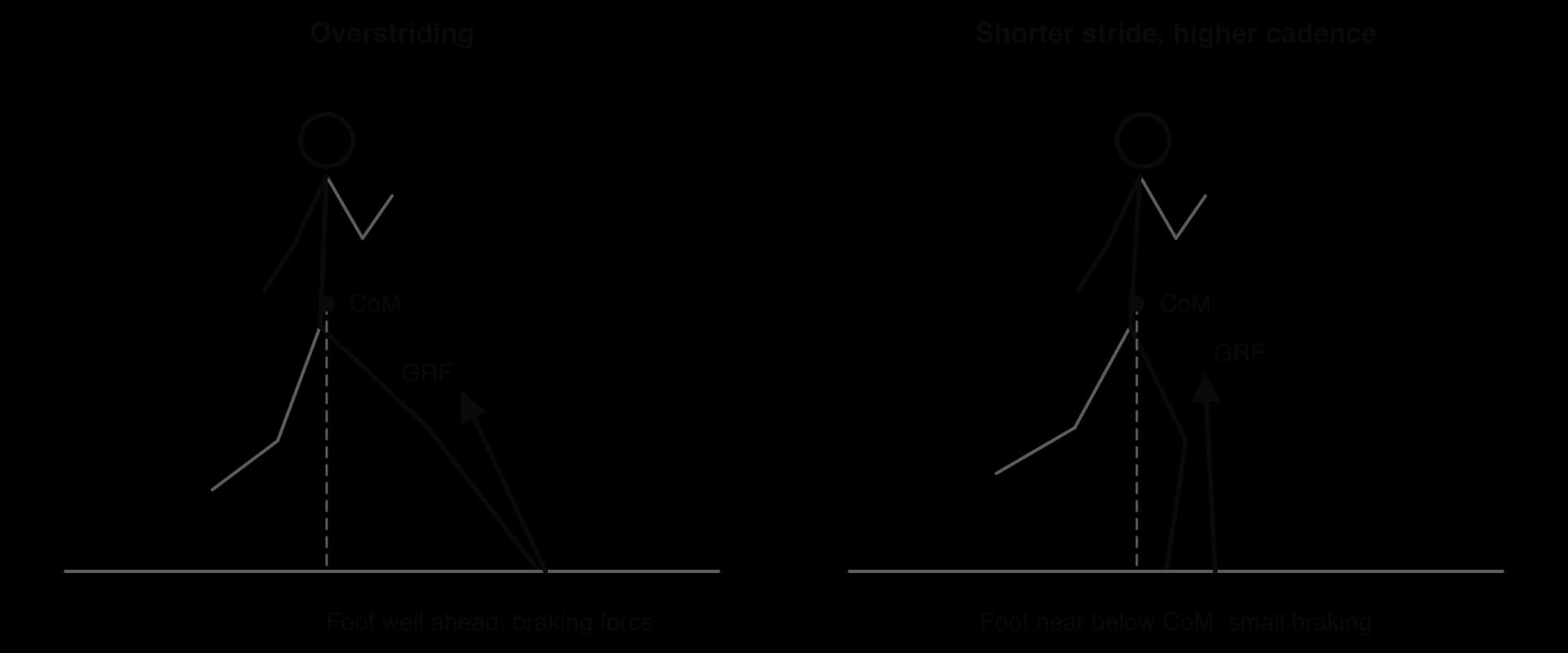
Heel striking itself is not necessarily harmful; many healthy and fast runners land on the heel. The problem is overstriding combined with a stiff knee. Shortening the stride by raising the step rate (cadence) at the same running speed brings the foot closer to below the body. In a laboratory study of 45 recreational runners, increasing step rate by 5% reduced the energy absorbed at the knee, and increasing it by 10% also reduced loading at the hip.
Sources: Heiderscheit et al., Medicine and Science in Sports and Exercise, 2011.
In dynamic knee valgus, the knee moves inside the line from hip to foot as the leg takes load. It is seen on video analysis of many non-contact ACL injuries, and it is linked with weak hip muscles, poor neuromuscular control and fatigue. It also increases load on the kneecap, so it is linked with patellofemoral pain.
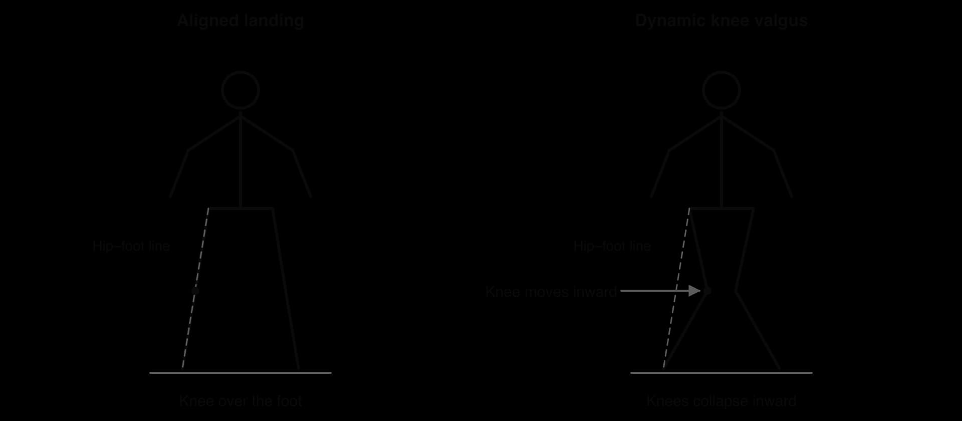
Whether valgus can be used to predict which athletes will be injured is less clear. A 2005 study of 205 female football, basketball and volleyball players found that the nine who later ruptured an ACL had landed in a drop-jump test with 8° more knee abduction and a 2.5 times greater knee abduction moment. A much larger study of 710 elite female football and handball players, published in 2016, recorded 42 new ACL injuries but found that the drop-jump test could not usefully identify who would be injured: among players without a previous ACL injury, none of the landing measures was associated with injury.
Sources: Hewett et al., American Journal of Sports Medicine, 2005; Krosshaug et al., American Journal of Sports Medicine, 2016.
In throwing and serving, force should flow from the legs through the trunk to the arm, following the principle of summation of joint forces. If the legs and trunk contribute too little, because of poor technique, weakness or fatigue, the shoulder and elbow must produce more of the speed themselves. The small muscles of the rotator cuff and the ligaments of the inner elbow are then overloaded, leading to shoulder pain and elbow injuries in baseball pitchers, javelin throwers and tennis players. Correction aims to restore the contribution of the legs and trunk, so that the arm is no longer overloaded.
Pronation is the natural inward roll of the foot that absorbs force after landing. For decades, “overpronation” was blamed for many running injuries, and runners were sold motion-control shoes to correct it. Research has not supported this belief for most runners. A one-year study of 927 novice runners in neutral shoes, who ran a total of more than 326,000 km, found that runners with moderately pronated feet had no higher risk of injury than those with neutral feet, and in fact had fewer injuries per 1,000 km. Whether very highly pronated feet carry extra risk remains uncertain.
Sources: Nielsen et al., British Journal of Sports Medicine, 2014.
Gait retraining uses feedback to help the runner feel and change the movement. The changes can be simple, such as running to a metronome set 5–10% above the usual step rate, or more specific, such as watching a screen that shows the angle of the thigh during stance. Feedback is given often at first and then gradually removed, so that the new pattern becomes automatic.
The evidence for gait retraining is promising. In a study of ten runners with patellofemoral pain, eight sessions of running on a treadmill with real-time feedback on hip adduction reduced the inward movement of the thigh and the drop of the pelvis, and the improvements in pain and function were still present a month later.
Stronger evidence comes from a randomised controlled trial. In Hong Kong, 320 novice runners received either two weeks of gait retraining with visual feedback to land more softly or the same amount of treadmill running without feedback. Over the following year, 16% of the retrained runners and 38% of the control group suffered a running injury, a 62% lower risk.
Sources: Noehren, Scholz and Davis, British Journal of Sports Medicine, 2011; Chan et al., American Journal of Sports Medicine, 2018.
Lumbar stress fractures cause more lost playing time than any other injury in professional cricket, and they are concentrated in young fast bowlers. Movement analysis has linked them to the mixed action, in which the hips and shoulders point in different directions during the delivery stride, so the lower back is twisted and bent sideways under high load. In a three-dimensional study of 50 male professional fast bowlers, a high percentage used a mixed action, defined by a shoulder counter-rotation of more than 30°.
Coaching can change this. In Western Australia, young fast bowlers in development squads received a yearly clinic and small-group coaching sessions over three years to reduce shoulder counter-rotation. Video analysis showed that counter-rotation fell, and MRI scans showed that the incidence and progression of degeneration in the lumbar discs were also reduced. A later systematic review found moderate evidence linking shoulder counter-rotation with low back pain, but judged the intervention studies to be of low quality.
Sources: Ranson et al., Journal of Sports Sciences, 2008; Elliott and Khangure, Medicine and Science in Sports and Exercise, 2002; Morton et al., British Journal of Sports Medicine, 2014.
Equipment can reduce the load on a tissue without changing the athlete’s technique, or help a new technique: a larger grip or softer strings on a tennis racket, a forearm brace for tennis elbow, a lighter or shorter implement for young throwers, cushioned or less cushioned running shoes, and orthotics for some foot problems. Equipment changes are quick and easy to apply, but they can move the load elsewhere and do not remove the underlying maladaptation.
Correcting a maladaptation is a motor learning problem: an automatic, well-practised pattern has to be replaced. The constraints-led approach offers one way to do this. Instead of only giving verbal instructions, the coach changes the constraints of the task or environment so that a better pattern emerges: running to a metronome, landing over a low hurdle that forces a softer, more flexed landing, or bowling with markers that encourage the hips and shoulders to stay aligned. Practice with varied conditions helps the new pattern hold up under fatigue and pressure.
Approach | Strengths | Limitations |
|---|---|---|
Gait retraining | Targets the cause; good evidence from a randomised trial (62% lower injury risk); cheap tools (metronome, mirror) | Small samples in many studies; change may not last without practice; shifting load may affect other tissues (e.g. forefoot striking loads the Achilles) |
Technique coaching (e.g. bowling action) | Reduces the specific harmful movement; can be built into normal coaching | Hard to change an automatic skill; may reduce performance at first; low-quality intervention studies |
Equipment changes | Quick; no need to relearn a skill | Does not change the maladaptation; evidence for some products (e.g. motion-control shoes) is weak |
Correcting biomechanical maladaptations is most effective when the faulty pattern is clearly linked to the tissue that hurts, the change is coached with feedback and practised until it is automatic, and the change is combined with sensible load management and strength training. Because technique is only one of several risk factors, correcting it lowers the risk of overuse injury without removing it.
Anterior cruciate ligament injuries have become one of the most discussed problems in women’s football. Before the 2023 FIFA Women’s World Cup, the absent players included Beth Mead and Leah Williamson of England, Vivianne Miedema of the Netherlands, Catarina Macario of the United States, Marie-Antoinette Katoto and Delphine Cascarino of France, and Janine Beckie of Canada, all with ACL injuries. A few high-profile cases do not show whether the injury is common, so researchers rely on injury surveillance: recording every injury and every hour of exposure in a defined group of players.
The UEFA Women’s Elite Club Injury Study followed 596 players from 15 elite European clubs over four seasons, from 2018/2019 to 2021/2022. Medical staff recorded every injury that caused a player to miss training or matches, and the exact hours of training and match play. ACL injuries were uncommon, 33 out of 1,527 injuries, but they caused the greatest burden of time lost of any injury because each one kept a player out for most of a year.
Measure | Value |
|---|---|
Players, teams, seasons | 596 players, 15 clubs, 4 seasons (2018/2019 to 2021/2022) |
All injuries | 1,527 (6.7 per 1,000 hours) |
ACL injuries | 33 (2% of injuries), in 30 players from 11 clubs |
ACL incidence: training / match | 0.1 / 0.6 per 1,000 hours (rate ratio 9.4) |
Median days lost per ACL injury | 292 days |
Non-contact ACL injuries | 64% (21 of 33) |
Main non-contact mechanisms | Twisting or turning (10 of 21); jumping or landing (5 of 21) |
Expected ACL injuries per squad per season | About 0.7 |
Sources: Hallén et al., British Journal of Sports Medicine, 2024 (UEFA Women’s Elite Club Injury Study).
A comparison with men needs data collected in the same way for both sexes. A study of 57 clubs in Swedish men’s and women’s top leagues and European men’s professional leagues between 2001 and 2009 (2,329 players, 78 ACL injuries) found that the risk of ACL injury for female players was 2.6 times that for male players. Women were also much younger when injured (mean age 20.6 years, compared with 25.2), and after adjusting for age the female risk was still 2.4 times higher. ACL injury risk was about 21 times higher in matches than in training, and 58% of injuries occurred without contact.
Sources: Waldén et al., Knee Surgery, Sports Traumatology, Arthroscopy, 2011.
The data fit the risk factor model well. The internal factors include sex differences in anatomy and neuromuscular control, young age (and therefore less training history and strength), and previous ACL injury, which in another large study of elite female players nearly quadrupled the risk of a new one. The external factors include match play, which is more intense and unpredictable than training, congested schedules, boot and pitch conditions, and, for many players, limited access to strength and conditioning. The inciting event is usually a twist, turn or landing without contact, when the knee collapses inward with the foot fixed.
Because many of these factors are modifiable, prevention is realistic. Neuromuscular warm-up programmes that teach landing and cutting technique and strengthen the hamstrings and hips reduce ACL injuries, and they form a central part of the prevention strategies covered under injury interventions. Equipment designed for women’s feet and better access to conditioning address external factors that have nothing to do with biology.
Sources: Al Jazeera, “Eight big names missing out on the 2023 Women’s World Cup”, 3 July 2023; Associated Press, “ACL injuries are keeping stars out of the Women’s World Cup”, 12 July 2023; Hallén et al., 2024; Waldén et al., 2011; Krosshaug et al., 2016.
Using the risk factor model, explain why ACL injuries in elite women’s football occur mainly in matches and without contact.
Suggest which of the risk factors in the case study could be modified, and by whom (player, coach, club, governing body, manufacturer).
Evaluate the claim that the higher ACL injury rate in female footballers is caused by biological sex differences.
Multiple-choice questions on the causes of injury usually test definitions and classification.
Classification questions often hinge on the mechanism, not the tissue.
Grading questions test whether the tissue and the extent of damage are both known.
Paper 1B style. Injury surveillance in elite women’s club football recorded injuries and exposure separately for training and matches over four seasons. Part of the data is shown.
Training | Match play | |
|---|---|---|
Exposure (hours) | 195,945 | 31,977 |
All injuries: incidence (per 1,000 h) | 4.8 | 18.4 |
ACL injuries (number) | 13 | 20 |
Sources: Hallén et al., British Journal of Sports Medicine, 2024.
Risk factor questions often ask for a link between a factor and the mechanism by which it raises risk.
Extended responses at HL usually ask for an evaluation that weighs evidence, not just a description of a method.
Injury occurs when the load on a tissue exceeds its capacity; capacity rises with progressive training and falls with fatigue, inactivity, injury and age.
Internal risk factors (age, sex differences, pregnancy, effects of training, congenital factors, previous injury) are characteristics of the individual; previous injury is the strongest single predictor.
External risk factors (protective equipment, equipment, surface, weather, rules, opponents, coaching) belong to the environment and are often modifiable.
Internal factors predispose an athlete, external factors make them susceptible, and an inciting event causes the injury; risk changes with every exposure.
Acute trauma results from a single sudden or excessive force; cumulative trauma from repeated loads with insufficient recovery.
Sprains affect ligaments, strains affect muscles; both are graded I–III. Fractures, stress fractures, dislocations, contusions, skin wounds and concussion complete the classification.
Concussion is a functional brain injury: scans are usually normal, so it is recognised from signs and symptoms.
Overuse injuries are often linked to technique, such as overstriding, dynamic knee valgus or a mixed bowling action (HL).
Gait retraining, technique coaching and equipment changes can correct maladaptations and lower risk, especially when combined with load management (HL).
I can outline internal and external risk factors, with sporting examples.
I can explain how risk factors interact using a risk factor model.
I can explain and evaluate possible reasons for the higher ACL injury rate in female athletes.
I can distinguish between acute and cumulative trauma using the load–capacity balance.
I can describe sprains, strains, fractures, dislocations, contusions, skin injuries and concussion, including grades.
I can outline the signs and symptoms of concussion.
I can calculate and interpret injury incidence per 1,000 hours.
I can explain how faulty technique causes common overuse injuries (HL).
I can evaluate gait retraining, technique coaching and equipment changes (HL).