Explain how the heart, blood vessels, blood and lungs transport oxygen and other substances, calculate cardiac output and minute ventilation, and explain how both systems respond to exercise in different people and activities.
Each is in the SEHS glossary, with every other term in the subject.
Tiny, thin-walled air sacs at the ends of the bronchioles, surrounded by capillaries, where oxygen diffuses into the blood and carbon dioxide diffuses out of it.
A small branch of an artery leading into a capillary network, whose muscular wall narrows or widens to control how much blood flows into the capillaries it supplies.
The difference between the oxygen content of arterial and mixed venous blood, usually in mL of oxygen per 100 mL of blood, showing how much oxygen the tissues extract.
A blood vessel that carries blood away from the heart under high pressure, with a thick wall of smooth muscle and elastic tissue and a relatively narrow lumen.
The enlargement of the heart, especially the left ventricle, caused by regular intense training, which increases stroke volume and maximal cardiac output as a healthy adaptation.
One of the two thin-walled upper chambers of the heart, which receives blood from the veins and pumps it into the ventricle below.
The force exerted by blood on the walls of the blood vessels, usually measured in the large arteries in mmHg and written as systolic over diastolic pressure, such as 120/80 mmHg.
Outline the structure of the heart, the blood vessels and the blood, and describe the pulmonary and systemic circulations.
Describe what the blood transports and how oxygen and carbon dioxide are carried.
Calculate cardiac output from heart rate and stroke volume, and minute ventilation from tidal volume and respiration rate.
Describe and explain the responses of heart rate, stroke volume, cardiac output and blood pressure to exercise.
Explain venous return, the Frank–Starling mechanism and the redistribution of blood during exercise.
Sketch and annotate the oxygen–haemoglobin dissociation curve and explain the Bohr shift during exercise.
Describe the structure of the respiratory system and explain how the alveoli are adapted for gaseous exchange.
Explain the mechanics of breathing at rest and during exercise.
Label a spirometer trace and describe how lung volumes and ventilation change during exercise.
Discuss how age, sex, body size, fitness, type of activity and intensity affect cardiovascular and respiratory responses.
Every working muscle fibre needs a steady supply of oxygen and fuel and a way to get rid of carbon dioxide, heat and other waste. Two systems work together to provide this transport. The cardiovascular system moves blood around the body, and the respiratory system brings oxygen into the blood and removes carbon dioxide from it.
Transport matters for both health and performance. For performance, the capacity of the heart and lungs to deliver oxygen sets the upper limit of endurance, which is why an elite rower can pump about twice as much blood per minute as an untrained adult. For health, the same systems explain why regular activity lowers resting
With a free account, Augment remembers where you stopped and what you have practiced.
The change in the distribution of cardiac output during exercise, in which vasodilation increases flow to working muscles and vasoconstriction reduces flow to less active organs such as the gut and kidneys.
The rightward shift of the oxygen–haemoglobin dissociation curve caused by more carbon dioxide, higher acidity and higher temperature, so haemoglobin releases more oxygen at a given pO₂.
A resting heart rate below 60 beats min⁻¹; in trained endurance athletes a normal result of a large stroke volume and high parasympathetic activity.
The smallest blood vessel, with a wall one cell thick and a lumen about the width of a red blood cell, where substances are exchanged between blood and tissues by diffusion.
The sequence of events in one heartbeat: contraction (systole) and relaxation (diastole) of the atria and ventricles, with the opening and closing of the valves.
The volume of blood pumped by the left ventricle per minute, equal to heart rate multiplied by stroke volume and measured in L min⁻¹; about 5 L min⁻¹ at rest.
The organ system made up of the heart, blood vessels and blood, which transports oxygen, carbon dioxide, nutrients, hormones, heat and waste products around the body.
A dome-shaped sheet of skeletal muscle separating the thoracic and abdominal cavities, which contracts and flattens to enlarge the thoracic cavity; the main muscle of inspiration.
The phase of the cardiac cycle in which heart muscle relaxes and the chambers fill with blood, with the semilunar valves closed and the atrioventricular valves open.
The lowest pressure in the arteries, reached while the ventricles relax and fill; about 80 mmHg at rest in a healthy young adult.
The passive net movement of molecules from a region of higher concentration or partial pressure to a region of lower concentration, needing no energy.
The maximum volume of air that can be breathed out after a normal expiration; about 1.2 L in a young adult man.
The principle that the more the ventricle is filled and stretched during diastole, the more forcefully it contracts, so greater venous return produces a greater stroke volume.
The movement of oxygen and carbon dioxide by diffusion between the alveoli and the blood, and between the blood and the tissues, down partial pressure gradients.
The percentage of the blood volume made up of red blood cells, typically higher in men than in women; raising it increases the oxygen-carrying capacity of the blood.
The iron-containing protein in red blood cells that binds reversibly with oxygen, loading it in the lungs and releasing it in the tissues; each molecule carries up to four oxygen molecules.
The number of times the heart beats per minute, measured in beats min⁻¹; typically 60–80 beats min⁻¹ at rest in an adult.
Flaps of tissue between the chambers of the heart and at the exits of the ventricles that open and close in response to pressure differences, so blood flows in one direction only.
The maximum volume of air that can be breathed in after a normal inspiration; about 3 L in a young adult man.
Two layers of muscle between the ribs: the external intercostals lift the ribs up and out in inspiration, and the internal intercostals pull them down and in during forced expiration.
The highest heart rate an individual can reach during maximal exercise; it falls with age and is often estimated as 220 minus age in beats min⁻¹.
The process by which respiratory muscles change the volume of the thoracic cavity, creating pressure differences between the lungs and the atmosphere that move air in and out.
The volume of air breathed in or out per minute, equal to tidal volume multiplied by respiration rate and measured in L min⁻¹; about 6–8 L min⁻¹ at rest.
The thick middle layer of the heart wall, made of cardiac muscle, which contracts rhythmically and involuntarily to pump blood.
An S-shaped graph of the percentage saturation of haemoglobin with oxygen against the partial pressure of oxygen, showing loading in the lungs and release at the lower pO₂ of the tissues.
The pressure exerted by one gas in a mixture of gases, proportional to its share of the mixture, written pO₂ for oxygen and pCO₂ for carbon dioxide.
The liquid part of the blood, about 90% water, in which nutrients, hormones, carbon dioxide, electrolytes, proteins and waste products are dissolved and transported.
A ring of smooth muscle where a capillary branches off an arteriole, which contracts to close the capillary and relaxes to open it, controlling blood flow into a capillary bed.
The circuit that carries deoxygenated blood from the right ventricle through the pulmonary arteries to the lungs and returns oxygenated blood through the pulmonary veins to the left atrium.
A biconcave, disc-shaped blood cell with no nucleus, packed with haemoglobin, whose function is to carry oxygen from the lungs to the tissues.
The volume of air that remains in the lungs after a maximal expiration, about 1.2 L in a young adult man; it cannot be measured with a simple spirometer.
The number of breaths taken per minute, also called breathing frequency; about 12–16 breaths min⁻¹ at rest in an adult.
The mechanism by which pressure changes in the chest and abdomen during breathing squeeze blood from abdominal veins into the veins of the chest and towards the heart.
The organ system of airways, lungs and breathing muscles that moves air into and out of the lungs and exchanges oxygen and carbon dioxide between the air and the blood.
The mechanism by which contracting skeletal muscles squeeze nearby veins and, with the valves in the veins, push blood back towards the heart.
An instrument that measures the volume of air inspired and expired over time, producing a trace from which lung volumes and capacities can be read.
The volume of blood ejected by the left ventricle in one contraction, measured in mL; about 70 mL at rest in an untrained adult.
The circuit that carries oxygenated blood from the left ventricle through the aorta to all body tissues and returns deoxygenated blood through the veins and venae cavae to the right atrium.
The phase of the cardiac cycle in which heart muscle contracts and pushes blood out of a chamber; ventricular systole ejects blood into the aorta and pulmonary artery.
The highest pressure in the arteries, reached when the left ventricle contracts and ejects blood; about 120 mmHg at rest in a healthy young adult.
The volume of air breathed in or out in one normal breath; about 0.5 L at rest in an adult, rising to 2–3 L in heavy exercise.
The volume of air in the lungs after a maximal inspiration, equal to vital capacity plus residual volume; about 6 L in a young adult man.
Forcefully breathing out against a closed airway, as when straining to lift a heavy load, which raises pressure in the chest and causes large rises in blood pressure.
A blood vessel that carries blood towards the heart under low pressure, with a thin wall, a wide lumen and, in the limbs, valves that prevent backflow.
The narrowing of veins by contraction of the smooth muscle in their walls, stimulated by sympathetic nerves, which reduces the blood stored in the veins and increases venous return.
The volume of blood returning to the right atrium through the veins per minute, aided by the skeletal muscle pump, respiratory pump, venous valves and venoconstriction.
One of the two thick-walled lower chambers of the heart, which pumps blood into the arteries: the right ventricle to the lungs and the left ventricle to the rest of the body.
The maximum volume of air that can be breathed out after a maximal inspiration, equal to tidal volume plus the inspiratory and expiratory reserve volumes; about 4–5 L in adults.
The cardiovascular system has three parts: the heart, which pumps; the blood vessels, which carry blood to and from every tissue; and the blood, which carries the substances being transported. Together they form a closed circuit, so the same blood returns to the heart again and again, about once a minute at rest.
Transport is only useful if the substances reach the right place at the right rate. During hard exercise the working muscles may need many times more oxygen than at rest, and the heat they produce must be carried to the skin. The table summarises what the blood carries and where it takes it.
Substance | How it is carried | From → to | Why it matters in exercise |
|---|---|---|---|
Oxygen | About 98–99% bound to haemoglobin in red blood cells; a small amount dissolved in plasma | Lungs → tissues | Needed by the oxidative system to resynthesise ATP |
Carbon dioxide | About 70% as hydrogencarbonate (bicarbonate) ions in plasma, about 20% bound to haemoglobin, 7–10% dissolved | Tissues → lungs | Produced by aerobic respiration; raises acidity if it accumulates |
Nutrients (glucose, fatty acids, amino acids) | Dissolved in plasma | Gut and liver, fat stores → muscles and other tissues | Fuel for ATP resynthesis and building blocks for repair |
Hormones | Dissolved in plasma | Endocrine glands → target cells | Epinephrine, insulin and others regulate heart rate and fuel use |
Heat | In the blood itself (water has a high heat capacity) | Working muscles → skin | Allows heat loss and prevents a dangerous rise in core temperature |
Waste (urea, lactate, excess water) | Dissolved in plasma | Tissues → kidneys, liver, other muscles | Removal and reuse of lactate; excretion of urea |
Several of these jobs link to other topics. Glucose and fatty acids are the fuels of the oxidative system; epinephrine is released by the adrenal glands and carried by the blood to the heart; and the heat carried to the skin is lost through thermoregulation. The carbon dioxide carried as hydrogencarbonate is part of the bicarbonate buffer that helps keep blood pH stable.
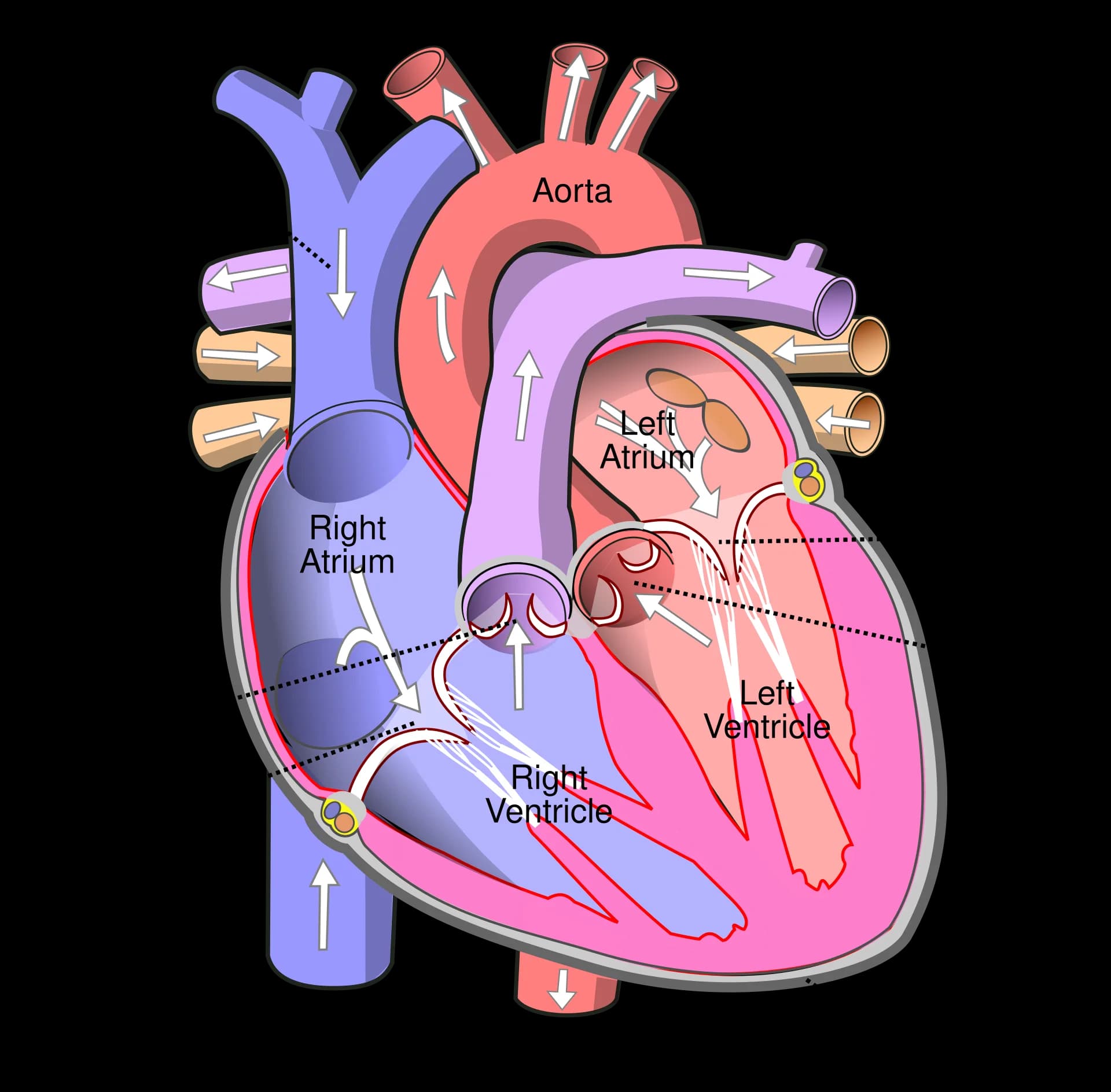
The heart is a hollow, muscular organ about the size of a clenched fist, lying in the chest between the lungs and slightly to the left. It is really two pumps side by side. The right side receives blood that has given up much of its oxygen and pumps it to the lungs; the left side receives oxygen-rich blood from the lungs and pumps it to the rest of the body.
Each side of the heart has two chambers. The upper chambers, the atria, receive blood returning in the veins. The lower chambers, the ventricles, pump blood out into the arteries. A wall of muscle called the septum separates the right and left sides, so deoxygenated and oxygenated blood do not mix.
The atria only need to push blood a few centimetres into the ventricles, so their walls are thin. The ventricles must generate much higher pressures, and the left ventricle, which pumps blood around the whole body, has the thickest wall of all, often about three times as thick as the wall of the right ventricle.
The wall of the heart is made of cardiac muscle, the myocardium. Cardiac muscle contracts rhythmically without tiring and without conscious control. Its contraction is started by the heart’s own pacemaker, the sinoatrial node, and then spread through the walls of the atria and ventricles by the conduction system.
The myocardium needs its own blood supply, delivered by the coronary arteries, which branch off the aorta just above the aortic valve. A blockage in a coronary artery cuts the oxygen supply to part of the heart muscle and causes a heart attack; this is one form of coronary heart disease.
Four valves keep blood flowing in one direction. Each valve is made of flaps of tissue (cusps) that are pushed open by blood flowing forwards and pushed shut when blood starts to flow backwards. Valves open and close passively, because of differences in pressure on either side of them; no muscle opens them.
Valve | Position | Prevents backflow from |
|---|---|---|
Tricuspid (right atrioventricular) valve | Between the right atrium and right ventricle | Right ventricle into right atrium |
Bicuspid (mitral, left atrioventricular) valve | Between the left atrium and left ventricle | Left ventricle into left atrium |
Pulmonary semilunar valve | At the exit of the right ventricle into the pulmonary artery | Pulmonary artery into right ventricle |
Aortic semilunar valve | At the exit of the left ventricle into the aorta | Aorta into left ventricle |
The tricuspid and bicuspid valves are attached by tendon-like cords to small muscles on the ventricle wall, which stop the cusps turning inside out when the ventricles contract. The “lub-dub” sound of the heartbeat is the sound of the valves closing: the atrioventricular valves first, then the semilunar valves.
Deoxygenated blood from the body returns through the superior and inferior venae cavae into the right atrium.
It passes through the tricuspid valve into the right ventricle.
The right ventricle pumps it through the pulmonary semilunar valve into the pulmonary artery, which carries it to the lungs.
In the lungs the blood picks up oxygen and loses carbon dioxide.
Oxygenated blood returns through the pulmonary veins to the left atrium.
It passes through the bicuspid (mitral) valve into the left ventricle.
The left ventricle pumps it through the aortic semilunar valve into the aorta, which distributes it to the whole body.
 YouTube
Blood passes through the heart twice on every complete circuit of the body, so humans have a double circulation. The two circuits run at the same time and carry the same volume of blood per minute, because whatever the right ventricle pumps out must eventually come back to the left ventricle.
The pulmonary circulation is short and has low resistance, so the right ventricle needs only a low pressure to drive blood through it. A low pressure also protects the delicate capillaries in the lungs and stops fluid being forced out of them into the air spaces.
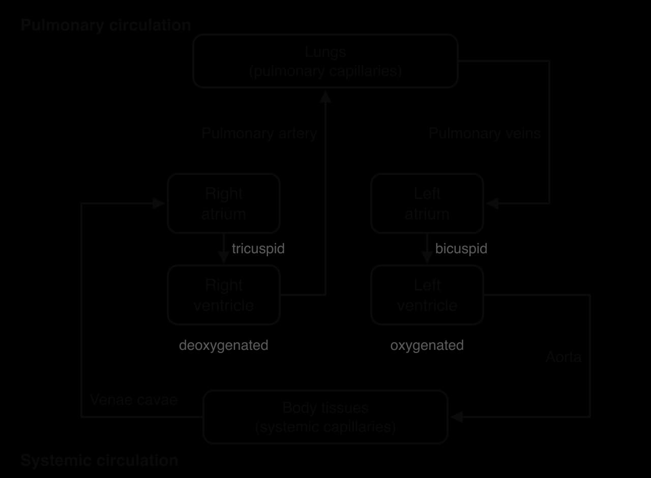
The systemic circulation is long and has many branches, so the left ventricle must generate a much higher pressure than the right. This difference in workload explains why the left ventricle wall is so much thicker. In exercise, both circuits carry more blood per minute, and the pulmonary circuit must keep pace so that all the extra blood is oxygenated.
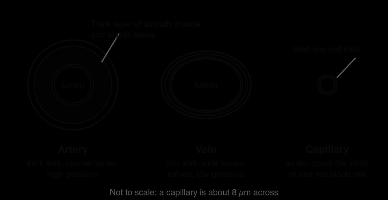
Blood leaves the heart in large arteries, which divide into smaller arteries and then into arterioles. Arterioles lead into networks of tiny capillaries, where exchange with the tissues takes place. Capillaries join into venules, venules into veins, and veins return blood to the heart. Each type of vessel has a structure that suits its job.
The elastic tissue in the walls of large arteries stretches each time the ventricles eject blood and then recoils, which smooths the flow and keeps blood moving between beats. This stretch and recoil is the pulse that can be felt at the wrist or neck.
Arterioles are the main control points of the circulation. When their smooth muscle contracts (vasoconstriction), the lumen narrows and less blood flows through; when it relaxes (vasodilation), more blood flows through. Changing the diameter of arterioles is how blood is redirected to working muscles during exercise.
Capillaries are so numerous that almost every cell lies close to one. Their thin walls give a very short diffusion distance, and blood moves through them slowly because their total cross-sectional area is huge, which gives time for exchange. Endurance training increases the number of capillaries around each muscle fibre.
Feature | Artery | Capillary | Vein |
|---|---|---|---|
Direction of flow | Away from the heart | Between arterioles and venules | Towards the heart |
Blood pressure | High, pulsating | Low, falling along its length | Very low |
Wall | Thick: smooth muscle and elastic tissue | One layer of flattened cells (endothelium) | Thin: little muscle and elastic tissue |
Lumen | Narrow relative to wall | Very narrow (about 8 µm) | Wide |
Valves | None (except at the exit of the heart) | None | Present, especially in the limbs |
Main function | Carry blood quickly at high pressure; arterioles control distribution | Exchange of gases, nutrients and waste with tissues | Return blood to the heart; act as a store of blood |
Venules are small veins that collect blood from capillary networks. At rest, about two-thirds of the blood volume is in the veins, so they act as a reservoir. During exercise, sympathetic nerves cause the smooth muscle in vein walls to contract (venoconstriction), which pushes some of this stored blood back towards the heart.
An adult has about 5 litres of blood, roughly 7–8% of body mass. If a sample is spun in a centrifuge, it separates into a pale yellow liquid, the plasma (about 55% of the volume), and the cells (about 45%), most of which are red blood cells.
Plasma volume matters for exercise. A larger plasma volume fills the heart more fully between beats, and it provides water for sweating. Endurance training increases plasma volume within a few weeks, while dehydration reduces it, which forces the heart to beat faster to maintain its output.
The biconcave shape gives each red blood cell a large surface area for its volume and lets it bend to squeeze through narrow capillaries. Red blood cells are made in the bone marrow, stimulated by the hormone erythropoietin (EPO) from the kidneys, and each lasts about 120 days.
Each gram of haemoglobin can carry about 1.34 mL of oxygen. With a typical concentration of about 150 g of haemoglobin per litre of blood, fully saturated arterial blood carries about 200 mL of oxygen per litre, nearly seventy times more than could be dissolved in the plasma alone. Iron is needed to make haemoglobin, so iron deficiency and anaemia reduce the oxygen-carrying capacity of the blood and impair endurance.
Component | Proportion of blood | Main function |
|---|---|---|
Plasma | About 55% | Transport of dissolved substances and heat |
Red blood cells | About 45% (most of the cell volume) | Carry oxygen bound to haemoglobin; carry some carbon dioxide |
White blood cells | Less than 1% | Defend the body against infection |
Platelets | Less than 1% | Cell fragments that help blood to clot and seal damaged vessels |
The proportion of blood volume made up of red blood cells is called the haematocrit. It is typically higher in men than in women, partly because testosterone stimulates EPO production. Raising haematocrit artificially, by injecting EPO or by reinfusing stored blood, increases oxygen delivery and is banned in sport because it improves endurance and makes the blood more viscous and dangerous to pump.
Each heartbeat is a sequence of contraction and relaxation that fills the chambers and then empties them. The sequence from the start of one beat to the start of the next is the cardiac cycle. At a resting heart rate of about 75 beats per minute, one cycle lasts about 0.8 seconds.
The two halves of the cycle have their own names. Contraction raises the pressure in a chamber and pushes blood out of it; relaxation lowers the pressure and allows the chamber to fill.
Atrial systole comes first and tops up the ventricles with the last part of their filling. Ventricular systole follows: as ventricular pressure rises above atrial pressure, the atrioventricular valves close; when it rises above the pressure in the arteries, the semilunar valves open and blood is ejected.
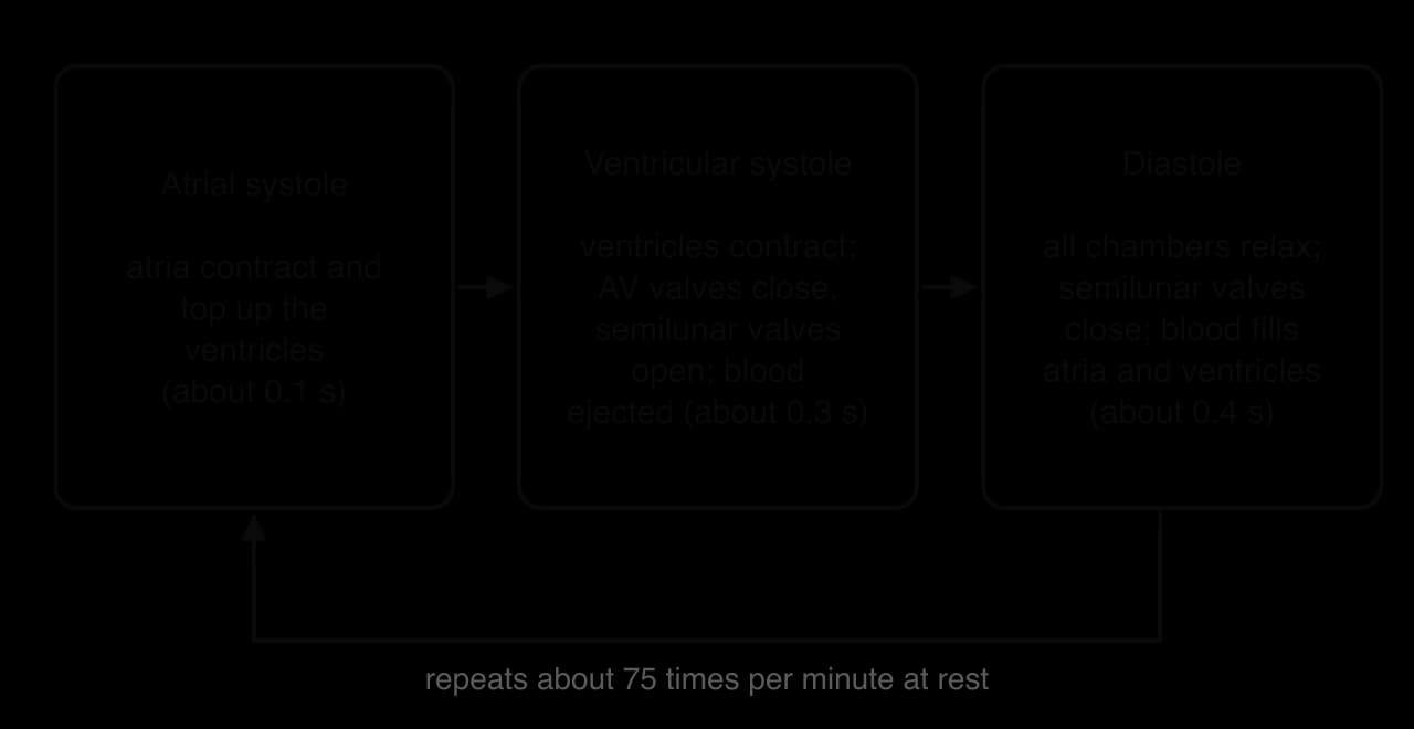
Diastole is also when the myocardium receives most of its own blood supply, because the contracting heart muscle squeezes the coronary vessels during systole. As heart rate rises in exercise, diastole shortens much more than systole. At about 180 beats per minute a cycle lasts only a third of a second, which leaves little time for filling. This shortening of filling time helps explain why stroke volume stops rising at high heart rates in untrained people.
The sinoatrial node fires an impulse that spreads across both atria, which contract together (atrial systole). The impulse is delayed briefly at the atrioventricular node, giving the atria time to empty, and then travels down the bundle of His and through the Purkinje fibres so that the ventricles contract from the apex upwards, squeezing blood up towards the arteries. The conduction system and how nerves and hormones change its rate are part of intrinsic and extrinsic control of heart rate.
The amount of blood the heart delivers depends on two things: how often it beats and how much it ejects with each beat. These are measured as heart rate and stroke volume, and multiplying them gives cardiac output, the most important single measure of the heart’s pumping capacity.
Heart rate is the easiest of the three to measure: by counting the pulse at the wrist (radial artery) or neck (carotid artery), or with a chest-strap or optical heart rate monitor. It is set by the sinoatrial node and adjusted by the sympathetic nervous system, which speeds it up, and the parasympathetic nervous system, whose vagus nerve slows it down.
Stroke volume is the difference between how full the ventricle is at the end of diastole and how much blood is left in it at the end of systole. It therefore rises when the ventricle fills more (more venous return), when it contracts more forcefully, or both.
Because stroke volume is measured in mL and cardiac output in L min⁻¹, the product must be divided by 1,000. A resting adult with a heart rate of 70 beats min⁻¹ and a stroke volume of 70 mL has a cardiac output of 4,900 mL min⁻¹, or 4.9 L min⁻¹. At rest, cardiac output is about 5 L min⁻¹ in almost everyone, trained or untrained, because it is set by the body’s resting oxygen demand.
Typical values for young adults are summarised in the table. The striking difference between trained and untrained people is at maximal exercise: maximal heart rate is similar, but trained endurance athletes have a much larger stroke volume, so their maximal cardiac output can be roughly 1.5–2 times higher.
Measure | Untrained, rest | Trained endurance athlete, rest | Untrained, maximal exercise | Trained endurance athlete, maximal exercise |
|---|---|---|---|---|
Heart rate (beats min⁻¹) | 60–80 | 40–60 | About 190–200 | About 180–195 |
Stroke volume (mL) | 60–80 | 90–110 | 100–120 | 160–200 |
Cardiac output (L min⁻¹) | About 5 | About 5 | About 20 | 30–40 |
Sources: typical values from OpenStax, Anatomy and Physiology 2e, 2022; Carlsson et al., Journal of Cardiovascular Magnetic Resonance, 2012; Gledhill, Cox and Jamnik, Medicine and Science in Sports and Exercise, 1994.
Heart rate does not simply jump to a new value when exercise starts. It follows a characteristic pattern before, during and after exercise, which can be recorded with a heart rate monitor and sketched as a graph.

Anticipatory rise (A). Just before exercise, heart rate rises above its resting value. Thinking about the coming effort activates the sympathetic nervous system and the release of epinephrine, an anticipatory rise that prepares the body before any movement has started.
Rapid rise (B). At the start of exercise, heart rate climbs quickly. Proprioceptors in the working muscles and joints signal movement to the cardiovascular control centre, which reduces parasympathetic (vagal) activity and increases sympathetic activity.
Steady state (C). During constant submaximal exercise, heart rate levels off within a few minutes, when the supply of oxygen to the muscles matches their demand. This steady state is reached sooner and at a lower heart rate in trained people.
Recovery (D and E). When exercise stops, heart rate falls rapidly as the stimulus from the muscles ends, then more slowly as circulating epinephrine is removed, temperature falls and the extra oxygen consumed after exercise is used to restore the body. A faster recovery is a sign of good aerobic fitness.
During progressive exercise, in which the intensity rises every minute or two, heart rate rises roughly in a straight line with intensity until it approaches its maximum. In very prolonged exercise, especially in the heat, heart rate can drift upwards even at a constant intensity. This cardiovascular drift happens as fluid is lost in sweat and stroke volume falls.
Maximum heart rate depends mainly on age. It falls by roughly one beat per minute each year in adulthood, mainly because the intrinsic firing rate of the sinoatrial node falls and it responds less to sympathetic stimulation. Training has little effect on it. The estimate 220 − age is popular because it needs no equipment, and it is used to set training intensities as a percentage of maximum heart rate.
The 220 − age formula was never derived from a large, carefully designed study. A meta-analysis of 351 studies with 18,712 participants, checked against laboratory tests of 514 healthy adults, found that maximum heart rate was better predicted by 208 − 0.7 × age, and that 220 − age underestimates the maximum of older adults. The same analysis found that the relationship did not differ between men and women or with habitual activity.
The main limitation of any age-based formula is individual variation: people of the same age can have very different maximum heart rates, so a training zone calculated from a formula may be too easy for one person and too hard for another. A directly measured maximum, from a supervised maximal test, is more valid but is tiring, needs motivation and may be unsafe for some people with heart disease. For most recreational exercisers, a formula combined with ratings of perceived exertion (RPE) is a sensible compromise; for elite athletes and clinical patients, a measured value is worth the effort.
Sources: Tanaka, Monahan and Seals, Journal of the American College of Cardiology, 2001.
Stroke volume rises at the start of exercise for two reasons. More blood returns to the heart, so the ventricles fill more during diastole; and sympathetic stimulation and epinephrine make the myocardium contract more forcefully, so more of that blood is ejected. The first of these effects is described by the Frank–Starling mechanism.
The mechanism works a little like an elastic band: within limits, the further the cardiac muscle fibres are stretched before contraction, the more strongly they recoil. This automatically matches what the heart pumps out to what flows into it. If venous return increases, the ventricle stretches more, contracts harder and ejects the extra blood, so blood does not back up in the veins.
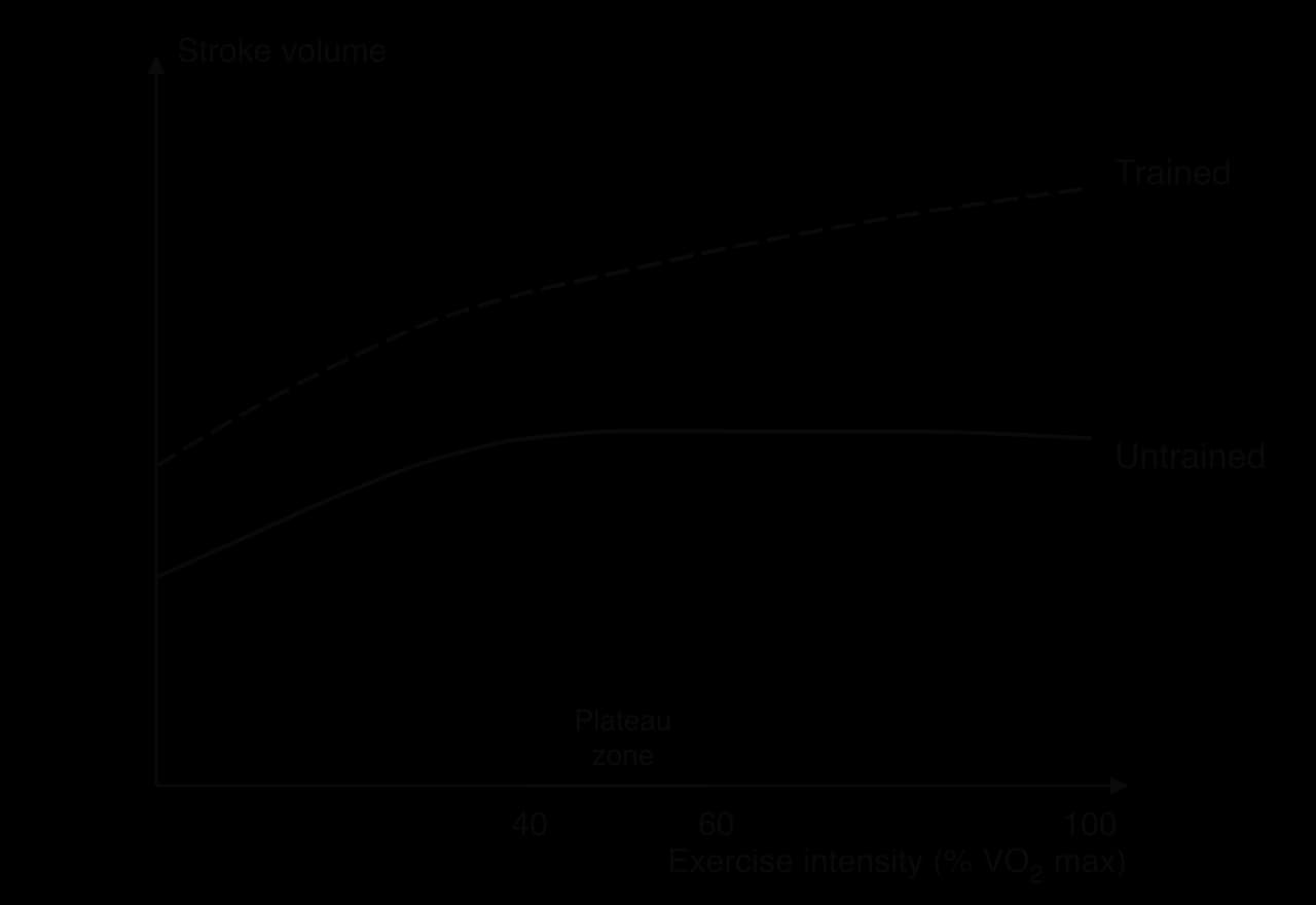
In untrained people, stroke volume usually reaches a plateau at about 40–60% of VO₂ max, so any further increase in cardiac output comes from heart rate alone. In a study of seven endurance-trained and seven untrained young men, the stroke volume of the untrained men levelled off at a heart rate of 120 beats min⁻¹, whereas that of the trained men kept rising up to their maximum heart rate. At 190 beats min⁻¹ the trained men’s ventricles filled 71% faster than those of the untrained men.
Sources: Gledhill, Cox and Jamnik, Medicine and Science in Sports and Exercise, 1994.
Above a certain heart rate, diastole is so short that the ventricles of an untrained person do not have time to fill any further, so the stretch on the myocardium and the stroke volume stop increasing. Endurance-trained hearts fill faster, because they have larger, more compliant ventricles and a larger blood volume, so they continue to fill well even when diastole is short. The study of trained and untrained men found that faster filling, more than faster emptying, explained most of the trained athletes’ advantage.
The heart can pump out only what returns to it. Blood in the veins is at a very low pressure, and in a standing person much of it must travel upwards against gravity from the legs. Several mechanisms keep it moving back to the heart.
The skeletal muscle pump. Contracting muscles squeeze the veins that run between them and push blood towards the heart.
The respiratory pump. Pressure changes in the chest and abdomen during breathing draw blood into the large veins of the chest.
Valves in the veins. Pocket-shaped valves allow blood to flow only towards the heart, so any squeeze moves blood in one direction.
Venoconstriction. Sympathetic nerves make the smooth muscle in vein walls contract, which reduces the volume of blood stored in the veins and pushes it towards the heart.
Gravity. Blood from above the heart (the head and neck) flows back with gravity’s help.
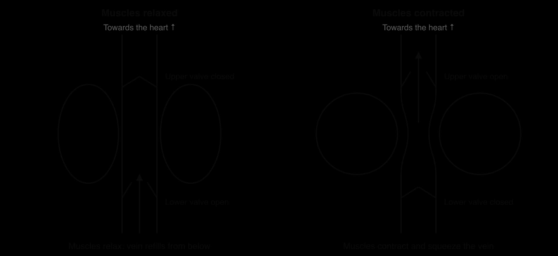
The skeletal muscle pump is most effective in rhythmic exercise such as running, cycling and swimming, which is one reason why stroke volume rises more in dynamic exercise than in static contractions. It also explains why stopping suddenly after hard exercise can make a person feel faint: the muscle pump stops, blood pools in the dilated vessels of the legs, venous return and cardiac output fall, and blood pressure drops. An active cool-down keeps the pump working while heart rate and blood flow return to normal.
Because breathing becomes deeper and faster in exercise, the respiratory pump becomes more effective at the same time as the skeletal muscle pump, and the two together raise venous return. Through the Frank–Starling mechanism, the extra filling then increases stroke volume.
Cardiac output rises in a nearly straight line with exercise intensity, from about 5 L min⁻¹ at rest to about 20 L min⁻¹ in an untrained young adult at maximal exercise, and to more than 35 L min⁻¹ in some elite endurance athletes. At low intensities both heart rate and stroke volume contribute; once stroke volume has levelled off, the remaining rise depends on heart rate. After exercise, cardiac output falls back towards its resting value as heart rate and stroke volume return to normal.
Blood flows from areas of high pressure to areas of low pressure. The pressure created by the left ventricle drives blood through the systemic circulation, and it is highest in the aorta and large arteries and falls steadily through the arterioles, capillaries and veins.
Arterial pressure rises and falls with each heartbeat, so it has a highest and a lowest value. A typical value for a healthy young adult at rest is about 120/80 mmHg. It is usually measured at the upper arm with an inflatable cuff, either by listening for sounds through a stethoscope or with an automatic monitor.
Systolic pressure depends mostly on how much blood the heart ejects and how forcefully, so it reflects cardiac output and the stiffness of the large arteries.
Diastolic pressure depends mostly on the resistance of the arterioles: the more they are constricted, the less easily blood drains out of the arteries between beats, and the higher the diastolic pressure stays.
Blood pressure that stays high at rest, at or above about 140/90 mmHg (some guidelines use 130/80 mmHg), is hypertension, a major risk factor for heart attack and stroke, and regular physical activity is one of its main treatments. Moment-to-moment changes in blood pressure are corrected by the baroreceptor reflex: baroreceptors in the aortic arch and carotid arteries detect the change, and the cardiovascular control centre adjusts heart rate and arteriole diameter to reverse it.
In dynamic (isotonic) exercise such as running, cycling or swimming, systolic pressure rises with intensity, often to 180–200 mmHg at maximal effort in a healthy young adult, because cardiac output rises. Diastolic pressure stays about the same or changes by only a few mmHg. This is because the arterioles in the working muscles dilate widely, which lowers the total resistance of the circulation and lets blood drain out of the arteries between beats as fast as it did at rest.
Heavy resistance exercise and static (isometric) contractions produce a different and much larger response. Contracting muscles squeeze their own blood vessels, which raises resistance, and lifters often hold their breath and strain against a closed airway (the Valsalva manoeuvre), which raises the pressure in the chest. Both systolic and diastolic pressure therefore rise steeply.
Direct measurements in experienced body builders, using a catheter in the arm artery, recorded a mean peak pressure of 320/250 mmHg during a double-leg press to failure, with one participant exceeding 480/350 mmHg. Even single-arm curls to failure produced a mean peak of 255/190 mmHg. Pressures fell during the lowering (eccentric) phase of each lift.
Sources: MacDougall et al., Journal of Applied Physiology, 1985.
These short spikes are harmless for most healthy people, but they explain why people with uncontrolled hypertension or heart disease are advised to avoid maximal lifts and breath-holding, and to use lighter loads with more repetitions while breathing out during the effort. Resistance training performed this way still lowers resting blood pressure over time.
At rest, the gut, liver and kidneys receive a large share of the cardiac output, and the skeletal muscles only about a fifth. During exercise the muscles need far more oxygen, but the heart cannot increase its output without limit. The body therefore redirects blood, a process called blood redistribution: more goes to the working muscles, the heart and the skin, and less to organs whose work can wait.
Two mechanisms work together. In the working muscles, the arterioles dilate in response to local chemical changes: lower oxygen, more carbon dioxide, more hydrogen ions (lower pH), more potassium and a higher temperature. In the gut and kidneys, the sympathetic nervous system makes the arterioles constrict. The control happens at the level of the arterioles and of the precapillary sphincters.
At rest only a fraction of the capillaries in a muscle are open at any moment. During exercise the precapillary sphincters relax and almost all of them open, which increases the surface area for exchange and shortens the distance oxygen must diffuse to reach each fibre.

The chart shows that the absolute flow to the brain stays the same, about 750 mL min⁻¹, because the brain must be protected at all times. The heart muscle receives three times more blood, matching its extra work. Skin blood flow rises to carry heat to the body surface, but in very hard or prolonged exercise in the heat, the muscles and the skin compete for a limited cardiac output, which is one reason why endurance performance suffers in hot conditions.
Redistribution has practical consequences. A large meal shortly before training means that the gut and the muscles compete for blood, which can cause discomfort and nausea. Reduced blood flow to the kidneys reduces urine production during exercise, which helps conserve water.
Oxygen is carried almost entirely by haemoglobin. How much oxygen haemoglobin holds depends on the partial pressure of oxygen around it: high in the lungs, where it loads oxygen, and low in the tissues, where it unloads it. The relationship is shown by the oxygen–haemoglobin dissociation curve.
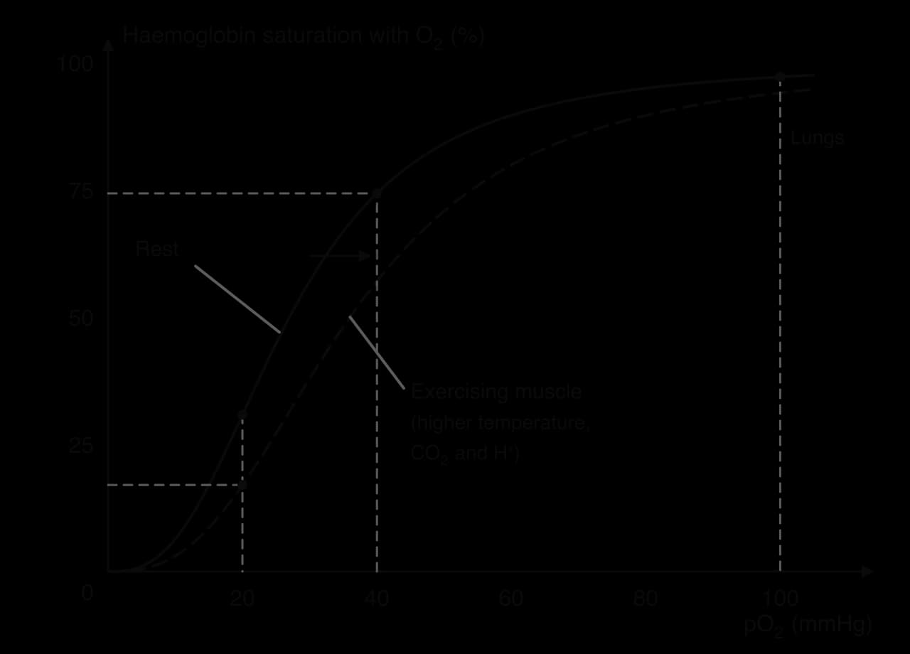
The S-shape has two useful consequences. The flat top of the curve means that haemoglobin stays almost fully saturated even if alveolar pO₂ falls a little, for example at moderate altitude. The steep middle section, between about 10 and 40 mmHg, matches the pO₂ found in active tissues, so a small drop in tissue pO₂ causes a large release of oxygen. The S-shape arises because once one oxygen molecule binds, the haemoglobin molecule changes shape and binds the next ones more easily.
During exercise, working muscle becomes warmer, produces more carbon dioxide and becomes more acidic as hydrogen ions accumulate. Each of these changes reduces haemoglobin’s attraction for oxygen, so the whole curve moves to the right.
The conditions that cause the Bohr effect are strongest in the working muscle, so more oxygen is unloaded where it is needed. In the lungs, carbon dioxide is breathed out and the blood is cooler, so the curve shifts back to the left and haemoglobin loads oxygen fully again.
At rest, arterial blood carries about 20 mL of oxygen per 100 mL and venous blood about 15 mL, so the a-vO₂ difference is about 5 mL per 100 mL: the tissues extract only a quarter of the oxygen delivered. During maximal exercise, venous blood returning from working muscle may contain very little oxygen, and the whole-body a-vO₂ difference rises to about 15–17 mL per 100 mL. Oxygen uptake equals cardiac output multiplied by the a-vO₂ difference, so a larger a-vO₂ difference, helped by the Bohr effect, more capillaries and more mitochondria, allows the muscles to use more oxygen from the same blood flow.
Heart rate, stroke volume, cardiac output, blood pressure and blood redistribution all vary between people and between activities. The same exercise can produce quite different responses in a 15-year-old girl, a 70-year-old man and an Olympic rower, and the same person responds differently to rowing, swimming and weightlifting.
As maximum heart rate falls with age, maximal cardiac output and VO₂ max tend to fall too. Arteries become stiffer with age, which raises systolic pressure at rest and during exercise. Children have smaller hearts and therefore smaller stroke volumes, so for a given cardiac output their heart rate is higher; their maximum heart rate is also high, often above 200 beats min⁻¹. Much of the decline in older adults reflects inactivity rather than age alone, and training improves cardiovascular function at any age.
On average, women have smaller hearts, a smaller blood volume and a lower haemoglobin concentration than men, even when body size is taken into account. Their stroke volume is therefore smaller, and to achieve the same submaximal cardiac output their heart rate is higher. The lower haemoglobin concentration means that each litre of blood carries less oxygen, which contributes to the lower average VO₂ max of women. These are average differences between groups with considerable overlap between individuals.
Larger people tend to have larger hearts, larger blood volumes and larger stroke volumes, and a higher absolute cardiac output. For this reason researchers often divide stroke volume and cardiac output by body surface area, giving the stroke volume index and cardiac index, so that people of different sizes can be compared fairly.
Regular endurance training produces an enlarged, more efficient heart with a larger left ventricular cavity and a larger stroke volume, together with a larger plasma volume and more capillaries in the muscles. Because resting cardiac output does not change, a larger stroke volume means a lower resting heart rate.
The most visible sign of an athlete’s heart is a low resting heart rate. Many endurance athletes have resting heart rates below 60 beats min⁻¹, and some below 40 beats min⁻¹.
Bradycardia in an athlete is a sign of fitness, but in an untrained person, or when it comes with dizziness or fainting, it can be a sign of disease of the conduction system and needs medical assessment. The same caution applies to the athlete’s heart itself, which must sometimes be distinguished from inherited heart disease.
Activity | Typical cardiovascular response | Why |
|---|---|---|
Dynamic leg exercise (running, cycling) | Large rise in cardiac output; systolic pressure rises, diastolic about constant | Large muscle mass, effective muscle pump, wide vasodilation |
Arm exercise (arm cranking, kayaking) | Higher heart rate and blood pressure than leg exercise at the same oxygen uptake; lower maximum | Smaller muscle mass, more static contraction of trunk muscles, smaller muscle pump |
Static or heavy resistance exercise | Large rise in both systolic and diastolic pressure; small rise in cardiac output | Contracting muscles squeeze blood vessels; Valsalva manoeuvre |
Swimming (horizontal, in water) | Lower heart rate and higher stroke volume than running at the same intensity | Horizontal position and water pressure help venous return; water cools the body |
The body position effect is easy to observe: heart rate is lower lying down than standing, because blood no longer has to return from the legs against gravity, venous return and stroke volume are higher, and the same cardiac output needs fewer beats. Swimmers also benefit from the pressure of the water on the legs and from easier heat loss.
Heart rate, cardiac output and systolic pressure all rise with intensity, and blood is redistributed more completely to the working muscles the harder the exercise. Stroke volume rises only up to moderate intensity in most untrained people. At low intensities a steady state is reached; at high intensities heart rate keeps rising until exercise stops or maximum heart rate is reached.
Factor | Health lens | Performance lens |
|---|---|---|
Low resting heart rate | Associated with good cardiovascular health; a fall in resting HR shows that an exercise programme is working | Shows a large stroke volume; leaves a large heart rate reserve for hard efforts |
Blood pressure response | Regular dynamic exercise lowers resting blood pressure; heavy lifting with breath-holding is risky in hypertension | Lifters learn to breathe out during the effort, which limits the pressure rise; after endurance training, blood pressure is lower at any given workload, so the heart works less hard |
Blood redistribution | Explains why heavy meals before activity cause discomfort; supports kidney function at rest | More blood to muscles means more oxygen for ATP resynthesis; heat makes muscles and skin compete |
Maximal cardiac output | Higher aerobic capacity in older age supports independence and reduces mortality risk | The main limit of VO₂ max and endurance performance |
The cardiovascular system can deliver only the oxygen that the lungs put into the blood. The respiratory system moves air between the atmosphere and the lungs, and provides a huge, thin surface where oxygen enters the blood and carbon dioxide leaves it. The oxygen is then used in cellular respiration in the mitochondria to resynthesise adenosine triphosphate (ATP).
Air enters through the nose or mouth and passes through a branching series of tubes that become narrower and more numerous at each division, like an upside-down tree. The upper airways also prepare the air: the nasal cavity warms and moistens it, and hairs and sticky mucus trap dust and microbes before they reach the delicate lungs.
Structure | Description | Function |
|---|---|---|
Nasal cavity | Space behind the nose lined with mucous membrane and hairs | Warms, moistens and filters incoming air |
Pharynx | Throat; shared passage for air and food | Carries air to the larynx |
Larynx | Voice box, containing the vocal cords; the epiglottis closes it during swallowing | Carries air to the trachea; prevents food entering the airways |
Trachea | Windpipe, held open by C-shaped rings of cartilage and lined with mucus and cilia | Carries air to the bronchi; cilia sweep mucus and trapped particles upwards |
Bronchi | Two branches of the trachea, one to each lung, supported by cartilage | Carry air into each lung |
Bronchioles | Many small branches with smooth muscle in their walls and no cartilage | Carry air to the alveoli; their diameter can change (they widen in exercise) |
Alveoli | Millions of tiny air sacs at the ends of the bronchioles, surrounded by capillaries | Site of gaseous exchange between air and blood |
The smooth muscle of the bronchioles is controlled by the autonomic nervous system. During exercise, sympathetic stimulation and circulating epinephrine relax it, so the bronchioles widen (bronchodilation) and air flows in and out more easily. In people with asthma the bronchioles narrow instead, which is why exercise can trigger symptoms and why inhalers that relax this muscle help.
 YouTube
The two lungs contain several hundred million alveoli. Their structure makes gaseous exchange fast and efficient, and each feature can be linked to the rate of diffusion.
A very large surface area. The huge number of alveoli gives an internal surface many times larger than the skin, so a large volume of gas can be exchanged at the same time.
Thin walls. The alveolar wall and the capillary wall are each one cell thick, giving a diffusion distance of less than a thousandth of a millimetre.
A dense capillary network. Each alveolus is wrapped in capillaries; the constant flow of blood carries oxygen away and brings carbon dioxide, which keeps the concentration gradients steep.
A moist lining. Gases dissolve in the thin film of fluid lining the alveoli before diffusing across the wall.
Ventilation. Breathing constantly replaces the air in the alveoli, keeping oxygen high and carbon dioxide low on the air side.
No energy is spent to move the gases across these walls. Each gas moves on its own from where it is more concentrated to where it is less concentrated.
For gases, concentration is described by partial pressure. Air is a mixture of gases, and each gas contributes part of the total pressure in proportion to how much of it there is. Oxygen makes up about 21% of dry air, so at sea level, where atmospheric pressure is about 760 mmHg, the partial pressure of oxygen is about 159 mmHg.

In the alveoli, pO₂ is about 104 mmHg, lower than in the atmosphere because the air there mixes with air that has already given up some oxygen. Blood arriving in the pulmonary capillaries has a pO₂ of only about 40 mmHg, so there is a gradient of 64 mmHg and oxygen diffuses rapidly into the blood. Carbon dioxide moves the other way, from about 45 mmHg in the blood to 40 mmHg in the alveoli. This gradient is much smaller, but carbon dioxide is about 20 times more soluble than oxygen and crosses the membrane easily.
At the working muscle the process is reversed. Arterial blood arrives with a pO₂ of about 100 mmHg, while the muscle fibres, using oxygen in their mitochondria, have a pO₂ of about 40 mmHg at rest and much lower during hard exercise. The harder the muscle works, the steeper the gradient and the faster oxygen diffuses in, which works together with the Bohr effect to increase the a-vO₂ difference.
Sources: partial pressures from OpenStax, Anatomy and Physiology 2e, 2022.
The same physics explains the challenge of altitude. At high altitude the air still contains 21% oxygen, but atmospheric pressure is lower, so the partial pressure of oxygen is lower. The gradient from alveolus to blood is smaller, haemoglobin loads less oxygen and endurance performance falls, a form of hypoxia.
The lungs have no muscle of their own. They are sealed inside the chest (thoracic cavity) and follow its movements, so air is moved by changing the volume of the chest. This process is the mechanics of breathing. When the volume of a fixed amount of gas increases, its pressure falls; when the volume decreases, the pressure rises. Air always flows from higher to lower pressure.
The main muscle of breathing is a dome-shaped sheet of muscle below the lungs.
The diaphragm is helped by the muscles between the ribs. The two layers of these muscles have opposite effects on the rib cage.
During inspiration at rest, the diaphragm contracts and flattens, and the external intercostals lift the ribs and sternum upwards and outwards. The volume of the thoracic cavity increases, the lungs are pulled outwards with it, and the pressure inside the lungs falls slightly below atmospheric pressure. Air flows in until the pressures are equal.
Expiration at rest is passive. The diaphragm and external intercostals relax, the elastic tissue of the lungs recoils and the ribs fall back under their own weight. The volume of the thoracic cavity decreases, the pressure in the lungs rises slightly above atmospheric pressure, and air flows out. No muscle contraction is needed, which saves energy during quiet breathing.
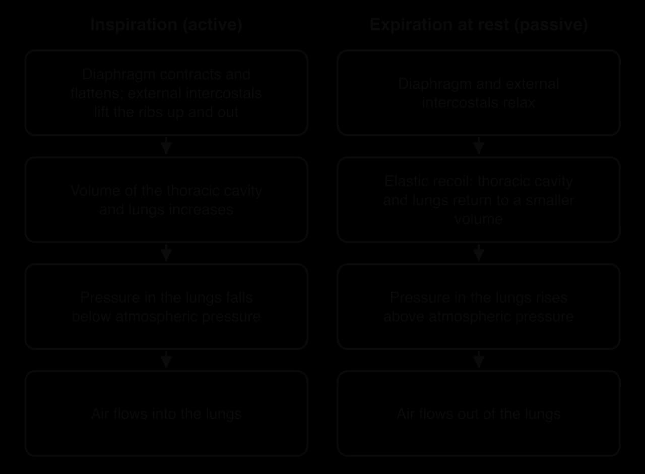
During exercise, breathing must become deeper and faster. Extra muscles, the accessory muscles of inspiration, contract to lift the rib cage higher, and expiration becomes active, so air is pushed out more forcefully and quickly.
Phase | At rest | During exercise |
|---|---|---|
Inspiration (active at rest and in exercise) | Diaphragm contracts and flattens; external intercostals lift ribs up and out | Diaphragm and external intercostals contract more strongly; accessory muscles (sternocleidomastoid, scalenes, pectoralis minor) lift the ribs and sternum further, so the chest expands more |
Expiration | Passive: diaphragm and external intercostals relax; elastic recoil of lungs | Active: internal intercostals pull the ribs down and in; abdominal muscles (rectus abdominis, obliques) push the abdominal organs up against the diaphragm |
Result | Small tidal volume, about 12–16 breaths min⁻¹ | Larger tidal volume and faster breathing; greater minute ventilation |
Lung volumes are measured with a spirometer, an instrument that records the volume of air breathed in and out over time. The trace it produces shows the normal breaths and the extra volumes that can be breathed in or out with maximal effort. Combinations of two or more volumes are called capacities.
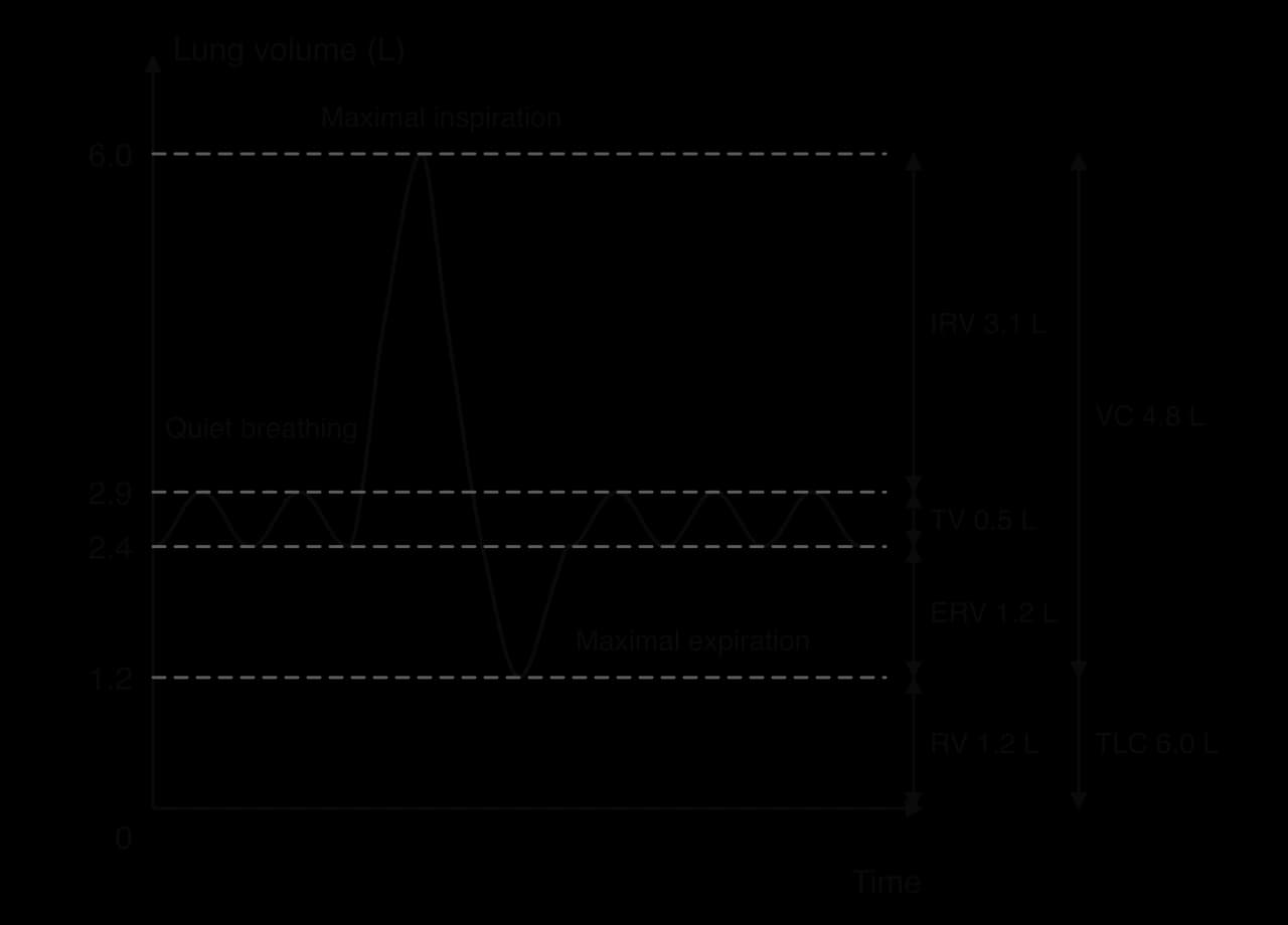
Above and below the tidal breath are the inspiratory reserve volume and the expiratory reserve volume, which can be used when breathing must increase.
The inspiratory reserve is large, so tidal volume can rise a great deal during exercise by using part of it.
Even after the most forceful expiration, the lungs do not empty completely. The air that remains keeps the alveoli open and allows gaseous exchange to continue between breaths.
Two capacities summarise the size of the lungs.
Vital capacity is easy to measure and is a common test of lung function. Adding the residual volume gives the total volume of the lungs, the total lung capacity.
Sources: typical lung volumes from OpenStax, Anatomy and Physiology 2e, 2022.
The volume of air moved per minute depends on how deeply and how often a person breathes. These are tidal volume and respiration rate, and their product is minute ventilation.
Respiration rate can be counted by watching the chest rise and fall. Minute ventilation needs a measure of tidal volume as well, so it is usually measured with a spirometer or a face mask connected to a gas analyser.
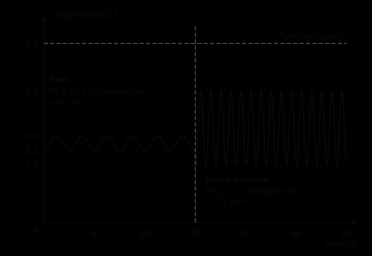
Ventilation follows a similar pattern to heart rate. It rises slightly before exercise (anticipatory rise), then increases sharply in the first seconds of exercise, mainly because of nervous signals from the motor cortex and from proprioceptors in the moving limbs. A slower rise follows over the next minutes, driven by chemical changes in the blood: rising carbon dioxide and hydrogen ions are detected by chemoreceptors, which signal the respiratory control centre in the brainstem. In submaximal exercise ventilation then levels off at a steady state. After exercise it falls rapidly and then more slowly.
At low and moderate intensities, minute ventilation increases mainly through a larger tidal volume, because deeper breaths are more efficient: each breath must first fill the airways (about 150 mL of “dead space” where no exchange occurs) before fresh air reaches the alveoli. As intensity rises further, tidal volume levels off at about half of vital capacity, and further increases come mainly from a faster respiration rate. During very hard exercise, ventilation rises faster than oxygen uptake, which helps to remove extra carbon dioxide and buffer the hydrogen ions produced alongside lactate.
Measure | Rest | Maximal exercise, untrained young adult | Maximal exercise, trained endurance athlete |
|---|---|---|---|
Tidal volume (L) | About 0.5 | About 2–2.5 | About 3 or more |
Respiration rate (breaths min⁻¹) | 12–16 | 40–50 | 50–60 |
Minute ventilation (L min⁻¹) | 6–8 | About 100 | 150–200 |
The table gives typical values; individuals vary widely, mainly with body size. The largest values are found in tall endurance athletes such as rowers and cross-country skiers.
Sources: Johnson, Saupe and Dempsey, Journal of Applied Physiology, 1992.
As with the heart, the respiratory response to exercise varies with the individual and the activity. The questions to ask are always the same: how large are the lungs and airways, how strong and fatigue-resistant are the breathing muscles, and how much oxygen does the activity demand?
Factor | Effect on lung volumes and ventilation | Explanation |
|---|---|---|
Age | Vital capacity and maximal ventilation fall in older adults; residual volume rises. Children have small volumes and breathe faster | Lung tissue and chest wall lose elasticity with age and the breathing muscles weaken; in children, small lungs mean small tidal volumes |
Sex differences | On average women have smaller lung volumes and narrower airways than men of the same height | Women reach expiratory flow limits more often and have a higher work of breathing at high ventilations |
Body size | Taller, larger people have larger lung volumes and higher maximal ventilation | Lung volumes scale mainly with height and chest size |
Level of fitness | Lung volumes change little; trained people ventilate less at a given submaximal workload and more at maximum | Training strengthens breathing muscles and improves oxygen use by muscles, so less ventilation is needed for the same work |
Type of activity | Rhythmic activities link breathing to movement (rowing, swimming); swimming restricts when breaths can be taken; holding the breath in lifting changes pressures | Breathing must fit the technique; water pressure on the chest makes inspiration harder |
Intensity of activity | Ventilation rises with intensity: first through tidal volume, then mainly through respiration rate | Greater production of carbon dioxide and hydrogen ions stimulates the chemoreceptors |
A study of endurance-trained men and women cycling to exhaustion found that expiratory flow limitation, where air cannot be breathed out any faster however hard the person tries, occurred in 90% of the women but 43% of the men. Above a ventilation of 90 L min⁻¹ the women’s work of breathing was on average twice that of the men, which the researchers linked to smaller lungs and narrower airways. This is one physiological difference between the sexes that persists even when both groups are highly trained.
Sources: Guenette et al., Journal of Physiology, 2007.
From the health lens, lung function is a strong indicator of general health. Smoking damages the alveoli and airways and reduces exercise capacity, while regular activity helps older adults keep their breathing muscles strong. People with chronic lung disease often find that their breathlessness, rather than their legs, limits exercise, and carefully planned training can improve how far they can walk.
Endurance athletes often have large hearts and very slow resting pulses. Because similar findings can occur in some forms of heart disease, doctors long debated whether they were a sign of damage. Imaging studies of large groups of athletes have shown that, in most cases, they are a healthy adaptation to training, and they give one of the clearest examples of how the cardiovascular system changes its structure to transport more oxygen.
A Swedish study used cardiovascular magnetic resonance (CMR), a scanning method that measures blood flow in the aorta without any catheter, to compare 60 professional athletes with age- and sex-matched healthy controls. The athletes, 30 women and 30 men aged 19–44, trained aerobically for at least 10 hours a week in triathlon, swimming, handball or football. Because the participants differed in size, the researchers expressed stroke volume and cardiac output per square metre of body surface area (stroke volume index and cardiac index).
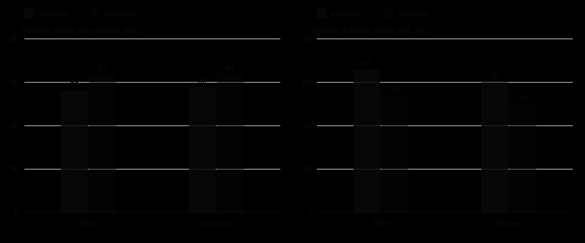
Group | Resting heart rate (beats min⁻¹), mean ± SD | Stroke volume index (mL m⁻²), mean ± SD |
|---|---|---|
Male athletes | 56 ± 7 | 66 ± 7 |
Male controls | 64 ± 10 | 54 ± 6 |
Female athletes | 58 ± 9 | 60 ± 9 |
Female controls | 64 ± 12 | 50 ± 7 |
The stroke volume index of the athletes was about 20% higher than that of the controls in both sexes, and their resting heart rate was 6–8 beats min⁻¹ lower; both differences were statistically significant. Their cardiac index, however, did not differ significantly from that of the controls (3.7 and 3.4 L min⁻¹ m⁻² in male and female athletes). This is exactly what the equation predicts: resting cardiac output is set by the body’s resting oxygen demand, and a heart that ejects more per beat needs fewer beats to deliver it.
The difference appears during exercise. The athlete’s heart starts from a larger stroke volume, which keeps rising up to maximal heart rate, so it reaches a much higher maximal cardiac output and supports a higher VO₂ max.
Training can thicken the wall of the left ventricle as well as enlarge its cavity, and a very thick wall is also a feature of hypertrophic cardiomyopathy, an inherited disease that is a cause of sudden cardiac death in young athletes. An Italian study measured the hearts of 947 elite athletes from 25 sports by echocardiography to find the upper limit of healthy thickening.
The thickest left ventricular wall among all 947 athletes was 16 mm.
Only 16 athletes (1.7%) had a wall of 13 mm or more, the range in which disease might be suspected; 15 of them were rowers or canoeists and one was a cyclist.
Every athlete with a wall of 13 mm or more also had an enlarged left ventricular cavity (55–63 mm across).
The researchers concluded that a wall thicker than 16 mm with a cavity that is not enlarged is unlikely to be caused by training and should be investigated as possible disease. Rowing and canoeing produced the largest hearts because they combine a high endurance demand with forceful contractions of a large muscle mass, loading the heart with both volume and pressure.
The athlete’s heart is an example of structure following function. Repeated endurance training increases the volume of blood returning to the heart and stretches the left ventricle; over months, the ventricle enlarges, and the Frank–Starling mechanism allows it to eject a larger stroke volume. A larger plasma volume and greater parasympathetic activity at rest contribute to the low resting heart rate. These changes are the main reason why endurance training raises maximal cardiac output and VO₂ max, a link to the training principles of specificity and reversibility: the enlargement partly reverses within weeks when training stops.
The evidence has limits. Both studies are cross-sectional: they compare athletes with non-athletes at one time and cannot show on their own that training caused the differences, since people born with larger hearts may be more likely to succeed in endurance sport. Longitudinal training studies, in which the same people are measured before and after training, are needed to show cause and effect. The CMR study measured people at rest only, and the echocardiography study was carried out in one country, so its limit of 16 mm may not apply equally to every population, including women, whose hearts are on average smaller.
Use the data in the table to calculate the cardiac index of the male controls, and compare it with the value for the male athletes.
Suggest why the researchers expressed stroke volume and cardiac output per square metre of body surface area.
To what extent does a low resting heart rate show that a person is fit? Consider both an athlete and an untrained adult with bradycardia.
How do specific qualities of long-term training, such as intensity and type of activity, influence the structures and functions of the cardiovascular system?
Sources: Carlsson et al., Journal of Cardiovascular Magnetic Resonance, 2012; Pelliccia et al., New England Journal of Medicine, 1991; Gledhill, Cox and Jamnik, Medicine and Science in Sports and Exercise, 1994.
Transport appears across all components of the exam. Paper 1A tests the structure of the heart, vessels and airways, the direction of blood flow, the dissociation curve and simple calculations. Paper 1B often uses data from a study of heart rate, blood pressure or ventilation. Paper 2 asks for labelled diagrams, graphs to sketch or annotate, calculations, and explanations of the responses to exercise.
Calculations follow the same pattern each time: the equation, the substitution with units, the answer with the correct unit and sensible significant figures.
Questions on the dissociation curve usually test the direction of the shift and its causes.
Calculation questions in Paper 1A are short and use simple numbers.
Breathing mechanics questions often contrast rest and exercise.
Paper 1B. In 1966, five healthy 20-year-old men took part in the Dallas Bed Rest and Training Study. Thirty years later, the same five men were tested again at age 50, and then after a 6-month endurance training programme. Their responses to maximal exercise are shown in the table (group means).
Measure at maximal exercise | 1966 (age 20, baseline tests) | 1996 (age 50, before training) | 1996 (age 50, after 6 months of training) |
|---|---|---|---|
VO₂ max (L min⁻¹) | 3.30 | 2.90 | 3.30 |
Maximal heart rate (beats min⁻¹) | 193 | 181 | 171 |
Maximal stroke volume (mL) | 104 | 121 | 129 |
Maximal cardiac output (L min⁻¹) | 20.0 | 21.4 | 21.7 |
Maximal a-vO₂ difference (mL O₂ per 100 mL blood) | 16.2 | 13.8 | 15.2 |
Sources: McGuire et al., Circulation, 2001 (two papers on the 30-year follow-up).
The next question checks whether the different responses of systolic and diastolic pressure can be explained.
Questions on the dissociation curve often combine a sketch with an explanation.
Short questions on venous return reward a named mechanism and how it works.
The cardiovascular system (heart, blood vessels, blood) transports oxygen, carbon dioxide, nutrients, hormones, heat and waste; humans have a double circulation, pulmonary and systemic.
The heart has four chambers and four valves; the left ventricle has the thickest wall because it pumps blood around the whole body.
Arteries have thick, muscular, elastic walls; capillaries have walls one cell thick for exchange; veins have thin walls, wide lumens and valves.
Cardiac output = heart rate × stroke volume; it is about 5 L min⁻¹ at rest and can rise to about 20 L min⁻¹ in untrained people and over 35 L min⁻¹ in elite endurance athletes.
Stroke volume rises with venous return (skeletal muscle pump, respiratory pump, venoconstriction) through the Frank–Starling mechanism and plateaus at moderate intensity in untrained people.
In dynamic exercise systolic pressure rises and diastolic pressure stays about constant; heavy resistance exercise raises both steeply.
Blood is redistributed to working muscles by vasodilation and away from the gut and kidneys by vasoconstriction.
The S-shaped dissociation curve shifts right in exercising muscle (Bohr effect), releasing more oxygen and widening the a-vO₂ difference.
Alveoli are adapted for diffusion; breathing depends on muscles changing thoracic volume and pressure; expiration is passive at rest and active in exercise.
Minute ventilation = tidal volume × respiration rate; tidal volume rises first, then rate; all responses vary with age, sex, body size, fitness, type and intensity of activity.
I can label the chambers, valves and great vessels of the heart and outline the route of blood through the pulmonary and systemic circulations.
I can compare the structure and function of arteries, capillaries and veins, and state the components of blood.
I can describe what the blood transports and how oxygen and carbon dioxide are carried.
I can calculate cardiac output and minute ventilation with the correct units.
I can sketch and annotate the heart rate response to exercise and explain the changes in stroke volume and cardiac output.
I can explain venous return, the Frank–Starling mechanism, blood pressure responses and blood redistribution.
I can sketch the oxygen–haemoglobin dissociation curve and explain the Bohr shift and the a-vO₂ difference.
I can explain how the alveoli are adapted for gaseous exchange by diffusion down partial pressure gradients.
I can explain the mechanics of breathing at rest and during exercise, and label a spirometer trace.
I can discuss how age, sex, body size, fitness, type and intensity of activity affect cardiovascular and respiratory responses.