Explain how negative feedback regulates blood pH, the heart, core temperature and blood glucose, what affects thermoregulation and, at HL, how the body responds and adapts to heat, humidity, cold and altitude.
Each is in the SEHS glossary, with every other term in the subject.
The physiological adaptations that develop with repeated or continuous exposure to a stressful environment such as heat, cold or altitude, reducing its strain on the body and improving performance in it.
An illness caused by rapid ascent to high altitude, usually above about 2,500 m, with headache, nausea, fatigue, dizziness and poor sleep, which usually ease within a few days.
Training or living at altitude, usually about 1,800–3,000 m, or in simulated low-oxygen conditions, to increase red cell mass and haemoglobin and so improve endurance performance.
The homeostatic control of blood glucose concentration by negative feedback, in which the pancreas releases insulin when glucose rises and glucagon when it falls.
A measure of the hydrogen ion concentration of the blood; the more hydrogen ions, the lower the pH. Normal arterial pH is about 7.35–7.45.
A type of fat tissue, plentiful in newborns and present in smaller amounts in adults, whose mitochondria release energy from fat directly as heat in non-shivering thermogenesis.
A substance in a body fluid that resists changes in pH by combining with or releasing hydrogen ions; the main blood buffer is hydrogen carbonate (bicarbonate).
Outline homeostasis, set points and normal ranges, and the roles of receptors, control centres and effectors.
Distinguish between negative feedback and positive feedback, and sketch a generic negative feedback loop.
Explain how carbon dioxide affects blood pH and how chemoreceptors and the respiratory control centre restore it during exercise.
Explain how intrinsic and extrinsic control of the heart, including the baroreceptor reflex, keeps blood pressure and blood gases in range.
Describe how heat is produced and gained by the body and lost by radiation, conduction, convection and evaporation.
Explain thermoregulation in hot and cold conditions, including the sweat response, vasodilation, vasoconstriction, shivering and non-shivering thermogenesis.
Discuss how training status, body composition, the environment and sex differences affect thermoregulation.
Explain how insulin and glucagon regulate blood glucose, and how exercise changes insulin release and glucose uptake.
Describe the acute responses and long-term adaptations to heat, humidity, cold and altitude, and how their impact depends on the activity (HL).
Evaluate strategies used to acclimatise to the environment and to support performance in it (HL).
The human body works properly only when the conditions inside it stay within narrow limits. Core temperature, the acidity of the blood, blood pressure and the concentration of glucose in the blood are all held close to their normal values, even though exercise and the weather push them away from those values all the time. The processes that keep them stable are called homeostasis.
With a free account, Augment remembers where you stopped and what you have practiced.
The transfer of heat by direct contact between the skin and a cooler or hotter solid, liquid or gas, such as cold water or a cold surface.
The part of a homeostatic system, usually in the brain or an endocrine gland, that compares information from receptors with the set point and signals effectors to correct any difference.
The transfer of heat to or from the skin by a moving fluid such as air or water; heat loss increases with wind speed or movement through water.
The temperature of the deep tissues of the body, such as the brain, heart and abdominal organs, normally kept at about 37 ± 1 °C.
A muscle or gland that receives a signal from a control centre and produces a response that changes a regulated variable.
Heat loss as water, mainly sweat, changes from liquid to vapour on the skin or in the airways; the main route of heat loss during exercise and the only one when air is hotter than skin.
The freezing of body tissue, usually of the fingers, toes, ears or nose, when skin temperature falls below freezing; severe frostbite can cause permanent tissue loss.
The adaptations to repeated exercise in hot conditions over about 1–2 weeks, including earlier and greater sweating, more dilute sweat, larger plasma volume and lower heart rate and core temperature at a given workload.
A heat illness in which the cardiovascular system cannot meet the demands of muscles and skin, usually with dehydration, causing fatigue, dizziness, nausea and sometimes collapse, with core temperature usually below 40 °C.
A life-threatening failure of thermoregulation in which core temperature rises above about 40 °C with central nervous system dysfunction such as confusion or collapse; it needs immediate whole-body cooling.
The self-regulating processes that keep the body's internal environment relatively stable, within narrow limits, so that cells and organs function optimally despite internal and external changes.
A fall in core temperature below 35 °C, when heat loss exceeds heat production, causing shivering, then confusion, loss of coordination and, if it continues, unconsciousness and death.
A shortage of oxygen reaching the body's tissues; at altitude it results from the lower partial pressure of oxygen, which reduces the saturation of haemoglobin with oxygen.
An altitude training strategy in which athletes live or sleep at moderate or simulated altitude to stimulate red blood cell production but train at low altitude to maintain training intensity.
A control mechanism in which a change in a regulated variable triggers a response that reverses the change, returning the variable towards its set point; the basis of homeostasis.
Heat production without muscle contraction, by raising the metabolic rate of tissues, especially brown adipose tissue, under the control of sympathetic nerves and hormones such as thyroxine and epinephrine.
The part of the total pressure of a gas mixture caused by oxygen, equal to the total pressure multiplied by the oxygen fraction; it falls with altitude and drives oxygen diffusion into the blood.
A control mechanism in which a change triggers a response that increases the change further until an outside event stops it, as in blood clotting and childbirth.
Lowering core and skin temperature before exercise in the heat, for example with ice slurry, ice vests or cold-water immersion, so that more heat can be stored before it limits performance.
The transfer of heat as infrared waves between the skin and surrounding objects without contact; the body loses heat to cooler surroundings and gains it from the sun and hot surfaces.
A specialised cell or group of cells that detects a change in a regulated variable and sends information about it to a control centre, usually as nerve impulses.
The amount of water vapour in the air as a percentage of the most the air could hold at that temperature; high relative humidity slows the evaporation of sweat.
The target value of a regulated variable, such as a core temperature of about 37 °C, around which control systems keep it fluctuating within a normal range.
Heat production by rapid, involuntary contractions of skeletal muscle, triggered by the hypothalamus when core temperature falls; nearly all the energy released becomes heat.
The secretion of sweat by eccrine sweat glands, stimulated by the sympathetic nervous system when core or skin temperature rises, so that heat is lost as the sweat evaporates.
A sensory receptor that detects temperature; central thermoreceptors in the hypothalamus detect blood temperature and peripheral thermoreceptors in the skin detect skin temperature.
The homeostatic regulation of core body temperature, in which the hypothalamus balances heat production and gain against heat loss to keep core temperature at about 37 ± 1 °C.
The narrowing of arterioles when the smooth muscle in their walls contracts, reducing blood flow to the tissue they supply, such as the skin when the body is cold.
The widening of arterioles when the smooth muscle in their walls relaxes, increasing blood flow to the tissue they supply, such as the skin when the body is hot.
An index of environmental heat stress combining the natural wet-bulb temperature (humidity and air movement), black globe temperature (radiation) and dry-bulb air temperature, weighted 0.7, 0.2 and 0.1 outdoors.
Cells are bathed in tissue fluid, which exchanges substances with the blood. This fluid is the cells’ internal environment. Enzymes, nerve impulses and muscle contraction all depend on its temperature, acidity, water content and concentration of nutrients and ions. If these drift too far, chemical reactions slow down or stop, and cells are damaged.
The word “relatively” is important. Homeostasis does not keep a variable perfectly constant. Each variable fluctuates around a target value, its set point, and stays inside a normal range that is safe for the body. Core temperature, for example, has a set point close to 37 °C but varies by about 0.5 °C over a normal day, being lowest in the early morning and highest in the late afternoon.
Changes that disturb the internal environment come from two directions. Internal changes come from the body itself: working muscles release heat, carbon dioxide and hydrogen ions, and use glucose. External changes come from the surroundings: hot or cold air, humid weather and the low oxygen pressure at altitude. Homeostatic mechanisms work all the time, at rest and during exercise, and respond to both.
Regulated variable | Typical normal value or range | Main control centre | Main effectors |
|---|---|---|---|
Core temperature | 37 ± 1 °C | Hypothalamus | Sweat glands, skin arterioles, skeletal muscle |
Arterial blood pH | 7.35–7.45 | Respiratory control centre (medulla) | Diaphragm and intercostal muscles; also the kidneys over hours to days |
Blood pressure (resting, adult) | About 120/80 mmHg | Cardiovascular control centre (medulla) | SA node, heart muscle, arterioles |
Blood glucose (fasting) | About 3.9–5.6 mmol L⁻¹ | Pancreas (islets of Langerhans) | Liver, skeletal muscle, adipose tissue |
Blood water and ion concentration | Plasma sodium about 135–145 mmol L⁻¹ | Hypothalamus | Kidneys (with ADH) |
The table shows that each regulated variable has its own receptors, control centre and effectors, but they all work on the same principle. The regulation of water and ions is part of water and electrolyte balance; the other four are blood pH, the heart, core temperature and blood glucose.
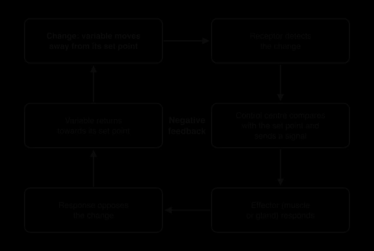
Every homeostatic system has three parts that pass information in a chain: a receptor senses the variable, a control centre compares it with the set point, and an effector acts to change it.
Receptors in homeostatic systems include thermoreceptors in the skin and hypothalamus, which detect temperature; chemoreceptors in the medulla, aorta and carotid arteries, which detect carbon dioxide, hydrogen ions and oxygen; and baroreceptors in the walls of the aorta and carotid arteries, which detect stretch caused by blood pressure.
The main control centres are in the brain. The hypothalamus controls temperature and water balance, while the medulla oblongata of the brainstem contains the respiratory control centre and the cardiovascular control centre. The pancreas acts as both receptor and control centre for blood glucose. Control centres send their signals through the autonomic nervous system (fast) or through hormones in the blood (slower but longer-lasting).
Effectors include skeletal muscle (shivering), smooth muscle in the walls of arterioles (vasodilation and vasoconstriction), cardiac muscle and the SA node (heart rate and force), sweat glands, the liver and the endocrine glands themselves.
Almost all homeostatic control works by negative feedback. When a variable moves away from its set point, the response of the effectors pushes it back in the opposite direction. As the variable returns towards the set point, the receptors detect less change, so the response becomes smaller. The system is “self-regulating” because the result of the response feeds back to switch the response off.
The loop in the diagram can be applied to any regulated variable by naming the specific receptor, control centre and effector. When core temperature rises during a run, thermoreceptors are the receptors, the hypothalamus is the control centre and sweat glands and skin arterioles are the effectors. The response (more heat loss) opposes the change (a rise in temperature). Each regulated variable usually has two opposing sets of responses, one for a rise and one for a fall, which gives finer control than a single response that is simply switched on and off.
In positive feedback the response increases the original change instead of reversing it, so the change gets larger and larger until some event outside the loop ends it. Positive feedback cannot keep a variable stable, which is why it is rare in homeostasis. It is useful only for processes that need to be completed quickly once they have started.
Two examples are well known. In blood clotting, platelets that stick to a damaged vessel release chemicals that attract and activate more platelets, so the clot grows rapidly until the wound is sealed. In childbirth, the pressure of the baby on the cervix causes the release of the hormone oxytocin, which strengthens contractions of the uterus, which increases the pressure further; the cycle ends when the baby is born.
Exercise can also show what happens when negative feedback is overwhelmed. If heat is produced faster than it can be lost, core temperature keeps rising. A higher temperature raises the rate of metabolism, which produces more heat. This vicious circle behaves like positive feedback and is one reason why heat stroke can develop rapidly once thermoregulation fails.
 YouTube
The acidity of a solution depends on its concentration of hydrogen ions (H⁺). The pH scale measures this: the more hydrogen ions, the lower the pH. Arterial blood is kept slightly alkaline, between pH 7.35 and 7.45. Enzymes, haemoglobin and the proteins that cause muscle contraction all work best within this range.
Because pH is a logarithmic scale, a fall of just 0.1 pH units means a rise of about 26% in hydrogen ion concentration. A pH below 7.35 is called acidosis, and a pH above 7.45 is called alkalosis. Both are dangerous if they are large or last a long time.
The main threat to blood pH during exercise is carbon dioxide (CO₂), a waste product of aerobic respiration in the mitochondria. CO₂ diffuses from the muscle cells into the blood, where most of it reacts with water to form carbonic acid (H₂CO₃). Carbonic acid then splits (dissociates) into hydrogen ions and hydrogen carbonate (bicarbonate) ions:
The double arrows show that the reactions are reversible. When CO₂ is added, the reactions move to the right, more H⁺ is produced and pH falls. When CO₂ is removed, for example by breathing it out, the reactions move to the left, H⁺ combines with HCO₃⁻ and pH rises. This is why ventilation and blood pH are so closely linked: the lungs control pH by controlling how much CO₂ stays in the blood.
During high-intensity exercise, a second source of hydrogen ions is added. Fast breakdown of glucose by the glycolytic system releases H⁺ in the muscle along with lactate. Some of these ions enter the blood, so pH can fall even when CO₂ is being removed efficiently.
Blood pH is regulated by negative feedback through the respiratory system:
During exercise, working muscles produce more CO₂ (and, at high intensities, more H⁺).
CO₂ and H⁺ concentrations in the blood rise, so pH falls.
Central chemoreceptors in the medulla detect the rise in CO₂ (through the H⁺ it forms in the fluid around the brain). Peripheral chemoreceptors in the aortic arch and carotid arteries detect the rise in CO₂ and H⁺, and a large fall in oxygen.
The chemoreceptors send nerve impulses to the respiratory control centre in the medulla.
The respiratory control centre increases the rate and force of the impulses sent through the phrenic nerve to the diaphragm and the intercostal nerves to the intercostal muscles.
Breathing becomes faster and deeper, so minute ventilation rises and more CO₂ is breathed out.
CO₂ and H⁺ in the blood fall and pH returns towards 7.4. The chemoreceptors are stimulated less and ventilation falls.
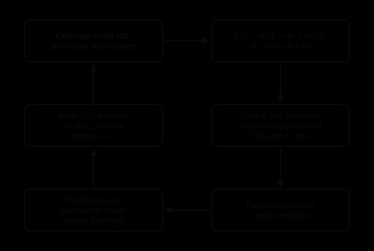
The loop explains a common observation after a sprint or a hard interval. Breathing stays fast and deep for several minutes after exercise stops, because the blood still contains extra CO₂ and H⁺ that have to be removed. A rise in breathing before exercise starts is a different process: it is an anticipatory response controlled by the brain, not by feedback from chemoreceptors.
Ventilation takes seconds to minutes to act. A faster first line of defence is chemical buffering. A buffer is a substance that takes up hydrogen ions when they are added and releases them when they are removed, so pH changes much less than it otherwise would.
Bicarbonate in the plasma combines with H⁺ to form carbonic acid, which then breaks down into CO₂ and water that can be breathed out. Proteins, including haemoglobin, also act as buffers inside red blood cells and muscle fibres. Over hours and days, the kidneys add a third, slower mechanism by excreting H⁺ in the urine and keeping or excreting bicarbonate.
The heart has to pump enough blood to keep blood pressure high enough to supply every organ, and to deliver oxygen and remove CO₂ at the rate the tissues need. Both depend on cardiac output, the volume of blood pumped per minute:
where heart rate (HR) is in beats min⁻¹ and stroke volume (SV) is the volume pumped per beat. The heart’s output is controlled in two ways, which together form a homeostatic system.
Intrinsic control comes from inside the heart. The sinoatrial node (SA node) in the wall of the right atrium is the pacemaker: it generates electrical impulses on its own, without any input from nerves. The impulse spreads across the atria, which contract, and reaches the atrioventricular node (AV node). After a short delay that lets the ventricles fill, it passes down the bundle of His and through the Purkinje fibres so that the ventricles contract from the apex upwards. These tissues are labelled on the standard diagram of the heart’s structure.
Intrinsic control also adjusts stroke volume. When more blood returns to the heart, the ventricle walls are stretched further and contract more forcefully, so more blood is ejected (the Frank–Starling mechanism). Without any nervous or hormonal input, the SA node would set a heart rate of roughly 100 beats min⁻¹; at rest, extrinsic control slows it below this.
Extrinsic control comes from outside the heart and adjusts the intrinsic rhythm to meet the body’s needs. The cardiovascular control centre in the medulla sends signals through two sets of autonomic nerves. Hormones from the adrenal glands add a slower, longer-lasting effect.
Extrinsic influence | Pathway | Effect on the heart | When it dominates |
|---|---|---|---|
Parasympathetic nervous system | Vagus nerve to the SA node and AV node | Slows the SA node: heart rate falls | At rest (resting HR is below the SA node’s own rate because of vagal “braking”); in recovery |
Sympathetic nervous system | Cardiac accelerator nerves to the SA node, AV node and ventricle walls | Raises heart rate and force of contraction, so stroke volume rises | During exercise, especially above about 100 beats min⁻¹; under stress |
Hormonal | Epinephrine and norepinephrine from the adrenal medulla, carried in the blood | Raise heart rate and force of contraction | Before and during exercise, under stress; effect lasts minutes |
At the start of exercise, the first increase in heart rate comes mainly from withdrawing parasympathetic (vagal) stimulation. As intensity rises, sympathetic stimulation and circulating epinephrine take over, raising both heart rate and stroke volume. The vagus nerve then becomes dominant again in recovery, which is why heart rate falls quickly in the first minute after exercise stops.
Blood pressure is the regulated variable that the heart’s extrinsic control protects most directly. Baroreceptors in the walls of the aortic arch and the carotid arteries are stretch receptors: the higher the blood pressure, the more the artery walls stretch and the more impulses the baroreceptors send to the cardiovascular control centre. The resulting negative feedback loop is called the baroreceptor reflex.
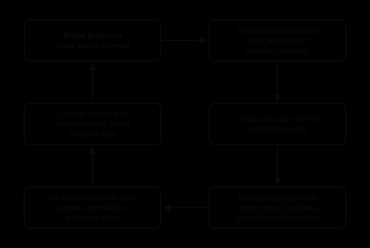
When blood pressure rises, the cardiovascular control centre increases parasympathetic stimulation and reduces sympathetic stimulation. Heart rate and force of contraction fall, so cardiac output falls, and arterioles in many tissues dilate, which reduces the resistance to blood flow. Blood pressure falls back towards normal. When blood pressure falls, for example when a person stands up quickly and blood pools in the legs, the opposite happens: heart rate and force of contraction rise and arterioles constrict.
Chemoreceptors feed into the same control centre. A rise in CO₂ or H⁺, or a large fall in oxygen, increases sympathetic stimulation of the heart, so cardiac output rises and blood flows faster through the lungs and muscles. In this way the heart helps to keep blood gases in range alongside the respiratory system.
During exercise, blood pressure rises, especially systolic pressure, yet heart rate keeps rising instead of being slowed by the baroreceptor reflex. This is not a failure of homeostasis. Signals from the motor areas of the brain (“central command”) and from receptors in the working muscles reset the baroreceptor reflex to work around a higher set point. The reflex still operates, preventing blood pressure from rising too far, but around a level that suits exercise. After exercise the set point returns to its resting level.
Of all the homeostatic systems, temperature regulation is the one most often pushed to its limits in sport. A marathon runner can produce enough heat to raise core temperature by about 1 °C every 5–10 minutes if none of it were lost, and an open-water swimmer in cold water can lose heat many times faster than in air at the same temperature.
The body can be pictured as a warm core (the brain and the organs of the chest and abdomen) surrounded by a cooler shell (the skin, fat and limb muscles). Core temperature is kept close to 37 °C, while the shell can vary much more: the skin of the hands may be below 20 °C on a cold day and above 35 °C during exercise in the heat.
The narrow range matters because enzymes and cell membranes are sensitive to temperature. A modest rise in muscle temperature during a warm-up is beneficial, because nerve conduction, enzyme activity and the release of oxygen from haemoglobin all speed up. A core temperature above about 40 °C, however, starts to damage proteins and impair the central nervous system, and a core temperature below 35 °C slows the heart and the brain.
All the energy released in metabolism eventually becomes heat. At rest, most of this heat comes from the liver, heart, brain and other organs. During exercise, skeletal muscle becomes by far the largest source, because muscle contraction is inefficient: only about 20–25% of the energy released from fuels is converted into mechanical work, and the other 75–80% is released as heat.
The body can also gain heat from its surroundings. Direct sunlight and hot ground add heat by radiation, and air or surfaces hotter than the skin add heat by convection and conduction. On a hot, sunny day the environment and metabolism together can add more heat than the body can lose, so core temperature keeps rising.
Heat moves from a hotter object to a cooler one. The body exchanges heat with its surroundings by four routes: radiation, conduction, convection and evaporation. The first three depend on the skin being warmer than its surroundings; the fourth does not.
At rest in a cool room, radiation is the largest route of heat loss. Outdoors in the sun, the direction reverses and the body gains a large amount of radiant heat, which is why shade matters so much for athletes and spectators.
Conduction needs contact, so it matters most when the skin touches cold water, snow, metal or the ground. When the fluid in contact with the skin is moving, the process becomes convection.
Conduction to air is small, because air conducts heat poorly, but conduction and convection to water are large: water conducts heat about 25 times better than air. This is why swimmers in cold water and runners in wet clothing lose heat so quickly. Convection also explains why a cyclist moving at 40 km h⁻¹ feels cooler than a runner at the same air temperature.
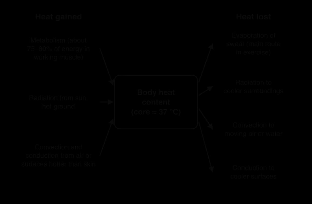
Evaporation removes a large amount of heat: about 2.4 kJ for every gram (millilitre) of sweat that evaporates from the skin. Because it does not depend on the skin being warmer than the air, it is the only way of losing heat when the air temperature is above skin temperature (about 33–35 °C). Its rate depends on the humidity of the air: the more water vapour the air already holds, the more slowly sweat evaporates.
The hypothalamus, at the base of the brain, acts as the body’s thermostat. It receives information from two sets of thermoreceptors: central thermoreceptors in the hypothalamus itself, which detect the temperature of the blood flowing through the brain, and peripheral thermoreceptors in the skin, which detect changes in skin temperature and give early warning of changes in the environment.
The hypothalamus compares the incoming information with its set point and sends signals through the sympathetic nervous system to the sweat glands and the smooth muscle of skin arterioles, through motor nerves to skeletal muscle, and through hormones to the body’s cells. It controls two sets of responses: one for heat and one for cold.
When core temperature rises, whether from exercise or a hot environment, the hypothalamus increases heat loss in two ways.
Sympathetic nerves stimulate the eccrine sweat glands, which are spread over the whole skin surface, to secrete sweat, a dilute solution of water, sodium, chloride and small amounts of other substances. Sweating starts within a few minutes of starting exercise and its rate rises with core temperature and exercise intensity. Sweat rates of 1–2 L h⁻¹ are common in endurance athletes in hot conditions.
The cost of the sweat response is fluid loss. Sweat comes from the plasma, so heavy sweating reduces blood volume, which leads to dehydration and contributes to cardiovascular drift during long exercise. The loss of sodium in sweat also affects electrolyte balance.
The hypothalamus reduces the sympathetic signals that keep skin arterioles narrow and activates nerves that widen them. The smooth muscle in the arteriole walls relaxes and the arterioles widen, so much more warm blood from the core flows through the skin. Heat passes from this blood to the skin surface, where it can be lost by radiation, convection and evaporation. Skin blood flow can rise from about 0.25 L min⁻¹ at rest in a cool room to several litres per minute in severe heat stress.
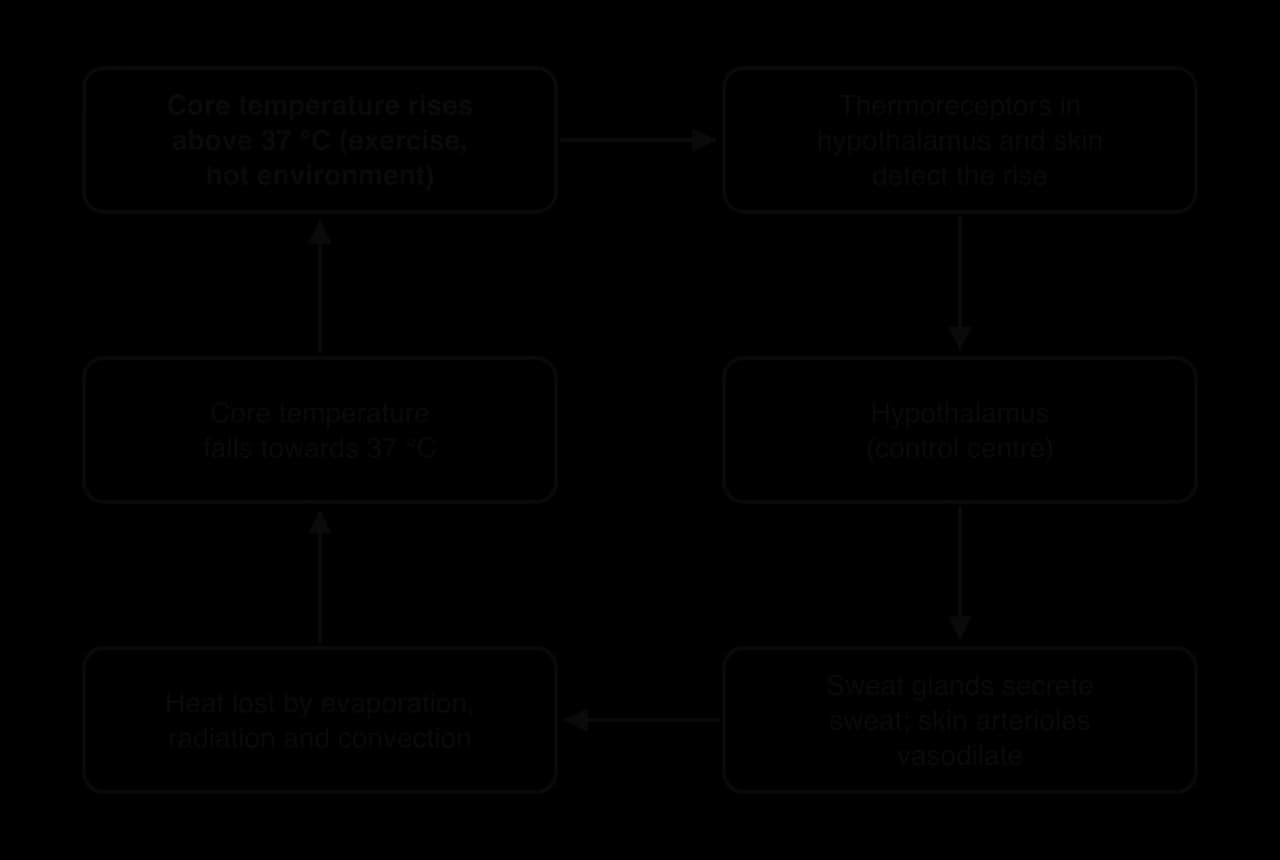
Heat stress therefore creates competition for blood. During exercise in the heat, cardiac output has to supply both the working muscles (for oxygen) and the skin (for cooling). Blood sent to the skin is not available to the muscles, and the large volume of blood held in the dilated skin veins reduces the blood returning to the heart. Stroke volume falls and heart rate rises to compensate, so the same pace feels harder in the heat than in cool conditions.
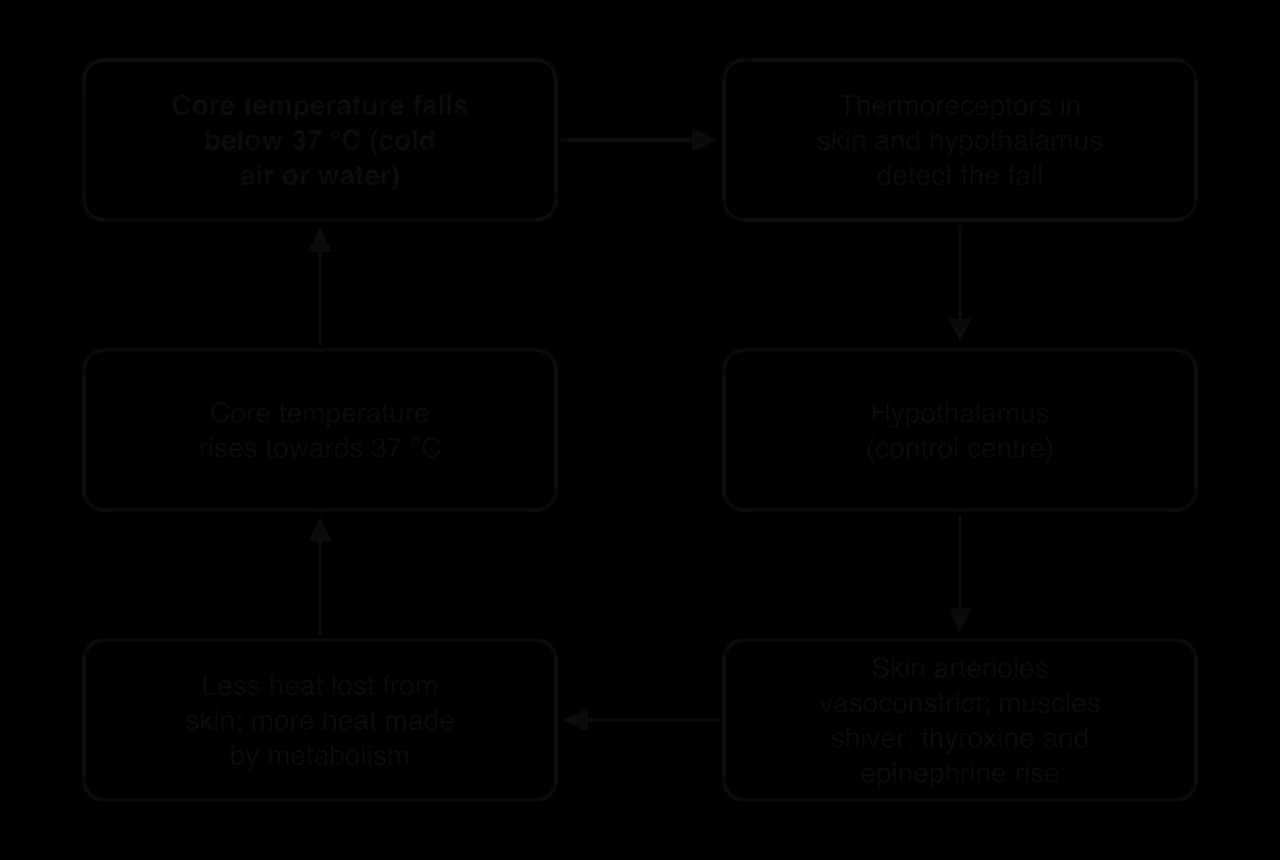
When core or skin temperature falls, the hypothalamus reduces heat loss and increases heat production.
Increased sympathetic stimulation makes the smooth muscle in skin arterioles contract. The arterioles narrow, so less warm blood flows to the skin and less heat is lost from the surface. The skin becomes pale and cool while the core stays warm, and the cooler shell of skin and fat acts as insulation.
Vasoconstriction protects the core at the expense of the hands, feet, ears and nose. In severe cold, skin temperature in these areas can fall low enough for tissue to freeze, which is why gloves, hats and warm footwear matter in winter sports.
If vasoconstriction is not enough, the hypothalamus triggers shivering: rapid, involuntary, rhythmic contractions of skeletal muscles that do no useful external work, so almost all the energy released becomes heat. Shivering can raise heat production to several times the resting rate, but it uses muscle glycogen and blood glucose, interferes with skilled movement and causes fatigue.
The body can also produce heat without muscle contraction. The hypothalamus increases sympathetic stimulation and the release of hormones that speed up metabolism in many tissues. Epinephrine and norepinephrine act within minutes. Thyroxine from the thyroid gland raises the basal metabolic rate over days to weeks of cold exposure. In brown adipose tissue, a special fat tissue found mainly around the neck, collarbones and spine, mitochondria release energy from fat directly as heat instead of using it to make ATP.
Brown adipose tissue is plentiful in newborn babies, who cannot shiver effectively. Adults have much less, and the amount varies between individuals; it increases with repeated cold exposure. Non-shivering thermogenesis is therefore a smaller source of heat in adults than shivering.
Thermoregulation depends on several body systems acting together, coordinated by the nervous system.
System | Role when hot | Role when cold |
|---|---|---|
Nervous | Thermoreceptors detect the rise; hypothalamus integrates; sympathetic nerves stimulate sweat glands and adjust skin arterioles | Thermoreceptors detect the fall; hypothalamus integrates; sympathetic nerves constrict skin arterioles; motor nerves trigger shivering |
Cardiovascular | Vasodilation of skin arterioles; higher heart rate keeps blood pressure up while blood is diverted to the skin | Vasoconstriction of skin arterioles keeps warm blood in the core |
Integumentary (skin, sweat glands, hair) | Sweat glands secrete sweat; skin surface is where heat is lost | Fat under the skin insulates; hair erector muscles raise body hair (of little use in humans) |
Muscular | Source of most heat during exercise; lower pace reduces heat production | Shivering produces heat |
Endocrine | Hormones such as antidiuretic hormone limit fluid loss in urine | Epinephrine, norepinephrine and thyroxine raise metabolic rate |
Behaviour adds a further layer of control. People seek shade, remove or add clothing, drink cold fluids and, in sport, slow down. Slowing the pace is a powerful response, because it reduces heat production at its source. Many researchers think that the drop in self-chosen pace in the heat is partly a protective, anticipatory response that limits the rise in core temperature, which links thermoregulation to the study of fatigue.
Two people doing the same exercise in the same conditions can end up with very different core temperatures. The main reasons are their training status, body composition and size, the environment, and differences between the sexes, including hormonal phases.
Endurance training improves thermoregulation, even in cool conditions, because regular exercise raises core temperature and so acts as a repeated heat stress. Compared with untrained people, endurance-trained athletes start sweating at a lower core temperature and sweat more at a given core temperature, so they lose heat earlier and faster. They also have a larger plasma volume and blood volume, which allows them to supply both the skin and the muscles while keeping stroke volume up. Training does not replace heat acclimatisation: athletes who train in a cool climate still gain further adaptations from training in the heat.
Body fat is a good insulator, because it conducts heat less well than muscle. A higher proportion of body fat helps in cold water, which is one reason why successful long-distance open-water swimmers often carry more fat than pool swimmers. In the heat it is a disadvantage, because it slows the movement of heat from the core to the skin, and extra mass increases the energy cost, and so the heat produced, of weight-bearing exercise.
Body size matters through the surface area to mass ratio. Heat is produced throughout the body’s mass but lost through its surface. A small, light person has a large surface area for each kilogram of mass, so they lose heat quickly; a large, heavy person has a small surface area for their mass and loses heat more slowly.
Large athletes, such as rugby forwards and American football linemen, are therefore at greater risk of overheating in hot weather. Children have a high surface area to mass ratio, so they gain heat quickly in hot environments (when air is hotter than skin) and lose it quickly in cold water. Children also sweat less than adults, so they rely more on vasodilation to lose heat.
Air temperature is only one part of heat stress. The other parts are:
Humidity. Relative humidity is the amount of water vapour the air holds compared with the most it could hold at that temperature. The more humid the air, the more slowly sweat evaporates, so heat loss falls even though the sweat rate stays high.
Wind (air movement). Moving air increases convection and evaporation. Running into a breeze helps cooling; a tailwind at the same speed as the runner removes this benefit.
Radiation. Direct sun and heat reflected from roads, running tracks and sand add a large heat load.
Water. Immersion in cold water removes heat far faster than air at the same temperature.
Because these factors act together, sports organisations measure heat stress with the wet bulb globe temperature (WBGT). Three thermometers are used: a natural wet-bulb thermometer covered by a wet wick (which reflects humidity and air movement), a black globe thermometer (which reflects radiant heat) and an ordinary dry-bulb thermometer (air temperature). Outdoors, they are combined as:
The weighting shows that humidity has by far the largest effect, because it decides whether sweat can evaporate. A WBGT reading is used to decide whether to modify a training session, add drinks and cooling breaks, or postpone or cancel an event. Its limitation is that it describes the environment only: it does not account for the athlete’s clothing, intensity of work, fitness or acclimatisation.
On average, men have higher maximal sweat rates than women. Much of this difference, and of differences in core temperature during exercise, is explained by body size and fitness rather than sex itself: when men and women of similar size, fitness and heat production are compared, their thermoregulation is similar. Women, on average, have a smaller body mass, a higher surface area to mass ratio and more body fat, which affect heat loss in the ways already described.
Hormonal phases also matter. After ovulation, in the luteal phase of the menstrual cycle, progesterone raises core temperature by about 0.3–0.5 °C and raises the temperature at which sweating and skin vasodilation begin. A woman exercising in the heat during the luteal phase therefore starts with a slightly higher core temperature and starts losing heat a little later. In the years around the menopause, falling and fluctuating oestrogen levels can disturb the hypothalamus’s temperature control, causing hot flushes: sudden episodes of vasodilation and sweating, in which the body loses heat even though core temperature has not risen much.
Factor | Health lens | Performance lens |
|---|---|---|
Training status | Regular activity improves heat tolerance in daily life and at work, for example in outdoor workers and older adults | Fitter athletes sweat earlier and have more plasma, so they can hold a higher pace in the heat |
Body composition and size | Children and people with obesity are at greater risk of heat illness; older and very lean people are more vulnerable in the cold | Large athletes overheat sooner; lean athletes cool quickly in cold water |
Environment | Heatwaves raise rates of heat illness and death, especially in older people | Humidity and sun determine scheduling, cooling breaks and pacing |
Sex and hormonal phase | Hot flushes around the menopause affect sleep and well-being | Higher luteal-phase core temperature may slightly reduce performance in the heat for some women |
Glucose is the main fuel for the brain and an important fuel for working muscles. Too little glucose in the blood starves the brain, causing confusion and, in severe cases, unconsciousness. Too much draws water out of cells and, over years, damages blood vessels, nerves and the kidneys. Blood glucose regulation keeps the concentration within a narrow range.
In a healthy adult who has not eaten for at least eight hours, plasma glucose is normally about 3.9–5.6 mmol L⁻¹ (70–100 mg dL⁻¹). After a meal containing carbohydrate it rises for an hour or two, then returns to this range. A fasting value of 7.0 mmol L⁻¹ (126 mg dL⁻¹) or more is one of the criteria used to diagnose diabetes, and a value below about 3.9 mmol L⁻¹ is classed as low (hypoglycaemia).
Sources: American Diabetes Association, diagnosis criteria, 2026.
The pancreas contains clusters of endocrine cells, the islets of Langerhans, that act as both receptor and control centre. Their beta (β) cells detect a rise in blood glucose and release insulin. Their alpha (α) cells detect a fall in blood glucose and release glucagon. The two hormones have opposite effects, so the system can correct a change in either direction.
When blood glucose rises (after a meal): insulin is released. It makes muscle, liver and fat cells take up glucose from the blood, and it stimulates the liver and muscles to store glucose as glycogen. Blood glucose falls back into the normal range, so insulin release falls.
When blood glucose falls (fasting, prolonged exercise): glucagon is released. It acts on the liver, which breaks down stored glycogen into glucose (glycogenolysis) and makes new glucose from non-carbohydrate sources such as amino acids, lactate and glycerol (gluconeogenesis). The liver releases this glucose into the blood, so blood glucose rises back into the normal range and glucagon release falls.
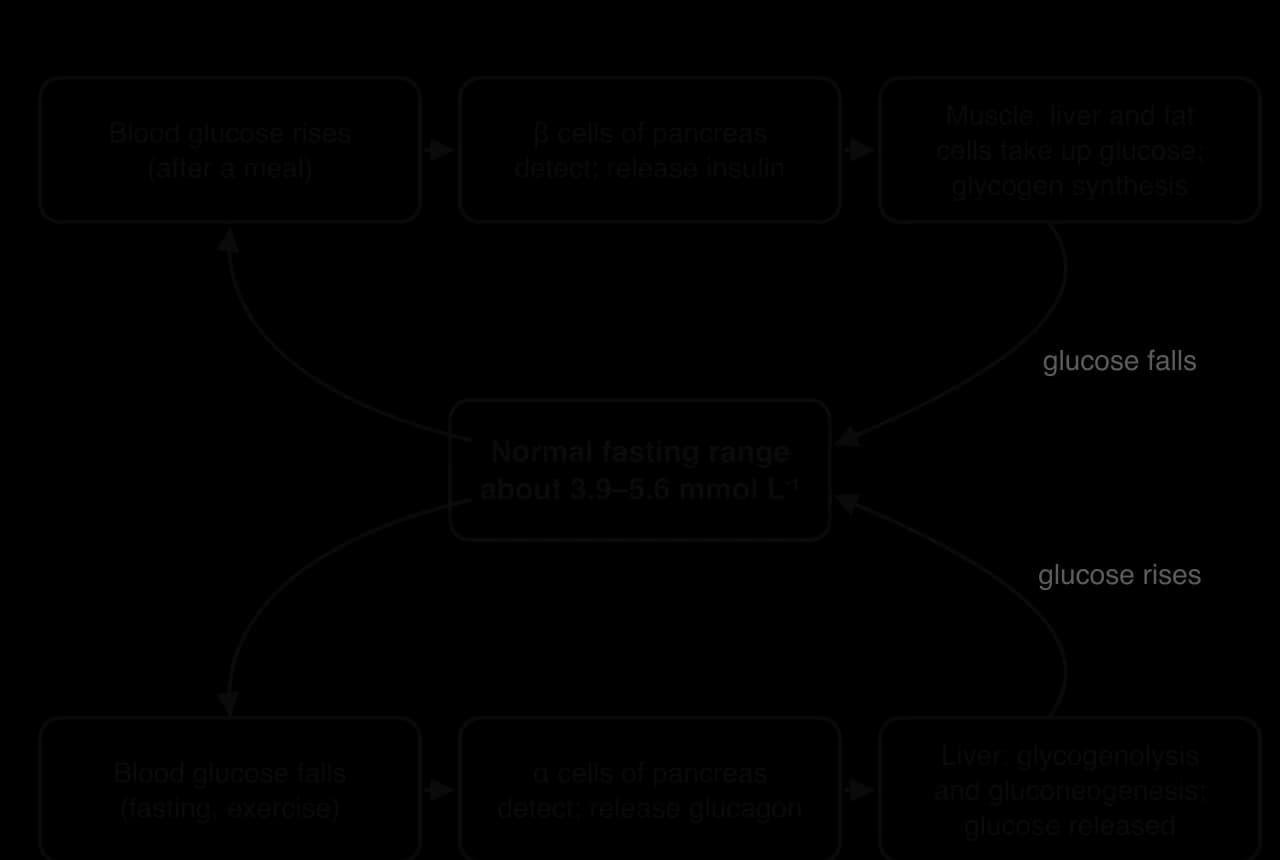
Exercise is a major challenge to blood glucose regulation, because working muscles can take up glucose many times faster than resting muscles. If insulin kept working at its resting level, blood glucose would fall quickly. Several responses prevent this:
Insulin release falls. Sympathetic nerves and circulating epinephrine and norepinephrine inhibit the β cells, so less insulin is released as exercise intensity and duration increase.
Glucagon release rises, especially in prolonged exercise, so the liver releases more glucose.
Epinephrine from the adrenal medulla also stimulates glycogenolysis in the liver and muscle, and the breakdown of fat for fuel.
The liver’s output of glucose rises to match much of the muscles’ uptake, so in most exercise blood glucose stays fairly stable. In very long exercise without carbohydrate intake, liver glycogen runs low and blood glucose can fall.
The muscles can still take up glucose even though insulin is low, because muscle contraction itself moves glucose transporter proteins (called GLUT4) from inside the muscle fibre to its membrane. These transporters let glucose enter the fibre. Contraction and insulin use different signalling routes to do this, so exercise increases glucose uptake independently of insulin.
After exercise, the muscles remain more sensitive to insulin for many hours, often up to two days, while they refill their glycogen stores. For this reason, regular physical activity improves glucose control and insulin sensitivity. It is one of the main treatments for type 2 diabetes, in which the cells respond poorly to insulin (insulin resistance): exercise lets glucose enter the muscles by a route that does not depend on insulin working properly.
The same regulation explains practical advice in sport. A large carbohydrate snack eaten 30–60 minutes before exercise raises insulin, and if exercise then starts while insulin is still high, insulin and muscle contraction together can briefly lower blood glucose, which some athletes notice as light-headedness in the first part of a session. Athletes with type 1 diabetes, whose pancreas produces no insulin, must adjust their injected insulin and carbohydrate intake around training to avoid hypoglycaemia, because injected insulin cannot be “switched off” as natural insulin is during exercise.
Blood glucose regulation also links the endocrine system to the energy systems: the hormones that control the release of glucose and fat into the blood decide which fuels are available to the oxidative and glycolytic systems, which in turn affects how long an athlete can keep up a given intensity.
Every homeostatic system has limits. In hot, humid air, at high altitude or in cold water, the environment pushes the internal environment away from its set points faster than at sea level in mild weather, and the body has to work harder to restore it. The body’s reaction has two time scales. An acute response is the immediate change during a single exposure, such as a rise in heart rate on the first run in the heat. A chronic adaptation is a lasting change in structure or function after repeated exposures, such as a larger plasma volume after two weeks of training in the heat.
The process of adapting to a new environment is called acclimatisation. Scientists sometimes distinguish acclimatisation, adaptation to a natural environment (for example, living and training in a hot country), from acclimation, adaptation to an artificial one (a heat chamber or an altitude tent). The adaptations are similar, and the single term acclimatisation is used for both here.
During exercise in the heat, the thermoregulatory responses (sweating and skin vasodilation) are larger and start sooner than in cool conditions. These responses bring physiological costs:
Cardiovascular strain. More blood flows to the skin and blood volume falls as sweat is lost, so venous return and stroke volume fall. Heart rate rises to maintain cardiac output, so heart rate at a given pace is noticeably higher, and maximal cardiac output and VO₂ max are reduced.
Fluid and electrolyte loss. Sweat rates of 1–2 L h⁻¹ or more lead to dehydration if fluid is not replaced, which further reduces plasma volume and raises core temperature. Sodium is lost in the sweat.
Metabolic changes. Muscle glycogen is used faster and more lactate is produced at the same intensity, so the lactate inflection point occurs at a lower pace.
A rising core temperature. If heat gain exceeds heat loss, core temperature rises towards levels at which the central nervous system reduces the drive to the muscles. This is hyperthermia, an important cause of fatigue in long events in the heat.
Humidity makes all of these worse, because it limits evaporation. In hot, dry air, a runner who sweats 1.5 L h⁻¹ may evaporate most of it; in hot, humid air much of the same sweat drips off, so the athlete loses the fluid without the cooling. Core temperature rises faster and sweat losses are larger for the same heat removed.
High core temperatures are common in elite endurance sport in the heat. At the 2016 UCI Road Cycling World Championships, held in hot conditions, ingestible pills showed that 34 of 40 riders studied reached a core temperature of at least 39 °C and 10 of them exceeded 40 °C, with a highest value of 41.5 °C. Because elite athletes can tolerate such temperatures during competition, a single core temperature reading cannot by itself diagnose heat illness.
Sources: Racinais et al., British Journal of Sports Medicine, 2019.
When the cardiovascular and thermoregulatory systems can no longer meet the demands of heat, exercise-related heat illness can develop. The two most important forms are heat exhaustion and heat stroke.
Heat exhaustion is the more common of the two and is not usually dangerous if the athlete stops and is cooled. Heat stroke is rarer but far more serious, because the brain and other organs are damaged by the high temperature itself.
Feature | Heat exhaustion | Heat stroke |
|---|---|---|
Cause | Cardiovascular strain and dehydration; blood volume too low to supply muscles and skin | Thermoregulation overwhelmed: heat gain far exceeds heat loss |
Core temperature | Raised, usually below 40 °C | Above about 40 °C |
Central nervous system | Normal: the athlete is alert and answers sensibly | Impaired: confusion, irrational behaviour, collapse, seizures or coma |
Skin | Pale, sweaty, cool or clammy | Often hot; may still be sweating in exertional heat stroke |
Treatment | Stop exercise, move to shade, lie down with legs raised, cool and give fluids | Medical emergency: cool immediately, ideally by whole-body cold-water immersion, then transport (“cool first, transport second”) |
Outcome | Usually recovers quickly | Can damage the brain, kidneys, liver and muscles; can be fatal if cooling is delayed |
Repeated exercise in the heat produces heat acclimatisation. Most adaptations develop over about 7–14 days of daily exposures of around 60–90 minutes, each raising core temperature, with the cardiovascular adaptations appearing in the first few days and the sweating adaptations taking longer.
Adaptation | How it helps |
|---|---|
Plasma volume expands (within about the first week) | More blood to supply both skin and muscles; higher stroke volume, so heart rate at a given pace falls |
Sweating starts at a lower core temperature | Heat loss begins earlier, so core temperature rises more slowly |
Higher sweat rate | More evaporative cooling, provided the sweat can evaporate |
More dilute sweat (less sodium) | Sodium conserved, helping to maintain plasma volume and electrolyte balance |
Lower resting and exercising core temperature | Larger margin before core temperature reaches a level that limits performance |
Skin blood flow begins at a lower temperature | Earlier transfer of heat from core to skin |
Less glycogen use and lower lactate at the same pace | Fuel saved; LIP at a higher pace than before acclimatisation |
Better thermal comfort and lower RPE | The same pace feels easier |
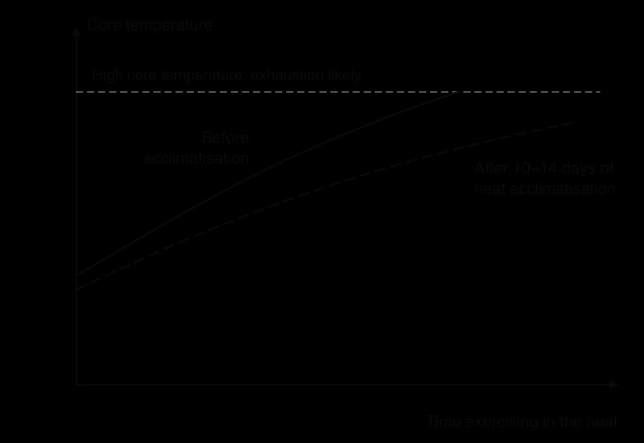
The adaptations fade if heat exposure stops, over a few weeks. Athletes therefore try to time the end of an acclimatisation block close to their competition. Fitter athletes acclimatise faster, because endurance training already provides part of the adaptation.
 YouTube
Heat acclimation training. Repeated sessions in the heat for 1–2 weeks before competition: training in a hot location, in a heat chamber, or by wearing extra clothing to raise core temperature in a cool climate. In “controlled hyperthermia” protocols, the exercise is adjusted to hold core temperature at a target (about 38.5 °C), which keeps the stimulus high as the athlete adapts. Hot-water immersion or sauna use after training is a cheaper alternative.
Pre-cooling and per-cooling. Lowering body temperature before exercise (pre-cooling) increases the amount of heat that can be stored before core temperature becomes limiting. Methods include drinking ice slurry (crushed ice mixed with a drink), wearing ice vests, cold towels and neck collars, and cold-water immersion. Per-cooling (cooling during exercise) uses cold drinks, ice slurry, ice towels and dousing the head and face with water.
Scheduling. Holding events in the coolest part of the day (early morning or night), and adding drinks and cooling breaks. The marathon and race walks at the Tokyo 2020 Olympics were moved to the cooler northern city of Sapporo to reduce the heat risk.
Hydration plans. Starting well hydrated and following an individual drinking plan, based on sweat rate measured in training, that limits body mass loss without overdrinking.
Clothing. Light-coloured, loose, breathable clothing that reflects radiation and lets sweat evaporate; minimal clothing where rules allow; hats or visors in the sun.
The evidence for pre-cooling and per-cooling is fairly strong in the laboratory. A meta-analysis of 28 studies in the heat (above 30 °C) found that cooling improved exercise performance by about 6.7% on average: about 5.7% for pre-cooling and about 9.9% for per-cooling, with combinations of methods most effective for pre-cooling. A larger meta-analysis of 59 studies later found that cooling helped more in constant-workload tests (cycling to exhaustion at a fixed intensity) than in self-paced tests such as time trials, which are closer to real races.
Sources: Bongers et al., British Journal of Sports Medicine, 2015; van de Kerkhof et al., Sports Medicine, 2024.
Strategy | Strengths | Limitations |
|---|---|---|
Heat acclimation training | Largest and longest-lasting benefit; reduces risk of heat illness; considered the most important single strategy | Takes 1–2 weeks; can add fatigue and reduce training quality; needs a hot venue, chamber or protocol; adaptations decay after a few weeks |
Pre-cooling | Cheap and quick; ice slurry and ice vests lower starting core temperature | Benefit is short-lived; ice slurry can cause stomach discomfort; too much cooling of muscles can reduce power in short events |
Per-cooling | Can be used throughout long events (drinks, dousing) | Depends on rules and aid stations; dousing adds little in humid air where evaporation is limited |
Scheduling | Removes much of the heat load at its source | Not under the athlete’s control; night events disturb sleep and routine |
Hydration plan | Limits fall in plasma volume and cardiovascular drift | Overdrinking risks hyponatraemia; sweat rates vary daily and between individuals |
Taken together, the evidence supports heat acclimatisation as the foundation, with cooling, hydration and clothing as additions. The best combination depends on the event (a marathon gains more from cooling and acclimatisation than a sprint), the athlete (fitness, body size, sweat rate, sex and previous heat exposure) and practical factors such as time, cost and the rules of the competition.
Cold is less often a problem in sport than heat, because exercise produces so much heat, but it is a serious risk in open-water swimming, mountain sports, winter sports and in endurance events in cold rain and wind. Cold water is especially dangerous because, as a much better conductor than air, it removes heat far faster than air at the same temperature.
Vasoconstriction of skin arterioles reduces heat loss but cools the hands, feet and face.
Shivering raises heat production but uses glycogen, increases oxygen demand and interferes with skilled movements.
Higher energy use and more fat and carbohydrate oxidation, partly from shivering and non-shivering thermogenesis, so fuel stores are used faster.
Breathing cold, dry air can irritate the airways and trigger narrowing of the airways (bronchoconstriction), which is common in cross-country skiers.
As muscles cool, their contraction speed, power and nerve conduction velocity fall, and the fingers lose dexterity, which affects catching, gripping and fine skills. A cold muscle is also stiffer. If heat loss continues to exceed heat production, core temperature falls and hypothermia develops. Exposed skin, especially of the fingers, toes, ears and nose, can freeze, causing frostbite.
Hypothermia affects the whole body, whereas frostbite is a local cold injury. Frostbite is most likely where vasoconstriction has already reduced blood flow and the skin is exposed to wind.
Clothing in layers: a base layer that moves sweat away from the skin, an insulating middle layer that traps still air, and a windproof, waterproof outer layer. Layers can be removed as the body warms, which limits sweating; wet clothing loses much of its insulation.
Covering the extremities: hats, gloves or mittens and warm socks reduce heat loss where vasoconstriction leaves the tissue coldest.
A thorough warm-up, kept close to the start of competition, raises muscle temperature for power and dexterity; warm clothing is kept on until the last moment.
Wetsuits in open-water swimming and triathlon add insulation and buoyancy; triathlon rules set water temperatures below which wetsuits are compulsory and above which they are banned.
Fuel and fluids: carbohydrate intake supports shivering and exercise; warm drinks help comfort but add little heat.
Avoiding wind and wet: wind increases convective heat loss (wind chill), and being wet increases conductive loss.
Humans show only limited acclimatisation to cold compared with heat. Repeated cold exposure mainly produces habituation: shivering starts at a lower skin temperature, the feeling of discomfort is reduced, and in some groups who work with their hands in cold water, blood flow to the hands is better maintained. An increase in brown adipose tissue and non-shivering thermogenesis has also been shown in some studies. These changes are small, so protective clothing and behaviour remain the main defence.
Air at the top of a mountain contains the same proportion of oxygen as at sea level, about 20.9%. What changes is the pressure. Barometric pressure is caused by the weight of the air above, so it falls with altitude, and the pressure of each gas in the air falls with it. The partial pressure of oxygen (pO₂) is the part of the total pressure caused by oxygen.
A lower pO₂ in the alveoli means a smaller pressure gradient for diffusion into the blood, so the haemoglobin in arterial blood is less saturated with oxygen. The shortage of oxygen in the tissues that results is called hypoxia. At moderate altitudes (about 1,500–3,000 m, where most altitude training takes place), the effect at rest is small, but during hard exercise it is large, because blood passes through the lungs quickly and has less time to load oxygen.
Hyperventilation. Peripheral chemoreceptors in the carotid and aortic bodies detect the low arterial oxygen and the respiratory control centre increases ventilation at rest and during exercise. This raises alveolar pO₂, but it also breathes out more CO₂, which makes the blood more alkaline (pH rises): an example of two homeostatic systems in conflict.
Higher heart rate at rest and at any submaximal workload, to deliver more blood and so more oxygen per minute to the tissues.
Lower plasma volume within the first days (fluid moves out of the blood and urine output rises), so stroke volume falls; this also raises the concentration of haemoglobin slightly.
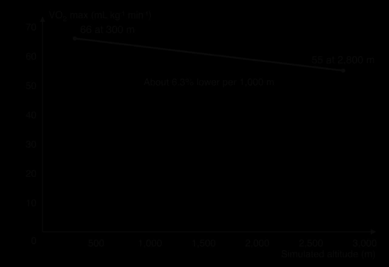
Lower VO₂ max, because less oxygen loads onto haemoglobin. Endurance performance falls, and the same pace feels harder.
Greater reliance on the anaerobic glycolytic system at a given workload, so lactate rises sooner.
Poor sleep, loss of appetite and greater fluid loss in dry mountain air.
The fall in VO₂ max starts at quite low altitudes in trained athletes. In one study, eight endurance-trained athletes ran to exhaustion in a hypobaric chamber at simulated altitudes from 300 m to 2,800 m in a randomised, double-blind design. Their VO₂ max fell in a straight line with altitude.
Some newcomers to altitude, especially above about 2,500 m and after a rapid ascent, develop acute mountain sickness. Its symptoms usually start 6–12 hours after arrival and include headache, nausea, tiredness, dizziness and poor sleep. It usually settles within a few days as the body acclimatises, but rarer, more serious forms involve fluid in the lungs or brain and need descent and medical care.
With continued exposure over weeks, the body acclimatises to altitude:
More erythropoietin and red blood cells. The kidneys detect low oxygen and release more erythropoietin (EPO) within hours. EPO stimulates the bone marrow to produce more red blood cells, so red cell mass and total haemoglobin increase over 3–4 weeks or more, and the blood can carry more oxygen.
Ventilatory acclimatisation. Ventilation stays high and the kidneys excrete bicarbonate in the urine, which corrects the rise in blood pH caused by hyperventilation.
Plasma volume recovers towards normal over weeks.
Changes in the muscles, such as greater capillary density and changes in mitochondria and enzymes, have been reported, mainly in people living at high altitude for a long time; their size in athletes on training camps is less certain.
Altitude training creates a dilemma. Living at altitude produces the useful adaptation (more red blood cells), but training at altitude forces athletes to run or ride more slowly, because VO₂ max is lower, so the quality of their training falls. Several approaches try to solve this:
Live high, train high: living and training at altitude, as at well-known training centres in Kenya, Ethiopia and the mountains of Colorado and Arizona. Simple, but training intensity is reduced.
Live high, train low: living or sleeping at altitude and travelling down to train at low altitude, so that the red cell adaptation and high-quality training happen together.
Simulated altitude: altitude tents, hypoxic rooms and hotel rooms with low-oxygen air let athletes “live high” while staying at sea level. They need many hours a day, often 12 or more, to have an effect.
Arrival timing for competition at altitude: a common recommendation is to arrive either within about 24 hours of the event, before the symptoms of acute mountain sickness and poor sleep develop, or at least two weeks before, to allow acclimatisation.
The best-known test of this approach randomly assigned 39 competitive runners to three groups for four weeks: living at 2,500 m and training at 1,250 m (live high–train low), living and training at altitude, or living and training near sea level. Both altitude groups increased VO₂ max by about 5% and red cell mass by about 9%, but only the live high–train low group improved its 5,000 m time, by about 13 seconds, because it kept up its sea-level training speeds.
Sources: Levine and Stray-Gundersen, Journal of Applied Physiology, 1997.
Later research showed that athletes differ widely in their response. Some increase red cell mass and performance markedly, while others show little change; these are responders and non-responders. Low iron stores limit the production of haemoglobin, so athletes are usually screened and given iron when needed before an altitude camp. Because the effect on performance is often small and the placebo effect of “having been to altitude” cannot easily be ruled out, the evidence remains debated.
Strategy | Strengths | Limitations |
|---|---|---|
Live high, train high | Full altitude stimulus; also prepares for competition at altitude | Training intensity reduced; risk of altitude sickness, poor sleep and weight loss; expensive travel |
Live high, train low | Red cell adaptation plus high-quality training; supported by a randomised trial | Needs suitable geography or travel between sites; individual responses vary widely |
Altitude tents and hypoxic rooms | No travel; dose can be controlled | Many hours per day needed; may disturb sleep; smaller effect than natural altitude in some studies |
Arrival timing | Free and practical | Does not improve performance; only limits the loss |
Sources: Robach et al., British Journal of Sports Medicine, 2012.
The same environment can help one event and harm another, because the effect depends on which energy system and which physical demands dominate.
Endurance events (long-distance running, cycling, race walking, triathlon) suffer most in the heat and at altitude. They rely on the oxidative system, so a lower VO₂ max, a higher heart rate, dehydration and a rising core temperature all reduce the pace that can be sustained.
Short explosive events (sprints, jumps, throws) are affected little by heat or altitude, because they rely on the phosphagen and glycolytic systems, which do not need oxygen during the event. Warm muscles contract faster and more forcefully, so warm weather can help. At altitude the thinner air also reduces drag, which helps sprinters, jumpers and cyclists over short distances.
Team and intermittent sports (football, rugby, tennis) are affected in between: repeated sprints are maintained, but players cover less high-intensity distance late in matches in the heat, and the recovery between efforts is slower at altitude.
Skill events can be affected by cold (loss of dexterity and power) and by fatigue in the heat, which impairs decision-making late in a match.
The 1968 Olympic Games in Mexico City, at 2,240 m, show both effects. World records were set in the men’s 100 m (9.95 s), 200 m (19.83 s) and 400 m (43.86 s), helped by lower air density. In contrast, the men’s 10,000 m was won in 29:27.4, about 108 seconds (6.5%) slower than the world record of 27:39.4 that stood at the time, and the 10,000 m and the marathon were won by athletes from Kenya and Ethiopia, countries where many athletes live and train at altitude.
Sources: Wikipedia, Athletics at the 1968 Summer Olympics and Men’s 10,000 metres world record progression, 2026.
The women’s marathon at the 2019 World Athletics Championships in Doha, Qatar, started at one minute to midnight on 27 September 2019 to avoid the daytime heat. Even so, the air temperature at the start was 32.7 °C with a relative humidity of 73%. Of the 68 women who started, only 40 finished: 28 (41%) did not finish. The winner, Ruth Chepng’etich of Kenya, ran 2:32:43, the slowest winning time in the history of the championships.
Scientists from Aspetar (Doha’s sports medicine hospital), Loughborough University and World Athletics studied the athletes in all the road events at these championships: the marathons and the 20 km and 50 km race walks. Core temperature was recorded with ingestible telemetry pills, skin temperature with thermal cameras, and athletes completed a questionnaire about their health and their heat preparation before the race. Across the road events, the roadside wet bulb globe temperature ranged from 23.5 °C to 30.6 °C.
Measure (all road events, Doha 2019) | Value |
|---|---|
Starters in the marathons and race walks | 305 |
Athletes who completed the health and acclimation questionnaire | 83 |
Athletes who did not finish (all starters) | 25% |
Mean peak core temperature during competition (n = 56) | 39.6 ± 0.6 °C (highest 41.1 °C) |
Mean skin temperature at start and end of races | 32.2 → 31.0 °C |
Performance compared with personal best | 3–20% slower (mean 13% slower) |
The results support the mechanisms of thermoregulation. Core temperature rose by about 1.5 °C during the races, but skin temperature fell, because air movement over moving athletes increased evaporation of sweat; this differs from laboratory studies at similar temperatures but without realistic air movement. Performance relative to personal best was closely linked to the WBGT of each race (R² = 0.89), showing the importance of humidity and radiation, not just air temperature.
Several findings show the limits of the core-temperature model on its own. Core temperature itself was not related to whether an athlete finished or needed medical care; skin temperature and the gradient between core and skin were. A recent episode of diarrhoea, reported by 9% of athletes, was a significant risk factor for an in-race medical event, probably because of dehydration and electrolyte loss. Thermoregulation interacts with fluid balance and general health, not just with temperature.
As evidence, the study has clear strengths: it measured elite athletes in real competition, which laboratory studies cannot do, with objective measures of core and skin temperature. Its limitations are those of an observational field study. Only 83 of 305 starters completed the questionnaire and only 56 swallowed a pill, so the sample may not be representative, and conditions differed between races and athletes. The study shows associations, such as the link between WBGT and performance, rather than proving causes.
Of the athletes who completed the questionnaire, 63% had done 5–30 days of heat acclimation before the championships.
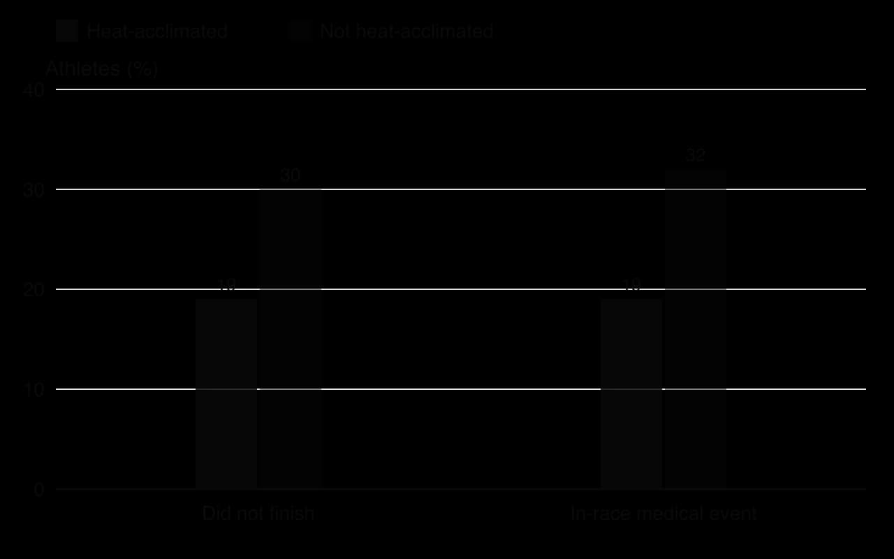
These athletes ranked better (average position 18 compared with 28, p = 0.009) and had a lower peak core temperature (39.4 °C compared with 39.8 °C, p = 0.044) than athletes who had not. Their rates of non-finishing (19% vs 30%) and medical events (19% vs 32%) were also lower, but with this sample size these differences could have arisen by chance (p = 0.273 and p = 0.179). Most athletes also planned to use pre-cooling, and ice vests were the only method associated with a lower core temperature before the race.
These results are consistent with the adaptations of heat acclimatisation, but they do not prove that acclimation caused the better outcomes. Athletes chose whether to acclimatise, so the acclimated group may also have been better prepared, better funded or fitter (confounding variables), and acclimation was self-reported. A randomised controlled trial, in which matched athletes are allocated to acclimation or no acclimation before the same race, would test the effect more directly, although it would be hard to arrange at elite level.
Explain why the women’s marathon in Doha was run more slowly than the same athletes’ races in cool conditions, referring to evaporation, cardiac output and core temperature.
Suggest why skin temperature, rather than core temperature, was related to whether athletes finished.
Evaluate whether the Doha data show that heat acclimation causes better performance in the heat (HL).
Sources: Racinais et al., British Journal of Sports Medicine, 2021 and 2022; Aylwin et al., Journal of Applied Physiology, 2023; World Athletics championship results via Wikipedia, 2026.
Homeostasis appears in every part of the written papers. Paper 1A tests definitions, the order of the parts of a feedback loop and the direction of responses. Paper 1B often uses data from studies of exercise in the heat, with core temperature, sweat loss or body mass change. Paper 2 asks you to explain feedback loops step by step, to sketch and annotate them, and, at HL, to evaluate strategies for heat and altitude.
Data-based questions on homeostasis often use real or hypothetical results from studies of exercise in the heat or at altitude.
A second question tests the direction of hormonal responses during exercise.
A third question tests the routes of heat loss.
Paper 1B-style. Researchers measured elite marathon and race-walk athletes at the 2019 World Athletics Championships in Doha, held in hot, humid conditions. Core temperature was measured with an ingestible pill before and after the race. Body mass was measured before and after the race to estimate fluid loss. Some athletes wore ice vests before the race. The table shows some of the results (mean ± SD).
Measure | Group | Result |
|---|---|---|
Core temperature before the race | All measured athletes (n = 56) | 37.7 ± 0.3 °C |
Core temperature after the race | All measured athletes (n = 56) | 39.6 ± 0.6 °C |
Core temperature before the race | Used ice vest / did not use ice vest | 37.5 ± 0.4 °C / 37.8 ± 0.3 °C (p = 0.024) |
Change in body mass during the race | Men / women | −2.8 ± 1.5% / −1.3 ± 1.6% (p < 0.001) |
Sources: Racinais et al., British Journal of Sports Medicine, 2021.
Questions on blood pH usually expect the full chain of cause and effect.
Thermoregulation questions often contrast responses to heat and cold.
A sketch question checks that the loop is complete and labelled.
Questions on the heart test the difference between the two forms of control.
Heat illness questions usually ask for clear differences.
Altitude questions often combine a calculation with an explanation.
The HL extended response usually asks for an evaluation of strategies.
Homeostasis keeps the internal environment within narrow limits around set points; receptors detect changes, control centres compare them with the set point and effectors respond.
Negative feedback reverses a change and is the basis of homeostasis; positive feedback amplifies a change and is rare (blood clotting, childbirth).
CO₂ forms carbonic acid, which releases H⁺ and lowers pH; chemoreceptors and the respiratory control centre increase ventilation to remove CO₂, and bicarbonate buffers H⁺.
The SA node sets the heart’s intrinsic rhythm; sympathetic and parasympathetic nerves and epinephrine adjust it, and the baroreceptor reflex keeps blood pressure in range.
Core temperature is kept at about 37 ± 1 °C by the hypothalamus; 75–80% of the energy released in muscle becomes heat, and evaporation of sweat is the main route of heat loss in exercise.
Heat triggers sweating and skin vasodilation; cold triggers vasoconstriction, shivering and non-shivering thermogenesis.
Training status, body composition and size, humidity, wind and radiation (combined in the WBGT), and sex and hormonal phase all affect thermoregulation.
Insulin lowers and glucagon raises blood glucose; during exercise insulin falls, glucagon and epinephrine rise, and contraction increases glucose uptake without insulin.
Heat, humidity, cold and altitude cause acute responses and, with repeated exposure, adaptations such as plasma volume expansion (heat) and more red blood cells (altitude); endurance events suffer most (HL).
Heat acclimatisation, cooling, hydration, clothing, scheduling, layering, warm-up, live high–train low and arrival timing support performance, with effects that vary between individuals (HL).
I can outline homeostasis and the roles of receptors, control centres and effectors, using the terms set point and normal range.
I can distinguish between negative and positive feedback, with an example of each, and sketch a generic negative feedback loop.
I can explain how CO₂ lowers blood pH and how chemoreceptors and ventilation restore it.
I can explain intrinsic and extrinsic control of the heart and the baroreceptor reflex.
I can describe heat production and the four routes of heat exchange, and calculate heat lost by evaporation.
I can explain the thermoregulatory responses to heat and to cold and draw both loops.
I can discuss how training status, body composition, the environment and sex differences affect thermoregulation, and calculate a WBGT.
I can explain blood glucose regulation by insulin and glucagon, and how exercise changes it.
I can describe the acute responses and adaptations to heat, humidity, cold and altitude, and calculate a partial pressure of oxygen (HL).
I can evaluate strategies for acclimatising and performing in heat, cold and altitude (HL).