Explain solvation and osmosis in terms of solute concentration, predict effects on cells with and without walls, analyse tissue data with error bars, and (HL) calculate water potential from Ψs and Ψp.
Each is in the Biology glossary, with every other term in the subject.
The shrinkage of an animal cell, such as a red blood cell, in a hypertonic solution as it loses water by osmosis, giving it a shrivelled outline with spiky projections.
A state in which movement or change continues in opposite directions at equal rates, so that there is no net change; for example water entering and leaving a cell in an isotonic solution.
A line drawn through a plotted mean on a graph, extending above and below it, that shows the variability or uncertainty of the data, usually ±1 standard error or ±1 standard deviation.
Describes a plant cell whose contents no longer press against the cell wall because it has lost water, so that it has no turgor pressure; flaccid tissue is soft and limp.
Describes a solution with a higher concentration of solutes than another solution, such as the cytoplasm of a cell; water moves by osmosis into it from the less concentrated solution.
Describes a solution with a lower concentration of solutes than another solution, such as the cytoplasm of a cell; water moves by osmosis out of it into the more concentrated solution.
The point at which the plasma membrane of a plant cell is just beginning to pull away from the cell wall as the cell loses water; turgor pressure has fallen to zero.
Water moves into and out of cells by osmosis, driven by differences in the concentration of dissolved solutes. The gain or loss of water by cells determines whether a plant stands upright or wilts, whether a red blood cell keeps its shape, and what fluid can safely be given to a patient through a drip.
The movement of water depends first on how water dissolves substances, and then on the solute concentrations on each side of a membrane. Cells with and without walls respond very differently to the same solution, and multicellular organisms, freshwater protists and hospitals all have ways of keeping cells in surroundings that do not harm them.
36 exam-style questions on this subtopic, written in the style of the IB papers. The first has its markscheme open. Counted on
Which row describes the attractions that form between water molecules and the ions when solid sodium chloride dissolves in water?
Row | Na+ ions are attracted to | Cl− ions are attracted to |
|---|---|---|
A | hydrogen atoms (δ+) of water molecules | oxygen atoms (δ−) of water molecules |
B | hydrogen atoms (δ−) of water molecules | oxygen atoms (δ+) of water molecules |
C | oxygen atoms (δ−) of water molecules | hydrogen atoms (δ+) of water molecules |
D | oxygen atoms (δ−) of water molecules | oxygen atoms (δ−) of water molecules |
The official deck for this part of the syllabus.
What is a solution?
State six examples of aqueous solutions in living organisms.
With a free account, Augment remembers where you stopped and what you have practiced.
The bursting of a cell when its plasma membrane breaks, for example when an animal cell takes in water by osmosis in a hypotonic solution; in red blood cells it is called haemolysis.
A sterile 0.9% w/v solution of sodium chloride in water, approximately isotonic with human blood plasma, used for intravenous fluids, washing wounds and storing tissues.
The total concentration of all solute particles (molecules and ions) in a solution, measured in osmoles or milliosmoles per litre (mOsm/L); osmolality is measured per kilogram of water.
The shrinking of the protoplast of a plant cell away from its cell wall when the cell loses water by osmosis in a hypertonic solution.
The component of water potential due to hydrostatic pressure on water; positive under pressure, as in a turgid plant cell, and negative under tension, as in xylem (symbol Ψp).
The component of water potential due to dissolved solutes; zero for pure water and more negative as solute concentration increases (symbol Ψs).
The standard error of the mean: an estimate of how far a sample mean is likely to be from the true mean, equal to the standard deviation divided by the square root of the number of measurements.
The pressure exerted by the contents of a plant cell against its cell wall, which builds up as water enters by osmosis; the wall pushes back with an equal pressure.
The potential energy of water per unit volume, relative to pure water at atmospheric pressure and 20 °C, which is given the value zero; symbol Ψ, usually measured in kPa.
A solution forms when one substance, the solute, dissolves in another, the solvent. In living organisms the solvent is almost always water. Cytoplasm, blood plasma, tissue fluid, xylem sap, phloem sap and urine are all aqueous solutions, and nearly every metabolic reaction takes place between molecules that are dissolved in water.
Water is a good solvent because it is a polar molecule. The oxygen atom attracts the shared electrons in each O–H bond more strongly than the hydrogen atoms do. As a result the oxygen carries a partial negative charge (δ−) and each hydrogen carries a partial positive charge (δ+). Because the molecule is bent, these charges sit on opposite sides of the molecule, and each water molecule behaves like a tiny magnet with a negative end and a positive end.
Solvation is the process in which solvent molecules surround and interact with particles of solute, separating them from one another and spreading them through the solution. With water as the solvent, solvation depends on two kinds of attraction: hydrogen bonds between water and polar solute molecules, and attractions between the charged ends of water molecules and ions.
Many biological molecules contain polar groups, such as the hydroxyl groups (–OH) of sugars, the amino (–NH₂) and carboxyl (–COOH) groups of amino acids, and the –NH₂ and C=O groups of urea. The δ+ hydrogen atoms of water are attracted to the δ− oxygen or nitrogen atoms of these groups, and the δ− oxygen of water is attracted to their δ+ hydrogen atoms. These attractions are hydrogen bonds, the same kind of bond that forms between neighbouring water molecules.
When a crystal of glucose is placed in water, water molecules form hydrogen bonds with the hydroxyl groups on the surface of the crystal. Each glucose molecule is pulled away from its neighbours and becomes surrounded by water molecules. Because the energy released by forming many new hydrogen bonds with water is similar to the energy needed to break the bonds between glucose molecules in the crystal, glucose dissolves readily: about 900 g of glucose dissolves in one litre of water at 25 °C.
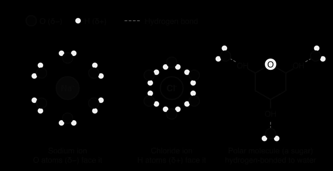
An ion is an atom or group of atoms with an electrical charge. When sodium chloride dissolves, the crystal separates into sodium ions (Na⁺) and chloride ions (Cl⁻). Each ion attracts the oppositely charged end of the water molecules around it:
Positive ions (cations) such as Na⁺, K⁺ and Ca²⁺ attract the δ− oxygen atoms of water, so the surrounding water molecules point their oxygen atoms towards the ion.
Negative ions (anions) such as Cl⁻, HCO₃⁻ and phosphate attract the δ+ hydrogen atoms of water, so the surrounding water molecules point their hydrogen atoms towards the ion.
The layer of oriented water molecules that surrounds a dissolved ion is called a hydration shell. The hydration shell keeps ions of opposite charge apart, so they do not rejoin to form a crystal, and it allows ions to be carried freely through the cytoplasm, blood and xylem sap.
The diagram of solvation shows that the orientation of water molecules in a hydration shell depends on the charge of the ion. Around Na⁺ the oxygen atoms face inwards; around Cl⁻ the hydrogen atoms face inwards. The same water molecules can therefore dissolve both positive and negative ions, which is why one solvent can carry the wide variety of ions found in cells.
Substances that dissolve in water because they are polar or charged are called hydrophilic ("water-loving"). Substances that are non-polar, such as fats, oils and steroids, cannot form hydrogen bonds with water or attract its charged ends. Water molecules hydrogen-bond to each other instead, and the non-polar molecules are pushed together. These substances are hydrophobic and are insoluble or only slightly soluble in water.
Differences in solubility shape how substances are transported in organisms. Glucose, amino acids and ions travel dissolved in blood plasma. Fats and cholesterol are carried in lipoprotein particles with a hydrophilic outer coat. Oxygen is non-polar and dissolves poorly in water, so most oxygen in blood is carried bound to haemoglobin in red blood cells. Solubility is an example of how a property at the level of molecules affects the functioning of whole organisms.
Solvation has a consequence for water itself. Water molecules held in hydration shells or hydrogen-bonded to solute molecules are less free to move than water molecules in pure water. The more solute particles there are in a solution, the larger the proportion of water molecules that interact with them. This explains why the concentration of dissolved solutes determines the direction in which water moves between solutions, the central idea of osmosis.
A partially permeable membrane allows some substances to pass through it but not others. The plasma membrane (also called the cell membrane) is partially permeable: water crosses it easily, through the phospholipid bilayer and through channel proteins called aquaporins, while ions and larger polar molecules such as sucrose cannot cross freely.
When two solutions are separated by a partially permeable membrane, water molecules move across the membrane in both directions by random motion. More water molecules cross from the solution with the lower solute concentration than cross back from the solution with the higher solute concentration. The result is a net movement of water from the less concentrated solution to the more concentrated solution. This passive net movement of water across a partially permeable membrane is called osmosis.
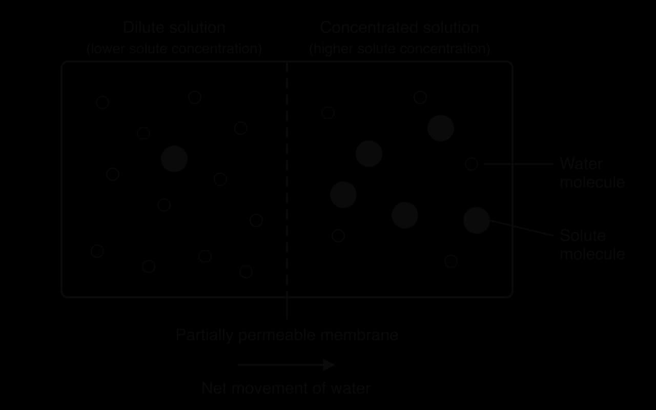
The diagram of two solutions separated by a membrane shows why this happens. On the dilute side, more of the water molecules that reach the membrane are free to cross. On the concentrated side, many water molecules are attracted to solute particles and fewer reach the membrane free to cross. Osmosis continues until the concentrations on the two sides are equal or until some other force stops it.
Osmosis is a form of passive transport. It needs no energy from ATP, because it depends only on the random kinetic energy of water molecules. In this respect it resembles simple diffusion, but osmosis is specifically the movement of water, the solvent, across a membrane that the solutes cannot cross.
Solute concentration can be expressed in several units, and each is used in different contexts:
Molar concentration, in mol dm⁻³ (moles of solute per cubic decimetre of solution, the same as mol L⁻¹). Laboratory sucrose and sodium chloride solutions are usually made up in mol dm⁻³.
Millimoles per litre (mmol/L or mmol L⁻¹), used in medicine for ions in blood: plasma sodium is normally 135–145 mmol/L.
Percentage by mass per volume (% w/v), grams of solute per 100 cm³ of solution: a 0.9% w/v sodium chloride solution contains 0.9 g of NaCl in every 100 cm³.
For osmosis, the important quantity is the number of dissolved particles rather than the mass of solute. One mole of glucose dissolves as one mole of molecules, but one mole of sodium chloride separates into one mole of Na⁺ and one mole of Cl⁻, which is two moles of particles. Osmolarity takes this into account.
Blood plasma has an osmolality of about 275–295 mOsm/kg. Most of this comes from Na⁺ and Cl⁻, with smaller contributions from glucose, urea, other ions and proteins. Two solutions with the same osmolarity have the same tendency to take up water, even if they contain different solutes.
Three terms compare the solute concentration of one solution with that of another. They are always relative: a solution is hypotonic or hypertonic only in comparison with something else, usually the cytoplasm of a cell or a body fluid.
Pure water and fresh water from a river or pond are hypotonic to the cytoplasm of almost every cell, because cytoplasm contains many dissolved ions, sugars, amino acids and proteins.
Seawater, with an osmolarity of about 1,000 mOsm/L, is hypertonic to human body fluids. This is why drinking seawater causes dehydration: the body must use more water to excrete the extra salt than the seawater supplied.
Every cell is surrounded by a plasma membrane that is permeable to water but much less permeable to most solutes. The cytoplasm and, in plant cells, the cell sap inside the vacuole, contain dissolved substances. A cell therefore behaves like the concentrated side of the osmosis diagram whenever it is placed in a more dilute solution, and like the dilute side when it is placed in a more concentrated one.
Environment compared with the cytoplasm | Solute concentration outside | Net movement of water by osmosis |
|---|---|---|
Hypotonic | Lower than inside the cell | Into the cell |
Isotonic | Equal to inside the cell | No net movement (dynamic equilibrium) |
Hypertonic | Higher than inside the cell | Out of the cell |
The table of environments summarises the three possible cases. In a hypotonic environment, water enters the cell, its volume increases and the cytoplasm becomes more dilute. In a hypertonic environment, water leaves the cell, its volume decreases and the cytoplasm becomes more concentrated. In both cases the change continues until the solute concentrations inside and outside become equal, or until another force, such as the pressure of a cell wall, prevents further entry.
In an isotonic environment the volume of a cell stays constant, but water does not stop moving. Water molecules continue to cross the plasma membrane in both directions at high rates. Because the solute concentrations are equal, the number of water molecules entering per second equals the number leaving per second, so there is no net change.
The time course of osmosis follows a predictable pattern. When plant tissue is first placed in a hypotonic solution, water enters quickly, because the difference in solute concentration is large. As water enters, the cytoplasm becomes more dilute and the cell wall begins to resist further expansion, so the rate of entry falls. Eventually the mass of the tissue stops changing because a new equilibrium has been reached.
The graph of mass change against time shows why investigations leave tissue in solution for a fixed, long period: if samples are weighed too early, the change in mass has not yet reached its final value, and samples left for different times cannot be compared.
 YouTube
The effect of solute concentration on water movement can be measured with pieces of plant tissue. Potato tuber is the usual choice because it is uniform, easy to cut into identical cylinders and has no air spaces large enough to affect its mass. Beetroot, carrot, sweet potato and apple can be used in the same way.
Prepare a series of sucrose or sodium chloride solutions, for example 0.0 (distilled water), 0.2, 0.4, 0.6 and 0.8 mol dm⁻³ sucrose, by diluting a stock solution.
Use a cork borer to cut cylinders from the same potato, then trim them with a scalpel to the same length (for example 40 mm). Remove any skin, which is less permeable than the inner tissue.
Blot each cylinder gently with paper towel, then measure its initial mass on a balance (to 0.01 g) and its initial length with a ruler (to 1 mm).
Place several cylinders (at least three, preferably five) in each solution, in labelled boiling tubes or Petri dishes, so that each cylinder is fully covered.
Leave all samples for the same time (for example 24 hours) at the same temperature.
Remove each cylinder, blot it in the same way, and measure its final mass and length.
Calculate the percentage change in mass (and length) for each cylinder, then the mean and a measure of spread for each concentration.
The independent variable is the concentration of the solution. The dependent variable is the percentage change in mass or length. Controlled variables include the type and source of tissue, the size and surface area of the cylinders, the volume of solution, the temperature and the time in solution. Blotting removes surface solution, which would otherwise add to the final mass without having entered the tissue.
In a hypotonic solution, water enters the cells of the cylinder by osmosis. Each cell swells until its wall resists further expansion, so the cylinder gains mass and becomes longer and firmer. In a hypertonic solution, water leaves the cells, so the cylinder loses mass, becomes shorter and becomes soft and bendy. At one concentration the cylinder neither gains nor loses mass. That solution is isotonic with the cytoplasm of the potato cells.
Cylinders never start with exactly the same mass. A larger cylinder gains or loses more water than a smaller one in the same solution, simply because it contains more cells. Percentage change expresses each change as a proportion of the starting value, so samples of different starting size can be compared fairly.
Each concentration is tested with several cylinders because individual pieces of tissue differ. One cylinder may contain more starch, or be cut slightly unevenly, or be blotted more firmly. Repeating each measurement and calculating the mean reduces the effect of these random variations. Repeats also show how much the results vary, which indicates the reliability of the measurements: if repeated cylinders give similar values, the mean can be trusted more.
The spread of a set of repeats is described by the standard deviation, which measures how far the individual values typically lie from the mean. A small standard deviation means the repeats are close together; a large one means they are widely scattered.
The standard deviation describes the individual measurements, but it does not say how precisely the mean itself is known. That is the job of the standard error.
Because the standard deviation is divided by , the standard error becomes smaller as more repeats are taken. Five repeats with a standard deviation of 2.0% give a standard error of ; twenty repeats with the same spread give . The formula does not have to be memorised, and calculators and spreadsheets calculate both values directly, but its meaning matters: more repeats give a more precise estimate of the mean.
Results are plotted with the independent variable (solution concentration) on the x-axis and the mean percentage change in mass on the y-axis. Each mean is plotted as a point, and its standard error (or standard deviation) is shown as an error bar extending above and below it.
Error bars are used to judge whether differences between means are likely to be real. When the error bars of two means do not overlap, the difference between them is probably not due to chance variation alone. When they overlap a lot, the difference may not be significant, and more repeats are needed before a firm conclusion can be drawn. In the worked example, none of the error bars overlap, so each increase in sucrose concentration caused a real decrease in mass.
Measuring length as well as mass gives a second, independent estimate of the isotonic point. Length is less sensitive than mass, because a 3 mm change in a 40 mm cylinder is close to the precision of a ruler, so mass data usually give the more reliable value. Changes in length also show that cells expand and shrink as water enters and leaves, not only that the tissue absorbs water on its surface.
Animal cells and many unicellular organisms are bounded only by a plasma membrane. The membrane is thin and flexible but cannot withstand much stretching, so the volume of the cell changes freely as water enters or leaves. A human red blood cell is the standard example. In blood plasma, which is isotonic to its cytoplasm, it keeps its normal biconcave disc shape.
In a hypotonic solution, water enters the red blood cell by osmosis. The cell swells and becomes spherical, and if water keeps entering, the plasma membrane breaks and the cell bursts. The bursting of a cell in this way is called lysis; when it happens to red blood cells it is called haemolysis, and the haemoglobin released turns the solution clear red.
In a hypertonic solution, water leaves the red blood cell by osmosis. The cell shrinks and its membrane forms spiky projections, a change called crenation. Crenated red blood cells are less flexible than normal ones and do not pass easily through narrow capillaries.
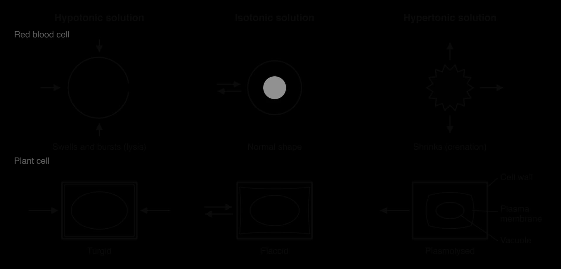
The upper row of the diagram of cells in three solutions shows the red blood cell; the lower row shows a plant cell in the same three solutions for comparison. In the isotonic solution, the two-way arrows represent the dynamic equilibrium: water crosses in both directions at equal rates.
Freshwater unicellular organisms, such as Paramecium and Amoeba, live in a hypotonic environment all the time. Pond water contains far fewer dissolved solutes than their cytoplasm, so water enters them continuously by osmosis. Without a cell wall to resist swelling, they would burst. They survive because they have contractile vacuoles, which collect excess water and expel it from the cell.
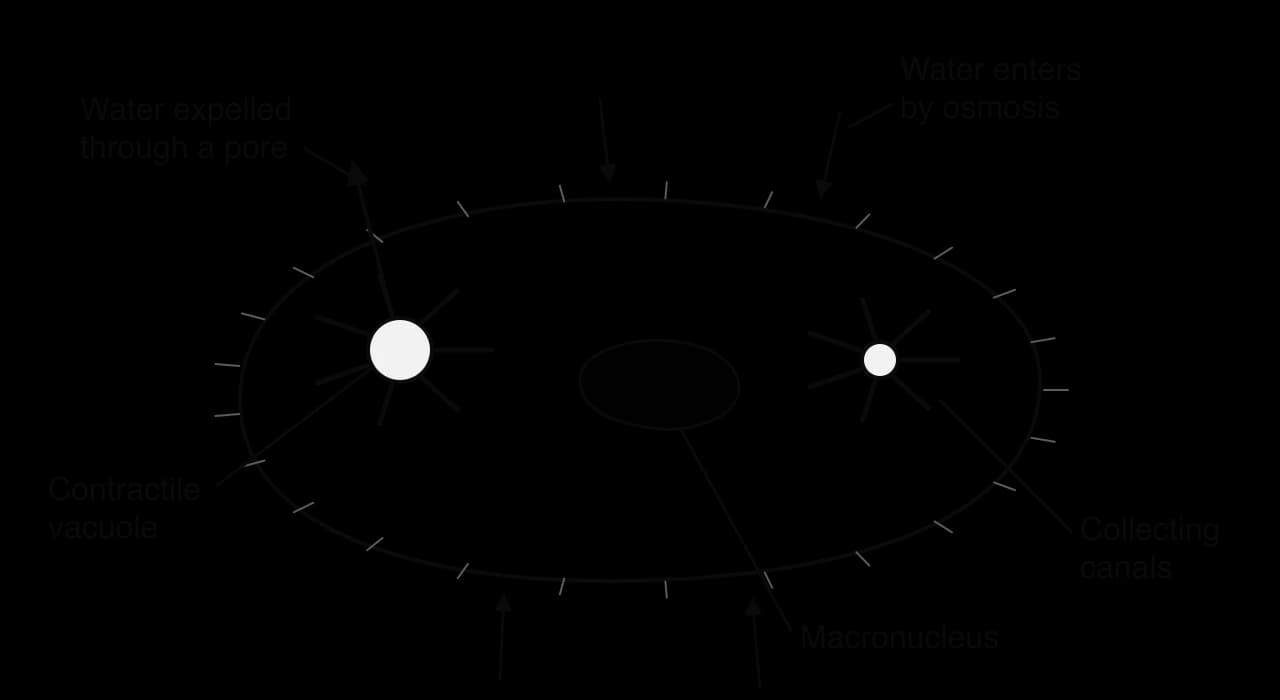
Water enters the cell across the plasma membrane by osmosis, all over its surface.
Radiating collecting canals around each contractile vacuole gather water from the cytoplasm. Proton pumps in the canal membranes move ions into the canals, and water follows by osmosis.
The vacuole fills and swells with water.
The vacuole contracts and empties its contents to the outside through a pore in the plasma membrane.
The cycle repeats every few seconds. In Paramecium the two vacuoles, one at each end of the cell, fill and empty alternately.
Removing water this way uses energy from ATP, which drives the proton pumps that load the collecting canals with ions; without this energy the cell could not get rid of the water that keeps entering by osmosis. The rate at which the vacuoles fill and empty varies with the environment: it is fastest in very dilute water and slows as the solute concentration of the medium rises. Many marine protists, whose cytoplasm is close to isotonic with seawater, have small or inactive contractile vacuoles or none at all, which supports the idea that their function is osmotic.
Cells in a multicellular animal are bathed by tissue fluid, which forms from blood plasma. If tissue fluid became hypotonic, body cells would take in water and swell; if it became hypertonic, they would lose water and shrink. Either change disrupts the cell. Swelling stretches the membrane and can cause lysis; shrinkage concentrates enzymes and substrates, changing the rates of metabolic reactions. Cells in the brain are especially vulnerable, because the skull leaves little room for the brain to swell.
Multicellular organisms therefore keep their tissue fluid isotonic with their cells, as part of homeostasis. In mammals the solute concentration of the blood is monitored by the hypothalamus and adjusted by the kidneys, which vary the volume and concentration of the urine (see D3.3 Homeostasis). This is an example of interaction between levels of organisation: a change in the composition of one body fluid affects every cell it bathes.
The importance of isotonic body fluids is shown by exercise-associated hyponatraemia, a condition in which the concentration of sodium ions in the blood plasma falls too low. Normal plasma sodium is 135–145 mmol/L. Endurance athletes who drink much more water than they lose in sweat dilute their plasma. Because Na⁺ is the main solute in plasma and tissue fluid, the tissue fluid becomes hypotonic to body cells.
In 2002, researchers took blood samples from 488 runners at the finish line of the Boston Marathon and recorded their weight before and after the race. Weight gain during a race can only come from drinking more fluid than is lost, so it was used as a measure of overdrinking.
Measurement at the finish | Result |
|---|---|
Runners sampled | 488 |
Hyponatraemia (plasma Na⁺ 135 mmol/L or less) | 13% of runners |
Critical hyponatraemia (plasma Na⁺ 120 mmol/L or less) | 0.6% of runners |
Women with hyponatraemia | 22% |
Men with hyponatraemia | 8% |
Main risk factor | Weight gain during the race (drinking more than was lost) |
Other risk factors | Finishing time over 4 hours; very low or very high body mass index |
Source: Almond et al., New England Journal of Medicine 352:1550, 2005.
The table of results shows that low plasma sodium was common among recreational runners: about one runner in eight was affected. The strongest predictor was weight gain, which shows that the cause was too much water rather than too much salt lost in sweat. Slower runners had more time to drink, and smaller runners (often women) needed less water to dilute their plasma, which helps to explain the other risk factors.
The biology follows directly from osmosis. Diluted plasma makes the tissue fluid hypotonic, so water enters body cells and they swell. In most tissues this causes few symptoms, but swelling of brain cells raises pressure inside the skull and can cause headache, confusion, seizures and, in severe cases, death. The study changed advice to runners: drink according to thirst rather than as much as possible. Treatment of severe cases uses intravenous solutions with a carefully controlled solute concentration.
Plant cells, fungal cells and most bacteria have a cell wall outside the plasma membrane. The plant cell wall is made mainly of cellulose fibres, which are very strong in tension. The wall is freely permeable: water and dissolved solutes pass through it easily. It does not control what enters the cell, but it does resist expansion.
Most mature plant cells have a large central vacuole filled with cell sap, a solution of sugars, ions and other solutes. Fresh water around the roots and the tissue fluid in a well-watered plant are hypotonic to cell sap. Water therefore enters the cell by osmosis, and the vacuole and cytoplasm swell, pushing the plasma membrane against the wall. The wall stretches only slightly and then pushes back. The pressure that builds up inside the cell is called turgor pressure.
Turgor pressure rises as water enters, until it is high enough to stop any further net entry of water. At that point the cell is fully turgid. A plant cell does not burst in a hypotonic solution, because the strong cellulose wall withstands the pressure. The same process in an animal cell causes lysis; the presence or absence of a wall is a clear case of form and function, where one structural difference changes the outcome of the same physical process.
Turgor pressure provides support. In non-woody (herbaceous) plants, such as lettuce, celery and young seedlings, the stems and leaves are held up mainly by turgid cells pressing against one another. When the plant loses more water by transpiration than it can absorb, its cells lose turgor and the plant wilts. Turgor pressure also drives cell expansion during growth, opens and closes stomata by changing the shape of guard cells, and causes rapid movements such as the folding of the leaves of Mimosa pudica when touched.
A cell that has lost turgor pressure, so that its contents no longer push against the wall, is described as flaccid.
In a hypertonic solution, water leaves the vacuole and cytoplasm by osmosis. The cell first becomes flaccid. As more water is lost, the protoplast (the plasma membrane and everything inside it) shrinks and pulls away from the cell wall, a change called plasmolysis.
The point at which the plasma membrane is just beginning to separate from the wall, usually at the corners of the cell, is called incipient plasmolysis. At incipient plasmolysis the turgor pressure has fallen to zero. In a plasmolysed cell the space between the wall and the shrunken protoplast fills with the external solution, which can pass freely through the wall.
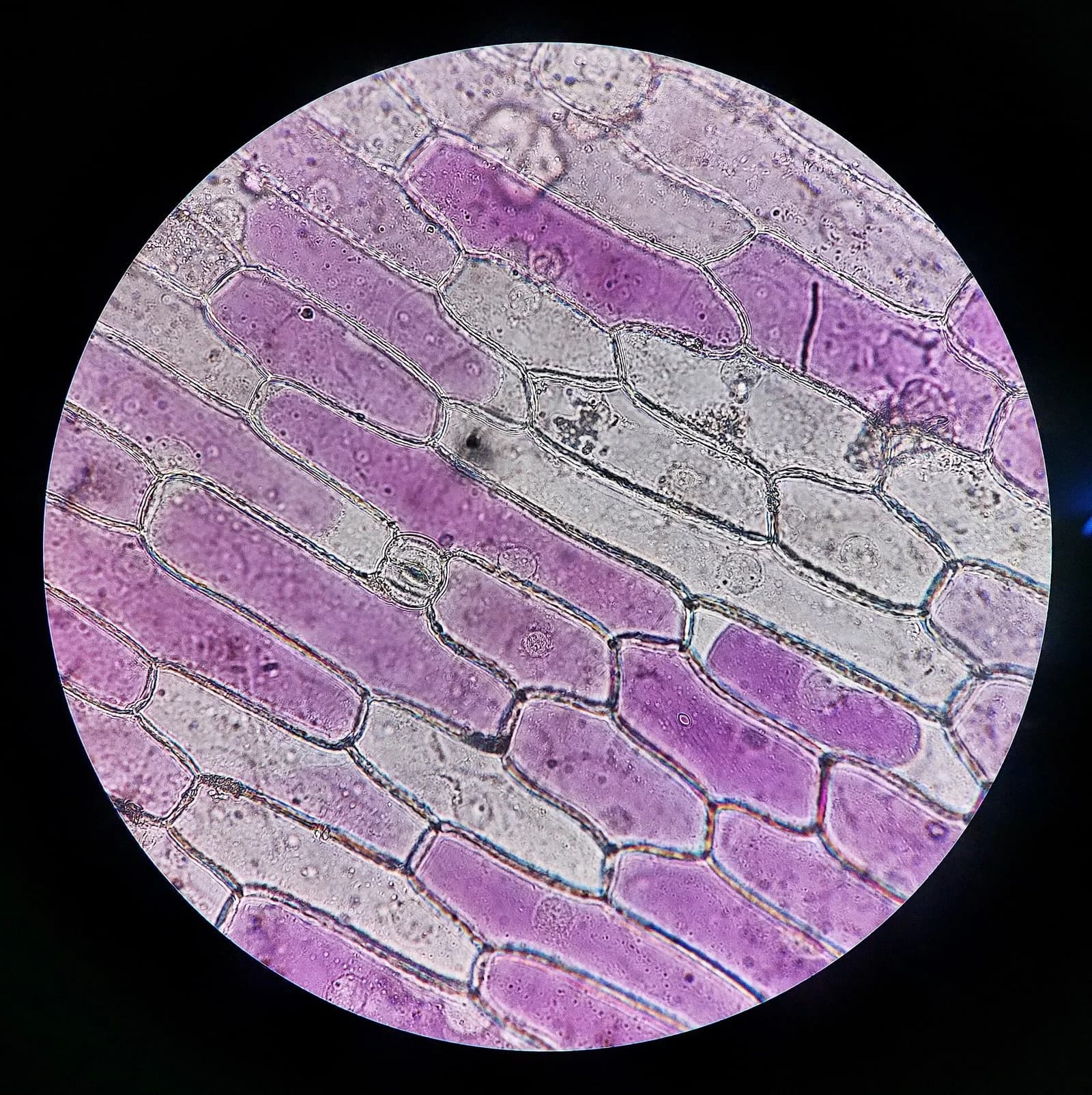
The micrograph of red onion epidermis is the standard demonstration of plasmolysis. The cell sap of red onion contains purple anthocyanin pigments, so the shrinking vacuole is easy to see. When a strip of epidermis is mounted in concentrated sodium chloride solution, the coloured protoplasts pull away from the walls within a few minutes. If the salt solution is replaced with distilled water, water re-enters and the cells recover turgor, a process called deplasmolysis. Severe or prolonged plasmolysis damages the membrane and kills the cell, because the plasma membrane is torn from its connections to the wall.
Plasmolysis also explains a traditional method of preserving food. Bacteria and fungi on salted fish or meat, or in jam with a high sugar concentration, are surrounded by a hypertonic solution. They lose water by osmosis and are plasmolysed, so they cannot grow and spoil the food.
Solution compared with the cell | Cell without a wall (red blood cell) | Cell with a wall (plant cell) |
|---|---|---|
Hypotonic | Water enters; swells and may burst (lysis, haemolysis) | Water enters; turgor pressure rises; turgid; does not burst |
Isotonic | No net movement; normal shape | No net movement; flaccid (no turgor pressure) |
Hypertonic | Water leaves; shrinks (crenation) | Water leaves; flaccid, then plasmolysed |
The comparison table shows an important difference in the isotonic solution. A red blood cell is in its normal state in an isotonic solution, but a plant cell in an isotonic solution is flaccid. Plant cells are healthiest in a hypotonic environment, where they stay turgid; animal cells are healthiest in an isotonic one.
Patients who are dehydrated, who have lost blood, or who cannot eat or drink after surgery are often given fluid directly into a vein through a drip. Intravenous (IV) fluid mixes immediately with blood plasma and comes into contact with red blood cells and the cells lining the blood vessels. If it were hypotonic, those cells would take in water and could undergo lysis; if it were hypertonic, they would lose water and crenate. IV fluids are therefore made isotonic with blood plasma.
The most widely used IV fluid is normal saline, a 0.9% w/v solution of sodium chloride in sterile water.
Fluid | Main solutes | Osmolarity / mOsm L⁻¹ | Relative to plasma |
|---|---|---|---|
Blood plasma | Na⁺, Cl⁻, HCO₃⁻, glucose, urea, proteins | about 275–295 (mOsm/kg) | — |
Normal saline (0.9% NaCl) | 154 mmol/L Na⁺ and 154 mmol/L Cl⁻ | about 308 | Approximately isotonic |
Distilled water | None | 0 | Hypotonic: never given into a vein |
3% NaCl ("hypertonic saline") | 513 mmol/L Na⁺ and 513 mmol/L Cl⁻ | about 1,027 | Hypertonic: used only for severe hyponatraemia |
Source: standard clinical reference values; osmolarities calculated from ion concentrations.
The table of IV fluids shows where the value of 0.9% comes from. One litre of normal saline contains 9 g of NaCl, which is 154 mmol. Each NaCl forms two ions, giving about 308 mOsm/L, close to the osmolality of plasma. Other common IV fluids, such as Hartmann's solution (also called Ringer's lactate), contain a mixture of ions closer to that of plasma but have a similar overall solute concentration. In contrast, 3% hypertonic saline is given slowly and under close monitoring to patients with severe hyponatraemia, so that water is drawn out of swollen brain cells.
An organ removed from a donor, such as a kidney, liver or heart, must be kept alive until it can be transplanted into the recipient. It is flushed with a cold preservation solution and stored in the same solution on ice. Cooling to about 4 °C slows metabolism, so the cells use less oxygen and ATP. The solution must be isotonic with the cells, or close to it, so that the cells neither take in water and swell nor lose water and shrink during storage.
Cooling creates an extra problem. At low temperature, the sodium–potassium pumps in the plasma membrane slow almost to a stop, so Na⁺ and Cl⁻ leak into the cells and water follows by osmosis. Cells stored in normal saline would gradually swell. The University of Wisconsin (UW) solution, developed by Folkert Belzer and James Southard in the 1980s, solved this by including large solutes, such as lactobionate and raffinose, that cannot cross the plasma membrane. These impermeant solutes keep the solution's solute concentration matched to that of the cells and prevent swelling. UW solution greatly extended the time a liver or kidney can be stored before transplantation and is still widely used.
Isotonic saline has many other medical uses: washing wounds and burns, rinsing the eyes, storing contact lenses, and keeping tissue samples and corneas for grafting moist. In each case the aim is the same, to avoid damaging cells by osmosis.
Water molecules have potential energy that can do work, for example by moving into a cell and pushing its contents against the cell wall. Water potential measures this potential energy. It is given the symbol Ψ (the Greek letter psi, pronounced "sigh"); some books write Ψw.
The absolute potential energy of water cannot be measured. Instead, water potential is always given relative to a standard: pure water, at atmospheric pressure and 20 °C, is defined as having a water potential of 0 kPa. Every other value is a difference from this standard. A solution with Ψ = −300 kPa has less potential energy per unit volume than pure water, and a solution under positive pressure can have a value above zero.
Energy per unit volume has the same units as pressure, which is why water potential is measured in pascals:
Values in biology are usually given in kilopascals (1 kPa = 1,000 Pa) or megapascals (1 MPa = 1,000 kPa). For comparison, atmospheric pressure at sea level is about 101 kPa.
Water moves spontaneously from a region of higher water potential to a region of lower water potential, in the same way that a ball rolls downhill from higher to lower gravitational potential energy. The movement releases potential energy and needs no ATP. It stops only when the water potentials on the two sides become equal, at which point water continues to move in both directions at equal rates.
Pure water has the highest water potential of any solution at atmospheric pressure. Dissolving solutes always lowers it, because water molecules attracted to solute particles in hydration shells and hydrogen bonds have less free energy and less tendency to move. A dilute solution therefore has a slightly negative water potential, and a concentrated solution has a more negative one.
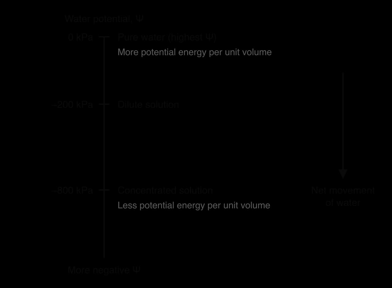
The water potential scale links the HL description to the SL one. A hypotonic solution has a higher water potential than the cell; a hypertonic solution has a lower water potential than the cell; an isotonic solution has the same water potential. Water moving from a dilute solution to a concentrated one is the same as water moving from higher to lower water potential.
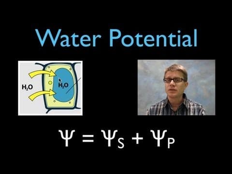
The water potential of a cell or solution depends on two factors: the concentration of dissolved solutes and the pressure acting on the water. Each is given its own potential, and water potential is their sum:
Here (or simply Ψ) is the water potential, is the solute potential and is the pressure potential. All three are measured in kPa, and the signs must always be kept.
Solute potential ranges from zero downwards. Pure water has . Adding any solute lowers it, and the more solute particles, the more negative it becomes. Solute potential can never be positive, because solutes always reduce the free energy of water. The cell sap of a typical plant cell usually has a solute potential between about −500 and −2,000 kPa, depending on the species and conditions.
Some plants survive in salty habitats by making their solute potential very low. Seawater has a water potential of about −2,500 kPa. Halophytes such as mangroves accumulate ions and other solutes in their root and leaf cells, lowering the water potential of their tissues below that of seawater. Water then still moves from the sea into their roots, from higher to lower water potential.
Pressure changes the potential energy of water. Pushing on water raises its potential energy; pulling on it (tension) lowers it.
Pressure potential has different values in different places:
Positive inside plant cells with walls. When water enters a plant cell, the wall pushes back on the expanding protoplast. This turgor pressure is a positive pressure potential, which raises the water potential of the cell. Values of +300 to +1,000 kPa are typical in turgid cells.
Zero in an open solution at atmospheric pressure, such as a beaker of sucrose solution, and at incipient plasmolysis or beyond, when the protoplast no longer presses on the wall. For a solution in an open container, .
Close to zero in animal cells, because the plasma membrane cannot withstand significant pressure. An animal cell takes in water until its solute potential matches the surroundings or it bursts.
Negative in xylem vessels of a transpiring plant, where water is pulled upwards under tension.
Evaporation of water from the walls of leaf cells during transpiration pulls water out of the xylem in the leaf veins. Because water molecules are held together by cohesion, the pull is transmitted down the continuous columns of water in the xylem to the roots. The water in the xylem is therefore under tension, a negative pressure potential. This is the basis of the cohesion–tension theory of water transport (see B3.2 Transport).

The water potential gradient through a plant shows why the tension matters. Water potential falls step by step from the soil, through root cells and xylem, to the leaf cells and finally the air, which has a very low water potential unless it is saturated with water vapour. At each step water moves from higher to lower water potential, so the whole flow is passive. The negative pressure potential in the xylem lowers its water potential enough for water to move into it from the root cells.
Xylem walls are thickened with lignin, which stops the vessels collapsing inwards under this tension. Under very dry conditions the tension can become strong enough to break a water column, forming an air bubble (an embolism) that blocks the vessel.
The diagram of a plant cell in three solutions follows one cell as the water potential of its surroundings is lowered. In every case water moves until the water potential of the cell equals that of the solution, so the cell values at equilibrium can be worked out from .
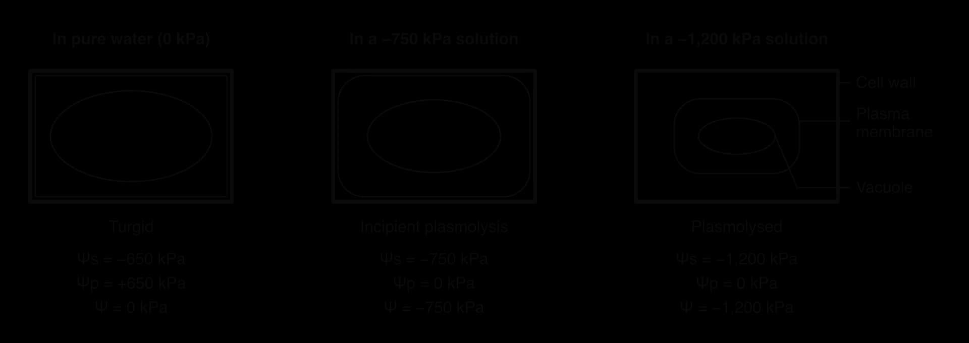
The solution has a higher water potential than the cell sap, so water enters the cell by osmosis.
As the protoplast swells against the wall, the pressure potential rises. The entry of water also dilutes the cell sap slightly, so the solute potential becomes a little less negative.
Both changes raise the water potential of the cell. Net entry of water stops when the cell's water potential equals that of the solution.
In pure water (Ψ = 0 kPa), equilibrium is reached when , so that . In the diagram, kPa. The cell is fully turgid.
The solution has a lower water potential than the cell, so water leaves the cell by osmosis.
The protoplast shrinks, so its pressure on the wall falls. The pressure potential decreases, and at incipient plasmolysis it reaches zero; the cell is flaccid.
With , the water potential of the cell equals its solute potential. If the solution's water potential is lower still, water continues to leave.
As water leaves, the cell sap becomes more concentrated and its solute potential becomes more negative. Water stops leaving when of the cell equals the water potential of the solution. The protoplast has pulled away from the wall: the cell is plasmolysed. In the diagram, the cell reaches kPa.
This model explains the results of the potato investigation. Cylinders gain mass in solutions with a higher water potential than the tissue, because their cells take in water until pressure potential rises enough to balance the difference. They lose mass in solutions with a lower water potential, as pressure potential falls to zero and then the cells plasmolyse. At the isotonic point the water potential of the solution equals the water potential of the tissue.
 YouTube
Two neighbouring cells in a tissue exchange water in the same way. Water moves from the cell with the higher water potential to the cell with the lower water potential, through the plasma membranes and the freely permeable walls between them. In a leaf, for example, cells near the xylem have a higher water potential than cells near the stomata, so water passes from cell to cell towards the surfaces where it evaporates.
Questions on water potential and osmosis appear in all parts of the assessment. Paper 1A multiple-choice questions ask candidates to predict the direction of water movement, identify drawings of turgid, plasmolysed, lysed or crenated cells, or (at HL) choose the correct water potential from Ψs and Ψp. Paper 1B and Paper 2 Section A use data from tissue experiments: calculating percentage change, plotting means with error bars, reading the isotonic concentration from a graph and evaluating the reliability of results. Paper 2 Section B can ask for extended explanations of osmosis in cells with and without walls, contractile vacuoles or the medical use of isotonic solutions.
The command terms most often used are calculate, deduce, predict, explain, compare and contrast and suggest. Answers at SL earn marks for describing water movement in terms of solute concentration (from lower to higher), and at HL for using water potential (from higher to lower) with correct signs.
The following question uses the results of an investigation with carrot root tissue.
NaCl concentration / mol dm⁻³ | Mean change in mass / % | Standard error / % |
|---|---|---|
0.00 | +12.0 | 0.8 |
0.10 | +4.4 | 0.7 |
0.20 | −3.1 | 0.9 |
0.30 | −8.6 | 1.0 |
0.40 | −12.9 | 1.1 |
I can explain solvation of polar molecules by hydrogen bonding and of ions by hydration shells.
I can state that water moves from a lower to a higher solute concentration, and use hypotonic, isotonic and hypertonic correctly.
I can predict net water movement into or out of a cell and explain dynamic equilibrium in an isotonic solution.
I can calculate percentage change and standard error, plot means with error bars and deduce an isotonic concentration.
I can describe lysis and crenation and explain how contractile vacuoles remove water from Paramecium.
I can explain why multicellular organisms keep tissue fluid isotonic, using hyponatraemia as an example.
I can explain turgor pressure, flaccidity and plasmolysis in plant cells.
I can explain why normal saline and organ-preservation solutions must be isotonic.
I can define water potential and explain why water moves from higher to lower water potential. (HL)
I can calculate water potential from solute and pressure potentials, including negative values in xylem. (HL)
I can explain changes in plant tissue in terms of Ψs and Ψp. (HL)
The oxygen atom of a water molecule carries a partial negative charge (δ−), so it is attracted to positive Na+ ions, while the partially positive (δ+) hydrogen atoms are attracted to negative Cl− ions; each ion becomes surrounded by a shell of water molecules. A reverses the attractions, B gives hydrogen and oxygen the wrong partial charges, and D wrongly has a negative ion attracted to a negatively charged atom.
The answer is C.
A cell is placed in a solution that has a higher solute concentration than its cytoplasm. Which term describes the solution, and what is the net movement of water?
A human cheek cell is placed in a solution that is isotonic with its cytoplasm. Which statements are correct?
I. Water molecules continue to cross the plasma membrane in both directions.
II. There is no net movement of water into or out of the cell.
III. The water concentration inside the cell is higher than outside the cell.