Distinguish the types of gene mutation and their effects, outline their causes, compare germ cell and somatic mutations, and link mutation to variation; at HL, gene knockout, CRISPR–Cas9 editing and conserved sequences.
Each is in the Biology glossary, with every other term in the subject.
A gene mutation in which one base in the DNA sequence is replaced by a different base.
A disease in which cells divide uncontrollably, forming malignant tumours that can invade tissues and spread; it results from mutations in genes that control cell division.
An agent that causes cancer; most carcinogens are mutagens that change genes controlling cell division.
A base or amino acid sequence that is identical or similar across a species or a group of species.
A gene-editing system in which a guide RNA directs the nuclease Cas9 to a complementary DNA sequence, where Cas9 cuts both strands so the gene can be disrupted or changed.
A gene mutation in which one or more nucleotides are removed from the base sequence of DNA.
An insertion or deletion of a number of nucleotides that is not a multiple of three, which changes the reading frame so every codon after the mutation is altered.
The deliberate alteration of the base sequence of DNA at a specific, chosen position in the genome, for example with CRISPR–Cas9.
A gene mutation is a change to the base sequence of a gene. Mutations are the reason that individuals differ genetically, the cause of many inherited diseases and of cancer, and the raw material on which natural selection acts.
Most mutations happen by chance, when DNA is damaged or copied incorrectly. Since the 2010s biologists have also been able to change genes deliberately, with great precision, using the gene-editing system CRISPR–Cas9. Knowing how natural mutations arise and what they do is the basis for understanding both.
42 exam-style questions on this subtopic, written in the style of the IB papers. The first has its markscheme open. Counted on
Genetic information varies between individuals and between species.
Outline what single-nucleotide polymorphisms (SNPs) are and how they arise.
4 marks
a. a SNP is a position in the base sequence where a single nucleotide differs between members of a species;
b. members of a species share most of their genome / SNPs are the commonest form of variation;
c. (arise by) base substitution mutations;
d. caused by errors in DNA replication;
e. OR caused by mutagens, e.g. UV radiation / chemicals;
f. mutations occur randomly / at any position;
g. mutation must occur in germ cells / gametes to be inherited;
h. a SNP may create a new allele / may have no effect if in non-coding DNA;
[4 max]
Describe how the polymerase chain reaction (PCR) and gel electrophoresis can be used to amplify and separate DNA.
4 marks
a. PCR makes many copies of a selected DNA sequence;
b. DNA is heated (to about 95 °C) to separate the strands;
c. cooled (to about 55 °C) so that primers bind to the ends of the target sequence;
d. heat-stable / Taq DNA polymerase extends the primers (at about 72 °C) using free nucleotides;
e. cycle is repeated many times, doubling the DNA each cycle;
The official deck for this part of the syllabus.
Define gene mutation.
State four reasons why gene mutations are important in biology.
With a free account, Augment remembers where you stopped and what you have practiced.
A technique for investigating the function of a gene by changing it so that it is inoperative, then observing the effect on the organism.
A change to the base sequence of a gene, by substitution, insertion or deletion of one or more nucleotides.
Differences in the alleles and base sequences carried by individuals of a population or species; mutation is its original source.
A cell that gives rise to gametes, or a gamete itself; mutations in germ cells can be inherited by offspring.
An RNA molecule with about 20 bases complementary to a target DNA sequence, which binds to Cas9 and directs it to cut at that sequence.
A base or amino acid sequence that has remained identical or very similar over long periods of evolution, in distantly related species.
A gene mutation in which one or more nucleotides are added to the base sequence of DNA.
A base substitution that changes a codon into one for a different amino acid, so one amino acid in the polypeptide is replaced.
A species studied in depth because it is easy to keep, breed and manipulate, so that discoveries about it can be applied to other organisms.
An agent that increases the rate of mutation, such as benzo[a]pyrene, nitrosamines, mustard gas, UV light, X-rays and gamma rays.
A base substitution that changes a codon for an amino acid into a stop codon, so translation ends early and the polypeptide is shortened.
A base substitution that changes a codon into another codon for the same amino acid, so the polypeptide is unchanged; also called a silent mutation.
Any body cell other than a germ cell; mutations in somatic cells are not passed to offspring but can cause cancer.
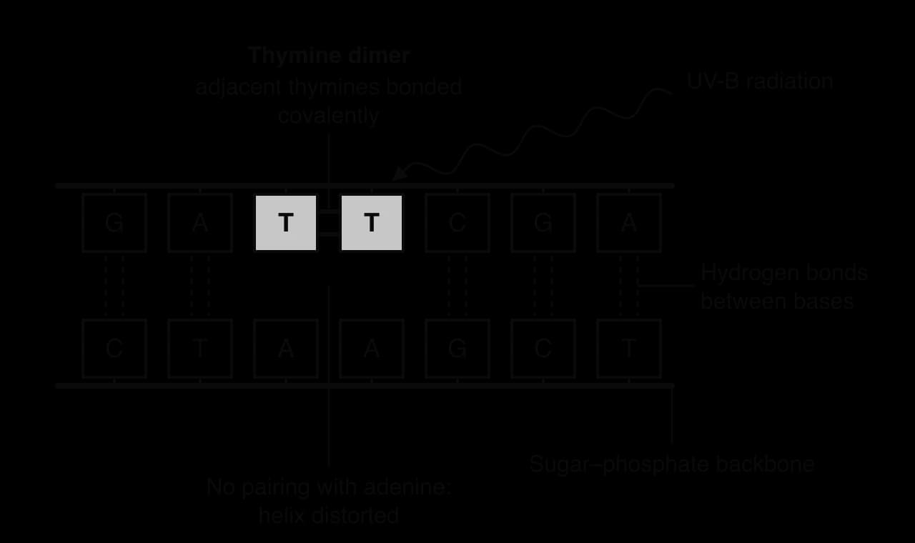
Two adjacent thymines on the same DNA strand joined by covalent bonds after absorbing UV radiation; the dimer distorts the helix and can cause errors in replication.
The function of a gene depends on its base sequence. A gene mutation changes that sequence at the molecular level, by altering, adding or removing one or more nucleotides. Because a gene codes for a polypeptide through the sequence of its codons, a change in the base sequence can change the polypeptide.
There are three basic types of gene mutation:
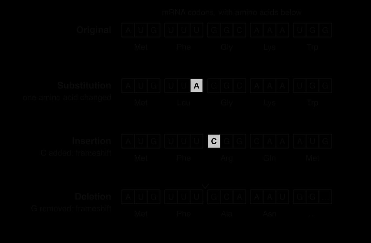
Base substitution: one base is replaced by a different base. The number of bases in the gene stays the same.
Insertion: one or more extra nucleotides are added to the sequence.
Deletion: one or more nucleotides are removed from the sequence.
The diagram of the four sequences shows the same short mRNA before and after each type of mutation, with the amino acids coded by each codon underneath. A substitution changes one codon only. An insertion or deletion of a single base shifts the way every following base is grouped into codons.
Mutations on this scale, affecting one or a few bases, are sometimes called point mutations or small-scale mutations. Larger changes, such as the loss or duplication of whole sections of a chromosome, also occur, whereas gene mutations are changes within single genes.
When a base substitution is passed on and becomes common in a population, the different versions of the sequence at that position are called a single-nucleotide polymorphism (SNP, pronounced "snip"). Any two unrelated humans differ at roughly one base in every 1000, mostly at SNPs, which adds up to several million differences across the genome. Every SNP began as a base substitution mutation in one individual at some time in the past.
A SNP can be in a part of the genome that codes for a polypeptide or in a non-coding part. Most SNPs are in non-coding DNA and have no effect on any polypeptide, although some affect how strongly a gene is expressed.
When a base substitution occurs in a codon of a gene, it has one of three effects on the polypeptide. Which one occurs depends on the codon table, and in particular on the degeneracy of the genetic code.
Type of substitution | Effect on the codon | Effect on the polypeptide | Example (mRNA codon) |
|---|---|---|---|
Same-sense (silent) | The new codon codes for the same amino acid | No change in amino acid sequence | GAA → GAG: glutamic acid → glutamic acid |
Missense | The new codon codes for a different amino acid | One amino acid replaced; the effect may be small or severe | GAG → GUG: glutamic acid → valine (sickle cell) |
Nonsense | The new codon is a stop codon | Translation stops early; the polypeptide is shortened and usually does not function | UGG → UGA: tryptophan → stop |
Same-sense mutations are possible because most amino acids have more than one codon. The codons for one amino acid usually differ only in the third base, so a substitution in the third base is often silent. Substitutions in the first or second base usually change the amino acid.
The effect of a missense mutation depends on where the new amino acid is and how different it is. Replacing an amino acid with a similar one on the surface of a protein may have no detectable effect. Replacing one in the active site of an enzyme, or one that changes how the protein folds, can make the protein useless. In sickle cell anaemia, a single missense mutation in the β-globin gene replaces a hydrophilic glutamic acid with a hydrophobic valine, with severe effects on the red blood cells (see D1.2 Protein synthesis).
The ribosome reads mRNA in codons of three bases from the start codon, without gaps. Inserting or deleting a number of bases that is not a multiple of three changes how all the following bases are grouped into codons, producing a frameshift mutation.
After a frameshift, every codon downstream of the mutation is different, so the amino acid sequence from that point on is completely changed. A stop codon is also likely to appear early in the new reading frame, because 3 of the 64 codons are stop codons. The result is usually a shortened polypeptide with an abnormal sequence that cannot fold into a functional protein. A frameshift near the start of a gene almost always destroys its function; one near the end may leave a partly working protein.
Insertions or deletions of exactly three bases, or a multiple of three, do not shift the reading frame. They add or remove one or more whole amino acids. The most common mutation causing cystic fibrosis, for example, is a deletion of three bases that removes a single phenylalanine from a membrane channel protein of 1480 amino acids, which is enough to stop the protein folding correctly.
Some mutations insert or delete hundreds or thousands of bases, or remove whole exons. These almost always stop the polypeptide from functioning, because large parts of the protein are missing or extra sequence is inserted into its structure. Many cases of Duchenne muscular dystrophy, for example, are caused by deletions of one or more exons from the very large gene for the muscle protein dystrophin.
 YouTube
Mutations arise in two ways: from errors made by the cell when it copies or repairs DNA, and from damage caused by agents in the environment called mutagens.
DNA polymerase occasionally inserts a nucleotide with the wrong base. Most of these errors are removed by proofreading and other repair enzymes, but a few remain, and they become permanent mutations when the DNA is replicated again. Errors also occur when cells repair damaged DNA, for example when the two ends of a broken double helix are rejoined and a few bases are lost. Even without any mutagens, every human is born with several dozen new mutations that were not present in either parent, most of them arising in the cells that formed the gametes, especially in the father.
A mutagen that causes cancer is a carcinogen. Most carcinogens are mutagens, because cancer develops from mutations in genes that control cell division. Mutagens are either chemicals or forms of radiation, and radiation is divided into ultraviolet and ionising radiation, as the table of examples shows.
Type of mutagen | Examples | How it damages DNA |
|---|---|---|
Chemical mutagens | Benzo[a]pyrene and nitrosamines in tobacco smoke; mustard gas | They react with bases and bind to them covalently, changing their shape and base-pairing, so wrong bases are inserted at replication |
Ultraviolet (UV) radiation | UV-B in sunlight; sunbeds | Causes covalent bonds between adjacent thymine bases (thymine dimers) on the same strand, which distort the helix and lead to errors in replication or repair |
Ionising radiation | X-rays, gamma rays, alpha particles from radioactive isotopes such as radon gas | High energy breaks chemical bonds in DNA, damages bases and can break both strands, leading to deletions and rearrangements |
Benzo[a]pyrene is one of the best-studied chemical mutagens. In the body it is converted into a reactive compound that binds to guanine in DNA. When the DNA is replicated, the damaged guanine is often paired incorrectly, so a G–C base pair is replaced by T–A. Such substitutions are found at particular positions in the TP53 gene in lung tumours from smokers. TP53 codes for a protein that normally stops damaged cells from dividing, so these mutations help explain how smoking causes lung cancer.
Nitrosamines, also present in tobacco smoke and formed from nitrites in some processed meats, add small groups (methyl or ethyl groups) to bases, which changes their base pairing in a similar way. Mustard gas, used as a chemical weapon in the First World War, forms covalent cross-links between guanines, including guanines on opposite strands, so the double helix cannot be separated for replication.
Ultraviolet radiation has too little energy to break DNA strands, but bases absorb it strongly. When two thymines lie next to each other on the same strand, UV-B can make covalent bonds form between them, joining them into a thymine dimer. The two thymines can no longer pair properly with adenines on the other strand, and the helix bends at that point. The cell usually cuts out and replaces the damaged section, but if replication reaches the dimer before it is repaired, DNA polymerase may insert the wrong bases opposite it.
Ionising radiation (X-rays, gamma rays and the alpha and beta particles emitted by radioactive isotopes) carries much more energy. It knocks electrons out of atoms and produces highly reactive free radicals from water in the cell, which attack DNA. The result can be altered bases, single-strand breaks or breaks in both strands. Double-strand breaks are the most dangerous, because when they are rejoined, bases are often lost or pieces of chromosomes are joined in the wrong order. Alpha particles cannot pass through skin, but radioactive isotopes that emit them are harmful when breathed in or swallowed: radon, a radioactive gas released by the decay of uranium in rocks such as granite, is the second most important cause of lung cancer after smoking in many countries.
Mutations can occur anywhere in the base sequence of a genome. A mutagen does not target a particular gene, and a copying error can happen at any position. Whether a particular base mutates in a particular cell is a matter of chance.
Mutation is random in position, but not every base is equally likely to change. Some positions mutate much more often than others, for example:
cytosine next to guanine (a CG sequence) is often methylated, and methylated cytosine can lose an amine group and become thymine; C → T substitutions at such sites happen at least ten times more often than other substitutions;
regions with short repeated sequences, such as short tandem repeats, are prone to insertions and deletions, because DNA polymerase can "slip" while copying them;
pairs of adjacent thymines are the usual targets of UV damage.
Randomness also means that mutations are not directed by need. No natural mechanism is known by which an organism can make a deliberate change to a particular base in order to change a trait. A population of bacteria exposed to an antibiotic does not produce resistance mutations because it needs them. In 1952 Joshua and Esther Lederberg showed this by replica plating: bacterial colonies resistant to a bacteriophage, or to streptomycin, were already present in populations that had never been exposed to it. Exposure to the phage or antibiotic only selected resistant mutants that were already present.
The consequences of a mutation depend on which cell it occurs in. Multicellular organisms have two kinds of cells in this respect.
Germ cells are the cells that give rise to gametes, in the testes and ovaries of animals, and the gametes themselves.
Somatic cells are all the other cells of the body, from skin and liver to muscle and nerve.

A mutation in a germ cell can be passed into a gamete. If that gamete is fertilised, the zygote carries the mutation, and so does every cell that develops from it, including the germ cells of the offspring. The mutation can then be passed on to later generations. Inherited diseases such as cystic fibrosis and sickle cell anaemia began as germ cell mutations in an ancestor.
Some inherited mutations do not cause disease directly but increase the risk of it. Women who inherit a harmful mutation in the BRCA1 gene, which codes for a protein used in DNA repair, have a lifetime risk of breast cancer of roughly 55–72%, compared with about 13% in the general population. Every cell of their body carries the mutation from conception.
A mutation in a somatic cell affects only that cell and the cells formed from it by mitosis. It is not passed to offspring and disappears when the individual dies. Most somatic mutations have no noticeable effect, because they are in non-coding DNA or in genes that the cell does not use.
A small number of somatic mutations affect genes that control the cell cycle, such as genes that stimulate cell division and genes that normally stop it (see D2.1 Cell and nuclear division). If a cell accumulates several such mutations, it may divide repeatedly without control and form a tumour. Cancer is therefore a consequence of mutations in somatic cells. Most melanomas, for example, are caused by UV-induced mutations in skin cells and are not inherited.
Because cancer usually needs several mutations in the same cell lineage, and somatic mutations accumulate throughout life, the risk of most cancers rises steeply with age. Exposure to mutagens such as tobacco smoke or UV light speeds up the accumulation of mutations, which is why it raises the risk.
The graph of incidence against age shows a curve that stays low through childhood and early adulthood and then rises more and more steeply. This pattern fits the model in which several independent mutations must build up in one cell before it becomes cancerous.
Feature | Mutation in a germ cell | Mutation in a somatic cell |
|---|---|---|
Cells affected in the individual | Gametes formed from that germ cell | The mutated cell and its descendants by mitosis |
Passed to offspring? | Yes, if the gamete is used in fertilisation | No (in animals) |
Cells affected in the offspring | Every cell | None |
Typical consequence | Inherited disease or variation in the next generation | Cancer, or no effect |
On 26 April 1986 an explosion and fire at the Chernobyl nuclear power plant in Ukraine released large amounts of radioactive material into the atmosphere. One of the most important was the isotope iodine-131, which has a half-life of about 8 days and emits beta and gamma radiation. It fell onto pasture across Ukraine, Belarus and western Russia, entered the milk of grazing cows and was drunk by people in the following weeks.
The thyroid gland takes up iodine from the blood to make the hormone thyroxine, so iodine-131 was concentrated in the thyroid. Children were most at risk: they drank more milk relative to their body size, their thyroid glands are small, so the same dose of radiation was delivered to a smaller mass of tissue, and their thyroid cells were dividing rapidly. The radiation damaged the DNA of thyroid cells, causing somatic mutations, some of which affected genes that control cell division.
Measure (people under 18 in 1986, in Belarus, Ukraine and the four most contaminated regions of Russia) | Value |
|---|---|
Thyroid cancers registered from 1991 to 2015 | about 20,000 |
Proportion estimated to have been caused by radiation exposure | about one quarter, with considerable uncertainty |
Source: United Nations Scientific Committee on the Effects of Atomic Radiation (UNSCEAR), white paper on thyroid cancer after Chernobyl, 2018.
A large rise in thyroid cancer among people exposed as children was clear by the early 1990s. The case shows several features of mutation together. Ionising radiation acts as a mutagen; the mutations are random, so only a small proportion of exposed people developed cancer; and the cancers arose from mutations in somatic cells, so they are not passed to the children of those affected.
It also shows why it is difficult to attribute an individual cancer to a mutagen. Many of the 20,000 thyroid cancers would have occurred without the accident, and increased medical screening after 1986 found many small tumours that might otherwise have gone undetected. An estimated quarter were caused by the radiation, but no test can show which individual cancers they are. Questions about causation like this can only be answered by comparing large populations, which is why epidemiology matters in studying mutagens.
Gene mutation is the original source of all new alleles, and therefore of all genetic variation. Meiosis and fertilisation shuffle existing alleles into new combinations, but only mutation creates alleles that did not exist before.
For an individual organism, most mutations are either neutral or harmful. A random change to a working gene is far more likely to disrupt it than to improve it, and most of the genome is non-coding, where many mutations have no effect. Only rarely does a mutation produce a new allele that improves survival or reproduction in a particular environment.
For a species over long periods of time, however, mutation is essential. Without new alleles there would be no variation for natural selection to act on, and no species could adapt to a changing environment. Beneficial mutations are rare, but in large populations over many generations enough arise to fuel evolution. Lactase persistence in humans is an example: a few mutations near the lactase gene, which allow adults to digest milk, have become common in populations with a long history of dairy farming. In this way mutation links change at the level of molecules to continuity and change in whole populations.
It is now cheap to test a person’s DNA. Companies sell tests directly to the public that read hundreds of thousands of SNPs from a saliva sample and report on ancestry, traits and the risk of some diseases. Such tests can give useful information about potential future health. In 2018, for example, one company was authorised in the USA to report three BRCA1 and BRCA2 variants that are common in people of Ashkenazi Jewish ancestry.
Without expert interpretation, however, this information can mislead. A test that reads only a few known variants may report "no variant detected" for a person who carries a different harmful mutation in the same gene, giving false reassurance. A result showing a raised risk does not mean that the disease will develop, and many diseases depend on many genes and on the environment. Results can cause anxiety, and they may show unexpected information about relatives. For these reasons, medical genetic testing is usually accompanied by a genetic counsellor who explains what the results do and do not show.
The function of many genes was first worked out by studying natural mutants: if a mutation that disables a gene causes a particular defect, the gene must be needed for the process that has failed. Gene knockout applies the same logic deliberately. A specific gene is changed so that it no longer works, and the organism is studied to see what has changed.
If a knocked-out gene codes for an enzyme in a metabolic pathway, the organism may be unable to make a product; if the gene is essential, the organism may not develop at all. Knockouts are usually studied in model organisms: species that are easy to keep and breed, have short generation times and are well understood, so that results can be applied to other species.
For several model organisms, libraries of knockout organisms are available, in which each line has a different gene inactivated. Researchers can order the line with the gene they are interested in.
Model organism | Knockout resource and its use |
|---|---|
Mouse (Mus musculus) | International projects are producing a knockout line for every protein-coding gene; mice are mammals, so they are used to study human disease genes |
Yeast (Saccharomyces cerevisiae) | A deletion collection covers nearly all of its roughly 6,000 genes; about 1,100 proved to be essential for growth |
Thale cress (Arabidopsis thaliana) | Collections of lines with insertions that disrupt almost every gene; used to study plant development and responses to the environment |
Fruit fly (Drosophila melanogaster) | Libraries of mutant and knockdown lines; used to study development and behaviour |
Zebrafish (Danio rerio) | Embryos are transparent and develop outside the mother, so the effect of a knockout on organ development can be watched directly |
Yeast data: Giaever et al., Nature 418:387, 2002.
The first knockout mice were made in the late 1980s, and Mario Capecchi, Martin Evans and Oliver Smithies shared the 2007 Nobel Prize in Physiology or Medicine for the method. Today knockouts can be made far more quickly with CRISPR–Cas9. Knockouts have limits: a gene may have no visible effect when knocked out because another gene can do the same job, and a mouse is not a human, so results must be confirmed in other ways.
Gene editing is the deliberate alteration of the base sequence of a gene at a chosen position in the genome. The most widely used gene-editing tool is CRISPR–Cas9. It comes from a system that bacteria use to defend themselves against viruses, and was adapted for gene editing in 2012 by Emmanuelle Charpentier, Jennifer Doudna and their colleagues, who showed that the Cas9 enzyme can be programmed to cut any chosen DNA sequence. Doudna and Charpentier were awarded the 2020 Nobel Prize in Chemistry.
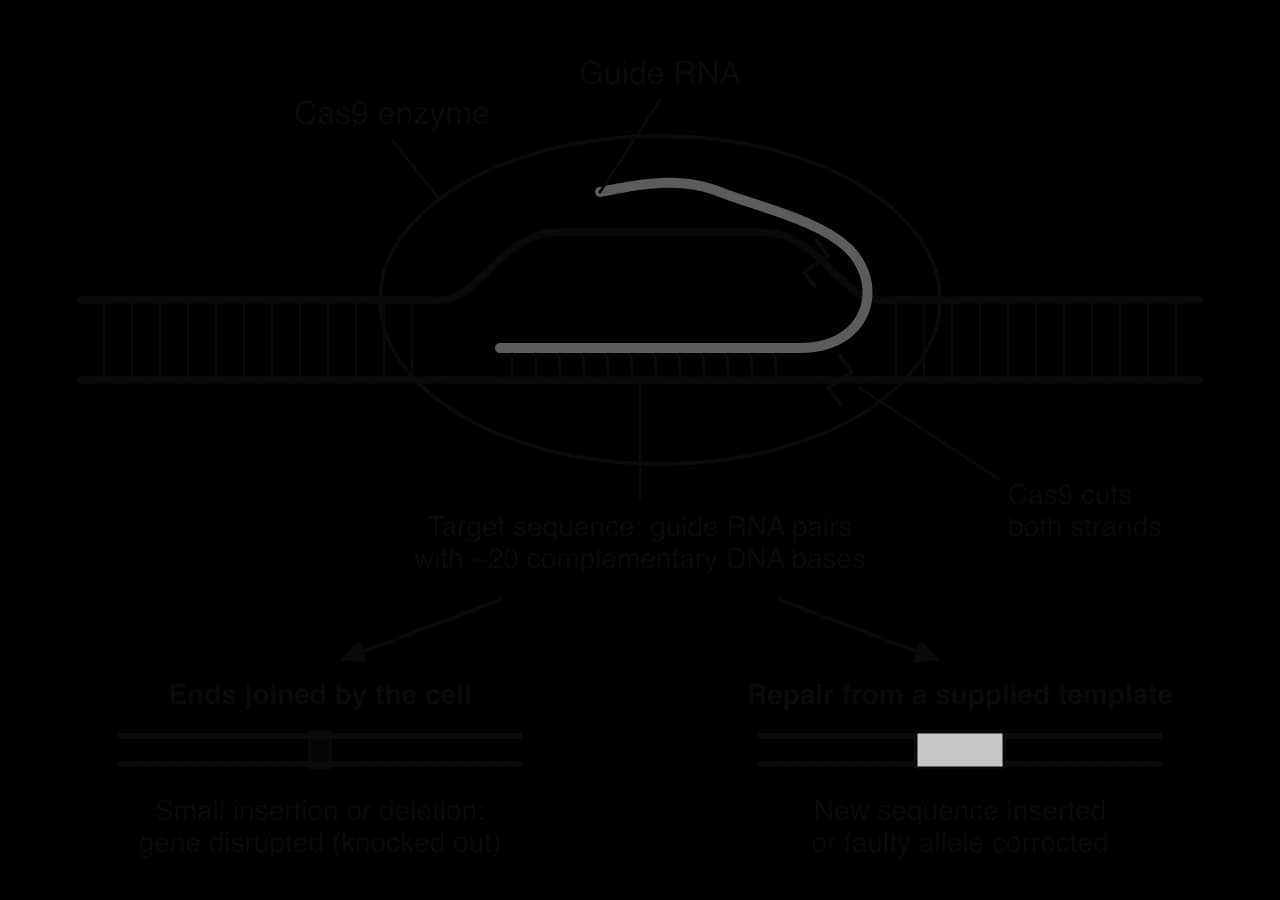
The system has two parts: the enzyme Cas9, a nuclease that cuts both strands of DNA, and a guide RNA that sets the position of the cut.
A guide RNA is designed with a sequence of about 20 bases complementary to the target sequence in the gene to be edited. One end of this RNA molecule binds to Cas9.
The Cas9–guide RNA complex is delivered into the cell.
The complex moves along the DNA. Where its guide RNA meets the complementary sequence, it forms hydrogen bonds with the bases of one DNA strand.
Cas9 cuts both strands of the DNA at the target, making a double-strand break.
The cell repairs the break. If the ends are simply joined, a few bases are often inserted or lost, which usually disrupts the gene (a knockout). If a piece of DNA with the desired sequence is supplied, the cell can copy it during repair, inserting a new sequence or correcting a faulty allele.
The first medicine based on CRISPR–Cas9, exagamglogene autotemcel (exa-cel, sold as Casgevy), was approved in the UK on 16 November 2023 for sickle cell disease and β-thalassaemia, and in the USA on 8 December 2023 for sickle cell disease and on 16 January 2024 for β-thalassaemia. The treatment leaves the sickle cell mutation in place and switches on the production of fetal haemoglobin, which is not affected by the mutation:
Blood stem cells are collected from the patient’s own bone marrow.
In the laboratory, CRISPR–Cas9 is used to cut and disrupt an enhancer of the gene BCL11A in these cells. BCL11A codes for a transcription factor that normally switches off fetal haemoglobin after birth, and the enhancer is needed for its expression in red blood cell precursors.
The patient receives chemotherapy to remove their remaining bone marrow stem cells, and the edited cells are returned by infusion.
Red blood cells made from the edited stem cells contain high levels of fetal haemoglobin, which does not polymerise, so the cells do not sickle.
In the clinical trials most treated patients had no further painful crises for at least a year. The treatment edits somatic cells only, so it is not inherited by the patient’s children. It is also very expensive and demanding for the patient, which limits who can receive it.
 YouTube
Some uses of CRISPR raise ethical issues that must be resolved before they are put into practice. The sharpest distinction is between editing somatic cells, as in Casgevy, which affects only the person treated, and editing germ cells or embryos, which would change the genes of future generations who cannot consent.
In November 2018 He Jiankui announced the birth of twin girls whose embryos he had edited to disable the CCR5 gene, which some HIV strains use to enter cells. The work was condemned worldwide: it was medically unnecessary, the safety of the edits was unknown, and the parents had not been properly informed. He was sentenced to three years in prison in December 2019. Other concerns include off-target cuts at similar sequences elsewhere in the genome, unequal access to expensive treatments, and the possible use of editing for enhancement rather than to treat disease.
Regulation of genome editing differs between countries: many prohibit heritable editing of humans, while the rules for research on embryos, for crops and for farm animals vary widely. Because research is international, there is an international effort to harmonise regulation. In 2021 the World Health Organization published recommendations on the governance of human genome editing, including a global registry of trials.
Comparing the same gene in different species shows that mutations accumulate over time, so the base sequences of most genes differ more between distantly related species than between close relatives. Some sequences, however, change very little.
A conserved sequence is one that is identical or similar across a species, or across a group of species.
A highly conserved sequence is one that is identical or similar over long periods of evolution, in species whose common ancestor lived hundreds of millions of years ago.
Histone H4, one of the proteins around which DNA is wound in nucleosomes, is a famous example. The H4 of pea plants and of cows differs at only 2 of its 102 amino acids, although plants and animals diverged more than a billion years ago. In the human genome there are also 481 "ultraconserved" sections of DNA, each over 200 base pairs long, that are completely identical in humans, mice and rats.
Two hypotheses have been proposed to explain why some sequences are conserved.
Functional requirements of the gene product. The protein or RNA has a structure that must be exactly right to work, for example because it binds tightly to DNA or to many other molecules. Almost any change would be harmful, so individuals carrying a mutation in the sequence survive and reproduce less well, and natural selection removes these mutations. On this hypothesis, mutations occur in the sequence at the usual rate but are removed by selection.
Slower rates of mutation. Some regions of the genome may mutate less often than others, for example because they are better protected or repaired more efficiently. If fewer mutations arise, the sequence changes more slowly.
The evidence mainly supports the first hypothesis. In conserved protein-coding genes, the third bases of codons, where most substitutions are same-sense, change much more than the first and second bases. If the region simply had a low mutation rate, all positions would change slowly. Instead, mutations are occurring at a normal rate and being removed by selection wherever they change the amino acid sequence. Both processes may contribute in some regions, and testing them is an active area of research.
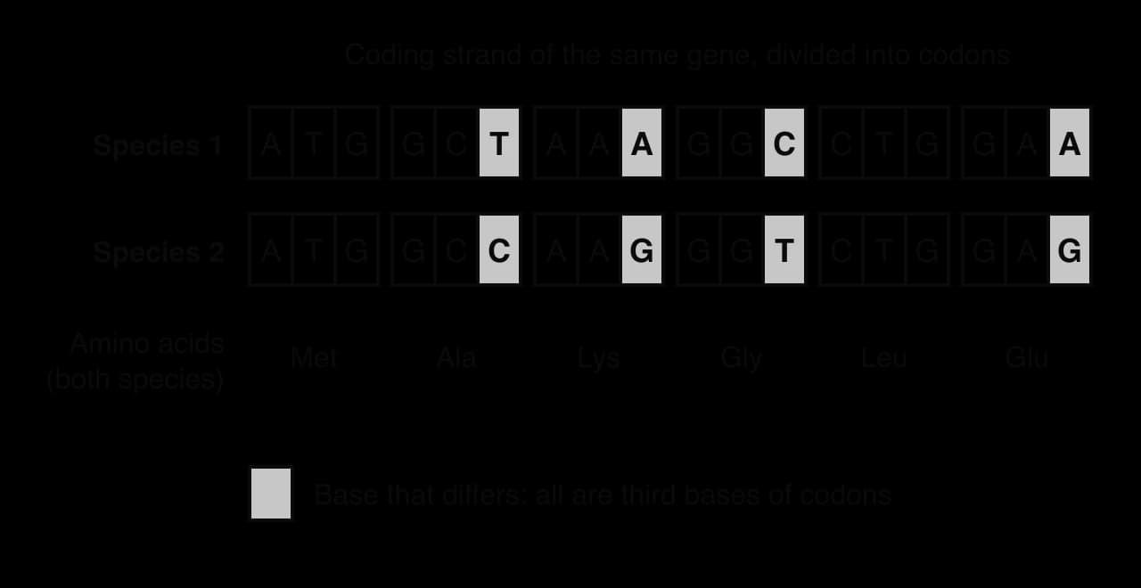
The comparison of the two coding sequences shows the pattern expected if selection keeps a sequence the same. Mutations have accumulated at third bases, where they are same-sense and do not affect the protein, but no differences have survived at first or second bases, where they would change an amino acid. The slower-mutation hypothesis would instead predict few differences at every position, including third bases. Highly conserved sequences are also useful to biologists: because they change so slowly, they can be compared across very distantly related organisms to work out evolutionary relationships (see A3.2 Classification and cladistics).
Mutations are assessed in Paper 1A through questions on the types of mutation, their effects and their causes. Paper 1B and Paper 2 Section A often give a short DNA or mRNA sequence before and after a mutation, with a codon table, and ask you to deduce the effect. Paper 2 Section B may ask you to explain the consequences of mutations in germ cells and somatic cells, or at HL to discuss the use of gene editing. Use the precise terms: substitution, insertion, deletion, frameshift, same-sense, missense, nonsense, germ cell, somatic cell.
I can distinguish between base substitutions, insertions and deletions.
I can explain why a base substitution may be same-sense, missense or nonsense, using degeneracy and SNPs.
I can explain how frameshift mutations and large insertions or deletions stop polypeptides from functioning.
I can name chemical mutagens and types of mutagenic radiation, and outline other causes of mutation.
I can explain what randomness in mutation means and why mutations are not directed by need.
I can compare the consequences of mutations in germ cells and somatic cells, including cancer.
I can explain why mutation is the source of genetic variation, and discuss commercial genetic tests.
I can outline gene knockout and name model organisms with knockout libraries. (HL)
I can explain how CRISPR–Cas9 edits a gene, describe Casgevy and discuss the ethics of gene editing. (HL)
I can evaluate two hypotheses for conserved and highly conserved sequences. (HL)
[4 max]
At least one mark must come from PCR and one from gel electrophoresis for full marks.
Explain the current and potential future uses of whole genome sequencing, and why these uses are becoming more widespread.
7 marks
a. whole genome sequencing determines the complete base sequence of an organism's genome;
b. sequencing has become much faster / automated;
c. cost has fallen greatly, e.g. from millions of dollars to hundreds per human genome;
d. more species / individuals can therefore be sequenced;
e. current: comparing genomes of species to deduce evolutionary relationships;
f. more similar base sequences indicate more recent common ancestry;
g. current: comparing genomes of individuals to find SNPs / alleles linked to diseases;
h. future: personalized medicine, choosing drugs / doses that suit an individual's genome;
i. future: predicting an individual's risk of disease so that it can be prevented / screened for;
j. future: sequencing tumour genomes to choose cancer treatments;
k. ethical concerns, e.g. privacy of genetic data / use by insurers, may limit uses;
[7 max]
One additional mark is available for the construction of the answer.
Changes over time leave a record in the base sequences of DNA and RNA.
Outline how gene mutations arise and how they change base sequences.
4 marks
Explain how the molecular clock is used to estimate when clades diverged, and why it can only give estimates.
7 marks
Describe the evidence for the classification of all organisms into three domains.
4 marks
Accuracy in copying genetic information is essential for living organisms.
Describe the roles of enzymes in the synthesis of the lagging strand in prokaryotes.
4 marks
Explain how DNA replication produces copies with very few errors.
7 marks
Outline the possible consequences of a base substitution mutation in a gene.
4 marks